INTRODUCTION
Facial cleft 0, described by Tessier, in 19761, can present with several alterations, including bifid nose, characterized by a wide
and low nasal dorsum and bifid tip, which occur due to anatomical changes such as
nasal bones and lateralized maxillary processes, horizontal upper lateral cartilages
(ULC) and flat and hypoplastic wing cartilages, with malformed and asymmetric nostrils2.
Surgical correction of the bifid nose is usually performed with chisel osteotomies
and structured rhinoplasty, using cartilaginous septal grafts to correct cartilaginous
anomalies3,4.
Recent studies have shown that piezoelectric devices provide greater control in nasal
osteotomies5,6. Some studies also emphasize the advantages of preservative rhinoplasty, such as
avoiding an open roof, middle third collapse, and internal valve insufficiency 7,8.
This article reports the treatment of bifid nose in a patient with Tessier No. 0 facial
cleft by preservative and piezo-assisted rhinoplasty.
CASE REPORT
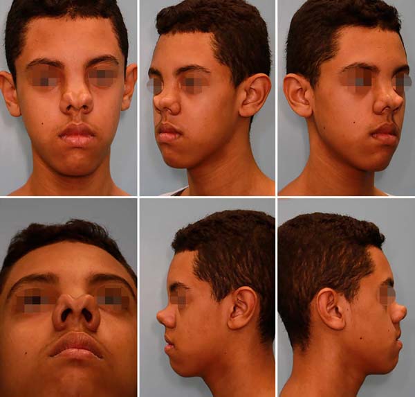
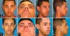
A thirteen-year-old boy with a bifid nose was referred to the plastic surgery department
of our institution complaining about the dorsum and the nasal tip. In addition to
the aesthetic complaint, the patient had psychological disorders due to social isolation
and school bullying (Figure 1).
Figure 1 - A-F. Male patient, 13 years old, with bifid nose: frontal, base, lateral and 45º.
Figure 1 - A-F. Male patient, 13 years old, with bifid nose: frontal, base, lateral and 45º.
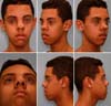
After computed tomography, he showed a bifid nasal tip, alar and ULC lateralized,
and distant nasal bones, defining the diagnosis of facial cleft No. 0, with no frontal
dysplasia findings (Figure 2).
Figure 2 - A. 3D soft tissue reconstruction in computed tomography; B-E. 3D bone reconstruction - frontal, left 45º, basal and right 45º.
Figure 2 - A. 3D soft tissue reconstruction in computed tomography; B-E. 3D bone reconstruction - frontal, left 45º, basal and right 45º.
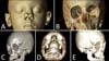
He underwent surgical correction with access through a diamond-shaped incision on
the nasal dorsum, removing the non-elastic skin from the same area and extending the
incision to the columella. An excess of abnormal soft tissues was identified in the
middle third of the nose, occupying an area above the septum (bifid) and between the
ULC, making them horizontal and distant. Although the alar cartilages were lateralized,
their domes were normoplastic and well defined (Figure 3).
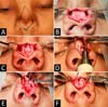
Figure 3 - A. Nasal dorsum skin to be resected; B. Medium wide third; C. Demarcation of excess soft tissue; D. Piezo-assisted resection of anomalous bone in the midline; E. Bone and soft tissue being resected; F. Horizontal and distant alar cartilages.
Figure 3 - A. Nasal dorsum skin to be resected; B. Medium wide third; C. Demarcation of excess soft tissue; D. Piezo-assisted resection of anomalous bone in the midline; E. Bone and soft tissue being resected; F. Horizontal and distant alar cartilages.
Despite being bifid, the nasal bones had a central spike that continued with the anomalous
soft parts of the middle third. Both structures were resected: soft parts with a blade
and bone part with piezoelectric, providing adequate exposure of the nasal bones and
upper lateral cartilages.
The nasal bones were medialized through lateral and medial osteotomies, performed
with a piezoelectric osteotome, to minimize damage to the structures.
The septal cartilage and ULC were preserved, without opening the cartilaginous roof,
preserving the individual units of the internal nasal valve described by Saban, in
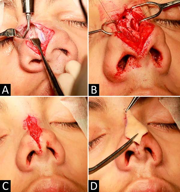
20187 and Ishida et al., In 19998. After detachment of zone K (separating the bone pyramid from cartilaginous), horizontal
“U” sutures were performed, approaching the ULC that was horizontal and apart, narrowing
the nose and raising the height of the nasal dorsum (Figure 4).
Figure 4 - A. Lateral osteotomy using a piezoelectric instrument; B. “U” points medializing the upper lateral cartilages; C. Soft tissue closure; D. Excess dry skin.
Figure 4 - A. Lateral osteotomy using a piezoelectric instrument; B. “U” points medializing the upper lateral cartilages; C. Soft tissue closure; D. Excess dry skin.
The treatment of the nasal tip was performed with interdomal points and between the
medial crosses, and the primary closure of the surgical wound was performed in two
planes, bringing together the soft parts preserved during the opening.
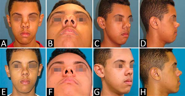
Standard postoperative care was performed, and the result after one year was satisfactory
for the patient (Figure 5).
Figure 5 - Front, base, side and 45º views: A-D. Preoperative; EH. Postoperative.
Figure 5 - Front, base, side and 45º views: A-D. Preoperative; EH. Postoperative.
DISCUSSION
The bifid nose is part of the spectrum of midline facial malformations (Tessier No.
0-14) and may occur in isolation or associated with deformities of the lips, palate,
and forehead9.
In this case, we present a 13-year-old patient with Tessier No. 0 facial cleft who
had an isolated bifid nose, whose treatment initially proposed was orthognathic surgery
and later rhinoplasty, but this was not accepted by the patient and family, who only
wanted rhinoplasty.
It was tactically opted for external access through a diamond-shaped incision in the
nasal dorsum, allowing broad access, dissection and visualization of redundant structures,
resection of bone, and anomalous soft parts in the midline, as well as the use of
piezoelectric instrumentation for osteotomies controlled.
The literature varies concerning the appropriate age and access to treat isolated
bifid noses. According to Saied and El-Sherbiny, in 20114, access in these cases varies according to the degree of the deformity; in cases
with a small separation of the domus, transcolumellar marginal access may be sufficient
for adequate correction10. On the other hand, cases with greater separation of the tip and cartilaginous back
require better visualization and resection of excess skin and soft tissues, with the
incision on the nasal dorsum being the access of choice2,3,4,9.
We opted for access through the back as the patient had excess skin and, in our view,
would not contract sufficiently after surgery if it were not resected. Despite the
unpredictability of healing, especially in patients with black skin, other studies
have shown similar maneuvers with good evolution2,3.
Regarding the tip, the alar cartilages were not fragile or hypoplastic, allowing treatment
with interdomal sutures. On the other hand, the ULC was released from the K zone and
medialized, without opening the cartilaginous roof, avoiding the violation of the
elements of the internal nasal valve7,8.
In 20187 and Ishida et al., in 19998, Saban point out the advantages of preservative rhinoplasty, such as preservation
of ULC and prevention of open roof and collapse of the middle third, which could occur
in bifid noses after classical rhinoplasty3,4,9.
Regarding bone treatment, Ortiz-Monasterio et al., in 198710, demonstrated that osteotomies should be routine in cases of bifid nose, regardless
of age. In this case, piezoelectric instrumentation was used for more controlled osteotomies
and resection of the bony dorsum to tune the upper nasal third. This technique allows
osteotomies with less ecchymosis and postoperative morbidity and more precise control
of the height of the osteotomy with less bone comminution5,6.
A limitation of the case is the persistence of nasal shortening. In order to lengthen
this type of nose, in our opinion, it would be necessary to use extended spacer grafts
attached to a columellar strut, in addition to dorsal grafts, but we chose not to
violate the septal cartilage to collect them at this time. This could be done in a
surgical review, but the patient is satisfied with the height of the back achieved
and does not admit further interventions.
Conservative and piezoelectric-assisted rhinoplasty is an alternative approach for
treating the bifid nose and has not yet been described in the literature. Long-term
follow-up and more cases are needed to prove its efficiency in treating bifid nose.
* All photos were authorized by the minor’s father, who signed the Free and Informed
Consent Term and the photos’ publication term.
REFERENCES
1. Tessier P. Anatomical classification of facial, crânio-facial and látero-facial clefts.
J Maxillofac Surg. 1976 Jun;4(2):69-92.
2. Kolker AR, Sailon AM, Meara JG, Holmes AD. Midline cleft lip and bifid nose deformity:
description, classification, and treatment. J Craniofac Surg. 2015 Nov;26(8):2304-8.
3. Ozturk S, Zor F, Isik S. Surgical correction of severe bifid nose. J Cleft Lip Palate
Craniofac Anomal. 2014;1(2):115-8.
4. Saied S, El-Sherbiny A. Algorithm for aesthetic reconstruction of the bifid nose in
tessier number 0 cleft. J Plast Reconstr Surg. 2011 Jul;35(2):187-90.
5. Gerbault O, Daniel RK, Kosins AM. The role of piezoelectric instrumentation in rhinoplasty
surgery. Aesthet Surg J. 2016 Jan;36(1):21-34.
6. Ilhan AE, Cengiz B, Eser BC. Double-blind comparison of ultrasonic and conventional
osteotomy in terms of early postoperative edema and study design and patient selection.
Aesthet Surg J. 2016 Abr;36(4):390-401.
7. Saban Y, Daniel RK, Polselli R, Trapasso M, Palhazi P. Dorsal preservation: the push
down technique reassessed. Aesthet Surg J. 2018 Feb;38(2):117-31.
8. Ishida J, Ishida LC, Ishida LH, Vieira JC, Ferreira MC. Treatment of the nasal hump
with preservation of the cartilaginous framework. Plast Reconstr Surg. 1999 Mai;103(6):1729-33;discussion:1734-5.
9. Miller PJ, Grinberg D, Wang TD. Midline cleft: treatment of bifid nose. Arch Facial
Plast Surg. 1999 Jul/Set;1(3):200-3.
10. Ortiz-Monasterio F, Fuente del Campo A, Dimopulos A. Nasal clefts. Ann Plast Surg.
1987 Mai;18(5):377-97.
1. Hospital das Clínicas, Faculty of Medicine, University of São Paulo, São Paulo,
São Paulo, SP, Brazil.
Corresponding author: Rodolfo Costa Lobato, Rua Doutor Melo Alves, 55, Cj 23, Cerqueira Cesar, São Paulo, SP, Brazil. Zip Code:
01417-010. E-mail: rodolfolobato49@yahoo.com.br
Article received: May 13, 2019.
Article accepted: June 22, 2019.
Conflicts of interest: none.