ABSTRACT
INTRODUCTION: The greater omentum was initially used in the repair of gastrointestinal defects in the 19th century; during the 20th century, it has been used extraperitoneally in the treatment of various disorders, in several surgical specialties. Despite the fact that the greater omentum was studies in detail in the 1960s, there are no reported comparative studies concerning the use of omental flaps extraperitoneally. The present study analyzed the adaptive features of the greater omentum in the extraperitoneal space, with the aim of identifying its surgical applicability.
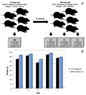
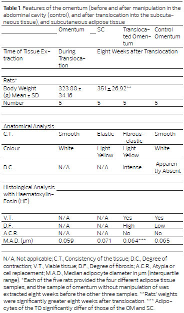
METHODS: A paired, controlled comparative study was conducted using 20 tissue samples from 5 obese female Sprague-Dawley rats (Rattus norvegicus). The following specimens from each animal were analyzed and compared, macroscopically and microscopically, using the hematoxylin-eosin (HE) technique: (1) omentum without manipulation; (2) intraperitoneally manipulated omentum; (3) extraperitoneally manipulated omentum; and (4) subcutaneous adipose tissue.
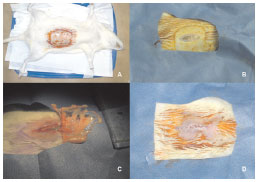
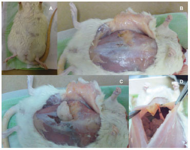
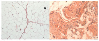
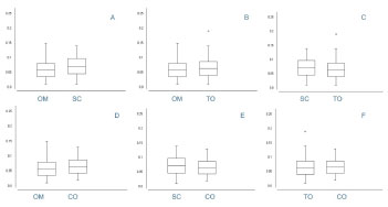
RESULTS: Macroscopically, the extraperitoneal omentum exhibited a more intense yellowish color and a higher degree of contraction than the control (intraperitoneal) omentum. The extraperitoneal omentum was similar in color to the adjacent subcutaneous adipose tissue. HE staining revealed a high degree of fibrosis and an average adipocyte size, similar to that in the control omentum, but lower than that in subcutaneous adipose tissue (p< 0.001).
CONCLUSION: The results of this study indicate that the extraperitoneal omentum was not able to promote tissue regeneration, as metaplasia of the translocated flap was not observed in the histological analysis. However, this structure may be used to correct small deformities, in the treatment of ischemic areas, as a carrier structure for surgical reconstruction and as a germination platform for the development of new organs.
Keywords: Omentum; Breast; Reconstruction; Epiploon; Metaplasia; Fat.


