ISSN Online: 2177-1235 | ISSN Print: 1983-5175

Previous Article Next Article

Original Article - Year2011 - Volume26 - Issue 3

ABSTRACT

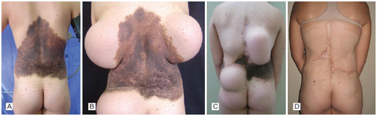
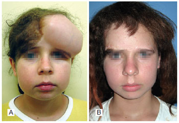
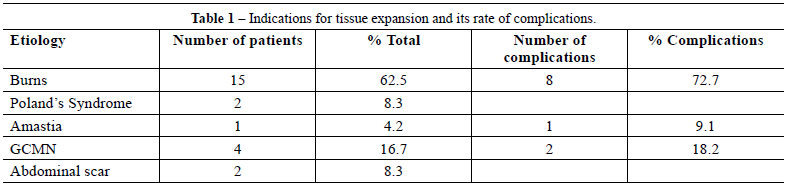
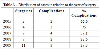
Background: The shortage of tissue for large defect reconstruction is a challenge for the plastic surgeon. Tissue expansion emerged in this context, and in the last 30 years has become one of the most widely used modalities in reconstructive surgery. Tissue expansion is a very versatile technique that can be performed in patients of all ages for the correction of different pathologies. The most common indications are burn sequelae and giant congenital nevus. The present study describes the indications and use of tissue expanders at the Hospital de Clínicas of Universidade Federal do Paraná. Methods: Patients who underwent tissue expansion for reconstructive surgery between January 2005 and December 2009 were retrospectively reviewed. Results: A total of 24 patients (70.8% female and 29.2% male) were analyzed. Ages ranged from 3 to 46 years old (average, 17.1 years). The most common indication for tissue expansion was the treatment of burn sequelae (62.5%), mainly in the head and neck. Alopecia was the second most prevalent indication (29.2%), followed by scar retraction in the neck (20.8%). Other indications were giant congenital melanocytic nevus (16.7%), Poland's syndrome (8.3%), abdominal scar (8.3%), and amastia (4.2%). Complications developed in 11 patients, and the highest incidence of complications, reported in 8 (72.7%) patients, was among those with burn sequelae as the primary pathology. The complications were infection, rupture, extrusion, wound dehiscence, and displacement of the expander. Conclusions: Tissue expansion is indicated for the treatment of several diseases among which burn sequelae is one of the most common indications.

Keywords: Tissue expansion devices. Tissue expansion. Reconstructive surgical procedures.

RESUMO

Introdução: A escassez de tecidos para reconstrução de grandes defeitos é um desafio ao cirurgião plástico. Nesse contexto, surgiu a expansão tecidual, que, nos últimos 30 anos, se tornou uma das modalidades mais utilizadas na cirurgia reparadora. A expansão é uma técnica muito versátil, que pode ser realizada em todas as idades e para correção de diferentes afecções. As principais indicações são sequelas de queimadura e nevo congênito gigante. Este estudo teve como objetivo demonstrar as indicações na utilização dos expansores tissulares e sua evolução em pacientes do Hospital de Clínicas da Universidade Federal do Paraná. Métodos: Estudo retrospectivo, incluindo pacientes submetidos a expansão tecidual para cirurgia reconstrutora, no período de janeiro 2005 a dezembro 2009. Resultados: Foram analisados 24 pacientes, sendo 70,8% do sexo feminino e 29,2% do sexo masculino. A idade variou entre 3 anos e 46 anos (média de 17,1 anos). A principal indicação de expansão tecidual foi o tratamento de sequelas de queimaduras (62,5%), principalmente na região da cabeça e do pescoço. Alopecia foi a indicação mais prevalente (29,2%), seguida por retração cicatricial em região cervical (20,8%). Outras indicações foram nevo melanocítico congênito gigante (16,7%), síndrome de Poland (8,3%), cicatriz abdominal (8,3%) e amastia (4,2%). Entre os pacientes avaliados, 11 evoluíram com alguma complicação, 8 (72,7%) dos quais tinham como doença primária sequela de queimaduras, demonstrando maior incidência de complicações em relação às outras indicações. As complicações encontradas foram: infecção, ruptura, extrusão, deiscência de sutura e deslocamento do expansor. Conclusões: A expansão tissular é indicada no tratamento de múltiplas doenças e uma das principais indicações continua sendo o tratamento de sequelas de queimaduras.

Palavras-chave: Dispositivos para expansão de tecidos. Expansão de tecido. Procedimentos cirúrgicos reconstrutivos.


INTRODUCTION

Tissue expansion is a technique used in reconstructive surgery that aims to increase the dimensions of a skin flap by stretching the skin with constant pressure applied through an expander. Through this technique, optimal esthetic results are achieved by replacing scar tissue with skin of similar texture and color, as well as ensuring a good functional outcome1,2. Tissue expansion has become one of the most commonly used treatment modalities in the last 30 years1. It is indicated for the treatment of various defects, of which the most common ones are congenital giant nevus and burn sequelae, especially in the head and neck.

The congenital melanocytic nevus is a pigmented injury arising from nests of melanocytes proliferating in the skin that is present at birth and affects approximately 1% of newborns3. These defects can be classified according to diameter, and are divided into small (<1.5 cm), medium (<1.5 cm to 20 cm), and giant (>20 cm) nevi.

The giant melanocytic nevus is a rare condition, with an estimated incidence of 1 in every 20,000 births4. Its etiology is unknown. In addition to the esthetic implications of this defect, it can be associated with malignancy (risk of 5% to 12%)4, and prophylactic excision is often recommended. Due to its large size, complete excision is generally not possible with a single surgical procedure, and tissue expanders may be required to obtain larger flaps (Figures 1 and 2).


Figure 1 - Patient with giant hairy nevus on the dorsum who underwent two rounds of expansion. A, preoperative period. B, first expansion, employing three expanders. C, second expansion, employing two expanders. D, final result.


Figure 2 - Patient with giant hairy nevus on the forehead and scalp. A, tissue expansion for resection of the frontal injury. B, final result.


Burns are a very prevalent problem throughout the world, and the fourth leading cause of death from trauma in the United States. In acute burns, after the immediate risk to life is reduced, there may be physical and psychological problems resulting from unsightly and dysfunctional scars. Various techniques are available for the correction of burn-related scars, including the use of expanders. Most often, a combination of techniques is required, and these are usually applied in more than one surgical procedure.

Although the versatility of tissue expanders is well known, high complication rates have been reported1. In the first reports on the use of tissue expansion, the complication rates reached 40%. However, in recent studies, the reported rate of complications has ranged from 13% to 20%. The main complications are hematoma, infection, exposure, extrusion, rupture of the expander, ischemia, and necrosis of the skin5. The highest rate of complications occurs in younger patients, especially those under 10 years old, and in expanders placed in the lower limbs, head, and neck.

This study aims to demonstrate the experience of our university service with the use of tissue expanders, describing the most common indications and the outcomes of patients treated at Hospital de Clinicas of Universidade Federal do Parana (UFPR).


METHODS

This is a retrospective, descriptive, and analytical study of patients who underwent tissue expansion for reconstructive surgery at Hospital de Clinicas of UFPR.

The medical records of patients who underwent tissue expansion between January 2005 and December 2009 were analyzed. All patients who underwent surgery for tissue expander implant within that period were included.

The data obtained included age, gender, disease, shape, and number of expanders placed, in addition to volume and time of expansion, number of surgeries performed, evolution, and complications.


RESULTS

The records of 24 patients (70.8% female and 29.2% male) were analyzed. Ages ranged from 3 to 46 years old (average, 17.1). The main indication for tissue expansion was burn sequelae treatment (62.5%), and the second most prevalent indication was alopecia (29.2%), followed by scar retraction in the neck (20.8%). Other indications were giant congenital melanocytic nevus (16.7%), Poland's syndrome (8.3%), abdominal scar (8.3%), and amastia (4.2%) (Table 1). Among the 24 patients examined, 32 procedures were performed and a total of 43 expanders were used.


Complications were reported in 11 patients and 14 expanders. The most common complications were infection (4 cases), rupture (3 cases), extrusion (3 cases), wound dehiscence (3 cases), and expander displacement (1 case). The complication resulted in poor outcome in five expanders, mostly due to infection. Two-thirds of the expanders associated with complications were used in the head and neck regions. In 8 (72.7%) patients who experienced complications, burn sequelae was the primary disease (four cases of alopecia: two on the neck, one on the back, and one on the trunk), indicating a higher incidence of complications in burn-related pathologies compared to other deformities.

The total number of surgeries required to complete the treatment, including surgeries for flap rotation and management of complications, ranged from one to six operations. Regarding age, 45.4% of complications occurred in patients 10 years old or younger (29% of patients). On the other hand, 18.2% of the complications occurred in patients who were over 20 years of age (29% of the study population) (Table 2).


Table 3 illustrates the increased use of expanders in recent years due to the purchase of expanders by the Unified Health System (SUS), which eliminated the need for patients to obtain it. No expander was reused, although there are reports of expander reuse in the literature6.


DISCUSSION

Neumann7 was the first to use tissue expansion for the treatment of burn-related deformities in 1957, and it is currently one of the most commonly used therapeutic techniques. Expanders have revolutionized reconstructive surgery, enabling the correction of defects through the use of skin flaps of the same color and texture, thus allowing the achievement of better functional and esthetic results. One of the advantages of the use of expanders is the ability to cover the defect with minimal donor site morbidity and good vascularization of the skin flap (superior to pedicle flaps)5. A combination of treatment modalities is often required to achieve better results in various types of deformities, especially in head and neck injuries. Injuries in this region are a big challenge because they involve multiple specialized anatomical structures, and the use of expanders is an effective alternative for reconstruction8. The combination of tissue expanders with resection surgery may be indicated7.

Although the versatility of tissue expanders is well known, high complication rates have been reported in some cases. In the first reports of tissue expansion, complication rates up to 40% were reported. However, recent studies have reported complication rates between 13% and 20%. The major complications are hematoma, infection, exposure, extrusion of the expander, ischemia, and necrosis of the skin1. In the present study, 32.5% of expanders were associated with complications. However, in 12.5% of patients, there was no completion of the proposed treatment. Complication rates varied according to the anatomical location of the expander, and were higher in the lower limbs due to impaired local vascularization and the amount of tissue available9,10. Cunha et al.11 observed higher rates of complications in the cervical region, which is in agreement with the present study. Age was another important factor affecting the rate of complications, and patients 10 years old and younger were found to be at a higher risk. Complications have also been associated with the surgeon's learning curve, as well as the cooperation of family members and the patient during the expansion5.


CONCLUSION

Tissue expansion is indicated for the treatment of several diseases, and one of the most common indications remains the treatment of burn sequelae. This method can be used to treat patients of all age groups, although the treatment of children between 0 and 10 years of age is associated with a higher rate of complications. The anatomical region is also an important factor, as the head and neck region is associated with a higher rate of complications.


REFERENCES

1. Rivera R, LoGiudice J, Gosain AK. Tissue expansion in pediatric patients. Clin Plast Surg. 2005;32(1):35-44,viii.

2. Dotan L, Icekson M, Yanko-Arzi R, Ofek A, Neuman R, Margulis A. Pediatric tissue expansion: our experience with 103 expanded flap reconstructive procedures in 41 children. Isr Med Assoc J. 2009;11(8):474-9.

3. Carneiro Jr LVF, Aguiar LFS, Pitanguy I. Tratamento cirúrgico do nevo melanocítico gigante. (Surgical treatment of the giant melanocytic nevus) Rev Bras Cir Plast. 2011;26(2):198-204.

4. Paschoal FM. Nevo melanocítico congênito. (Congenital melanocytic nevus) An Bras Dermatol. 2002;77(6):649-54.

5. Bozkurt A, Groger A, O'Dey D, Vogeler F, Piatkowski A, Fuchs PCh, et al. Retrospective analysis of tissue expansion in reconstructive burn surgery: evaluation of complication rates. Burns. 2008;34(8):1113-8.

6. Anger J. The reuse of expanders in developing countries. Plast Reconstr Surg. 1991;88(6):1114-5.

7. Neumann CG. The expansion of an area of skin by progressive distention of a subcutaneous balloon; use of the method for securing skin for subtotal reconstruction of the ear. Plast Reconstr Surg. 1957;19(2):124-30.

8. Nemetz AP, Costa TCD, Nemetz MA, Aguiar LFS, Trauczinski PA, Nery RA. Utilização de expansores teciduais na cirurgia reconstrutora de cabeça e pescoço. (Use of tissue expanders in head and neck reconstructive surgery) Rev Bras Cir Plast. 2007;22(4):219-27.

9. LoGiudice J, Gosain AK. Pediatric tissue expansion: indications and complications. J Craniofac Surg. 2003;14(6):866-72.

10. Bauer BS, Vicari FA, Richard ME. The role of tissue expansion in pediatric plastic surgery. Clin Plast Surg. 1990;17(1):101-12.

11. Cunha MS, Nakamoto HA, Herson MR, Faes JC, Gemperli R, Ferreira MC. Tissue expander complications in plastic surgery: a 10-year experience. Rev Hosp Clin Fac Med Sao Paulo. 2002;57(3):93-7.


1. Full professor, head of the Plastic and Reconstructive Surgery Service at Hospital de Clínicas of Universidade Federal do Paraná (UFPR), Curitiba, PR, Brazil.
2. PhD, associate professor III at the Plastic and Reconstructive Surgery Service at Hospital de Clínicas of UFPR, Curitiba, PR, Brazil.
3. Resident physician at the Plastic and Reconstructive Surgery Service at Hospital de Clínicas of UFPR, Curitiba, PR, Brazil.
4. Physician, general surgeon at Hospital de Clínicas of UFPR, Curitiba, PR, Brazil.

Correspondence to:
Renato da Silva Freitas
Av. General Carneiro, 181 - 9º andar - Alto da XV
Curitiba, PR, Brazil - CEP 80060-900
E-mail: dr.renato.freitas@gmail.com

Submitted to SGP (Sistema de Gestão de Publicações/Manager Publications System) of RBCP (Revista Brasileira de Cirurgia Plástica/Brazilian Journal of Plastic Surgery).
Received: July 21, 2011
Accepted: September 6, 2011

Study conducted at Hospital de Clínicas of Universidade Federal do Paraná (UFPR), Curitiba, PR, Brazil.


Previous Article Back to Top Next Article

Indexers

Licença Creative Commons All scientific articles published at www.rbcp.org.br are licensed under a Creative Commons license