


Case Report - Year 2020 - Volume 35 -
Lyell syndrome in 72% of the body surface
Síndrome de Lyell em 72% de superfície corporal
José Augusto Pupio Reis1,* ; Rita De Cássia Neves Noronha2; Carlos Cunha Oliveira2; Rogério De Oliveira Ribeiro2
ABSTRACT
Introduction: Lyell's syndrome is characterized by phlyctenas on more than 30% of the body surface and by involvement of the mucous membranes. It is related to the allergic reaction resulting from medications and has a high mortality.
Case report: A 28-year-old woman presented phlyctenas on 72% of the body surface after using various drugs. Despite a great extent, the patient progressed satisfactorily and was discharged without sequelae.
Conclusion: Early diagnosis, stratification, and management are essential to reduce mortality from the disease.
Keywords: Plastic surgery; Stevens-Johnson syndrome; Rash; Erythema multiforme; Skin diseases.
RESUMO
Introdução: A síndrome de Lyell é caracterizada por flictenas em mais de 30% de superfície corporal e acomete mucosas. Está relacionada à reação alérgica decorrente de medicamentos e apresenta alta mortalidade.
Relato de caso: Mulher de 28 anos apresentou flictenas em 72% de superfície corporal após uso de diversos fármacos. Apesar da grande extensão, a paciente evoluiu de maneira satisfatória e recebeu alta sem sequelas.
Conclusão: É fundamental o diagnóstico precoce, a estratificação e a conduta para reduzir a mortalidade da doença.
Palavras-chave: Cirurgia plástica; Síndrome de Stevens-Johnson; Exantema; Eritema multiforme; Dermatopatias
INTRODUCTION
Toxic epidermal necrolysis (TEN), also known as Lyell’s syndrome (LS), is an allergic reaction mediated by CD8 lymphocytes that evolves with epidermal necrosis due to keratinocyte apoptosis. The etiology is unknown, but the use of drugs triggers it. Due to the high mortality, knowledge of the condition, early diagnosis, stratification, and an adequate approach is essential for optimized patient management.
CASE REPORT
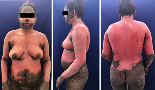
Patient MD, woman, 28 years old, mixed race, from the municipality of Laranjal do Jari in the state of Amapá. On January 26, 2019, the patient presented phlyctenas in the oral mucosa after using the medication allopurinol 200 mg per day for 17 consecutive days, 50 mg of diclofenac sodium, 30 mg of caffeine, 125 mg of carisoprodol, 300 mg of paracetamol every 12 hours. In the following 48 hours, the patient developed phlyctenas on 72% of the body surface. Involvement was also observed in the mucosa of the oral, vaginal, anal, and genitourinary tracts.
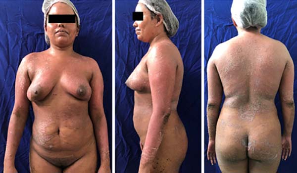
In the first 48 hours, her body temperature was 38ºC, and she was admitted to the Hospital Estadual de Laranjal do Jari on January 28, 2019. On February 1, 2019, she was referred to the Hospital de Emergência Osvaldo Cruz, where an evaluation was requested from the plastic surgery team (Figure 1). Upon entering the plastic surgery service, she was treated clinically, showing a satisfactory evolution, being discharged without the need for surgical treatment on February 10, 2019 (Figure 2). The SCORTEN scale was not calculated as it was not applied at the time of disease progression.


DISCUSSION
In 1922, Indian surgeon Albert Stevens and American pediatrician Frank Johnson described two cases of fever, conjunctivitis, and inflammation of the mucous membranes in children, with one case with total loss of vision. Despite the lack of knowledge of the cause at the time, it was the first description of what was later called Stevens-Johnson Syndrome (SJS). Back in 1956, Scottish dermatologist Alan Lyell described a condition in which disseminated epidermal necrosis occurred, which evolved with the formation of phlyctenas associated with a toxic febrile reaction. The pathology study was of apoptosis of keratinocytes, which the dermatologist named TEN1.
Despite the low incidence: 2 cases per million per year, mortality from the disease is high: 30%. It is a hypersensitivity reaction that affects the skin and mucous membranes and does not yet have a clear etiology. The leading cause of TEN is medication, and the primary triggers are allopurinol, aromatic anticonvulsants, sulfonamides, and non-steroidal anti-inflammatory drugs. It is also seen more frequently in patients with HIV, systemic lupus erythematosus, and patients undergoing bone marrow transplantation. On average, the exposure time to the trigger factor is two weeks, but there are reports of up to 48 hours2.
Didactically TEN can be divided according to the affected body surface. In this way, it is called SJS in cases where the participation is up to 10%; overlap syndrome when involvement varies between 10 and 30%; and TEN, also known as Lyell Syndrome (LS), when the participation exceeds 30%3. Due to the high mortality, the SCORTEN scale - Disease severity score was developed for the toxic epidermal necrolysis scale. On the scale, seven variables weigh one point each and are: age over 40 years; heart rate higher than 120 beats per minute; associated malignancy; epidermal detachment of more than 10% of the body surface on the first day; urea greater than 28 mg/dl; glucose higher than 252 mg/dl and bicarbonate less than 20 mEq /l. According to the score, mortality increases dramatically (Table 1). The scale should be applied on the first and third days of hospitalization to increase the predictive value4.
| Risk factors | |
| Age> 40 years | |
| Neoplasm | |
| Heart rate> 120bpm | |
| Epidermis detachment> 10% | |
| Urea> 28md / dl | |
| Glucose> 252mg / dl | |
| Serum bicarbonate <20mg / dl | |
| Mortality | |
| Scorten 0 or 1 | 3,2% |
| Scorten 2 | 12,1% |
| Scorten 3 | 35,3% |
| Scorten 4 | 58,3% |
| Scorten > 4 | 90% |

The initial manifestation can be confused with a flu-like syndrome for three days before the mucocutaneous manifestations. On physical examination, the Nikolsky and Asboe-Hansen sign can be seen: shear of the skin with light friction and lateral shedding of the skin after light pressure on the blister, respectively. The involvement of the urethral, genital, oral, and ocular mucosa is frequent and can precede skin lesions3.
Treatment consists of removing the causative agent and offering the necessary support to the patient, preferably in a burn treatment unit. There is no consensus on the best way to manage wounds, as some ointments can trigger a similar condition so that injuries can be treated with soap and water. Surgical debridement is also not a consensus. Systemic antibiotics should be reserved only for cases of infection, as they can also be causative agents of TEN. The use of intravenous corticosteroids has not shown benefits and may even delay the healing process and favor secondary infection. Plasmapheresis, although indicated by some authors, did not show a significant impact on mortality and length of stay. The use of cyclosporine has been indicated successfully, although the mechanism of action is not yet well defined. The use of immunoglobulins has shown good results and depends on the dose and early administration5.
REFERENCES
1. Lyell A. Toxic epidermal necrolysis: an eruption resembling scalding of the skin. Br J Dermatol. 1956 Nov;68(11):355-61. DOI: http://dx.doi.org/10.1111/j.1365-2133.1956.tb12766.x
2. Santos Neto FC, Piccinini PS, Andary JC, Sartori LDP, Cancian LT, Uebel CO, et al. Abordagem cutânea na necrólise epidérmica tóxica. Rev Bras Cir Plást. 2017;32(1):128-34.
3. http://www.scielo.br/scielo.php?script=sci_arttext&pid=S0365- 05962004000400009&lng=en http://dx.doi.org/10.1590/S0365-05962004000400009
4. Trent JT, Kirsner RS, Romanelli P, Kerdel FA. Use of SCORTEN to accurately predict mortality in patients with toxic epidermal necrolysis in the United States. Arch Dermatol. 2004 Jul;140(7):890-2. DOI: http://dx.doi.org/10.1001/archderm.140.7.890
5. https://www.scielo.br/scielo.php?pid=S0104-42302016000500468&script=sci_abstract http://dx.doi.org/10.1590/1806-9282.62.05.468
1. Universidade Federal do Amapá, Macapá, AP, Brazil.
2. Hospital de Emergência Osvaldo Cruz, Macapá, AP, Brazil.
Analysis and/or data interpretation, Conception and design study, Conceptualization, Data Curation, Final manuscript approval, Formal Analysis, Funding Acquisition, Investigation, Methodology, Project Administration, Realization of operations and/or trials, Resources, Software, Supervision, Validation, Visualization, Writing - Original Draft Preparation, Writing - Review & Editing
Corresponding author: José Augusto Pupio Reis Júnior Rodovia JK, KM 6, n 4440, Bairro Universidade, Macapá, AP, Brazil. Zip Code: 68903-419 E-mail: alieksei@gmail.com
Article received: February 22, 2019.
Article accepted: October 20, 2019.
Conflicts of interest: none.
 Read in Portuguese
 Read in English
 PDF PT
 Print
Print
 Send this article by email
 How to Cite
 Mendeley
 Pocket