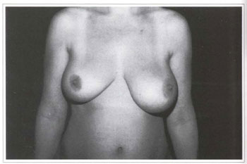
Fig. la - Preoperative period; frontal view.
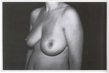
Fig. 1b - Preoperative period; lef lateral view.
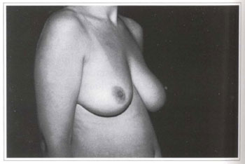
Fig. 1c - Preoperative period; right lateral view.
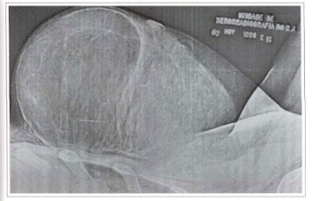
Fig. 2 - Detail of the x-ray showing the encapsulated tumor with defined contours and accurate limits, suggesting lipoma.
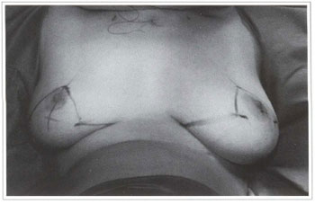
Fig. 3 - Preoperative marking according to Pitanguy's mammaplasty technique.
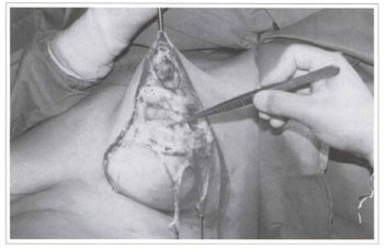
Fig. 4a - Intraoperative period; partial view of the tumor.
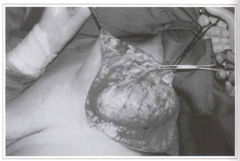
Fig. 4b - Resection of the tumor comprised of yellowish, shiny fatty tissue.
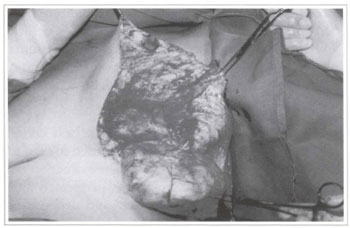
Fig. 4c - Resection of the tumor comprised of yellowish, shiny fatty tissue.
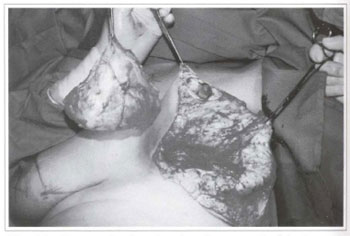
Fig. 4d - Panoramic view showing the removal of the large tumor.
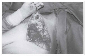
Fig. 4e - Space left after tumor removal.
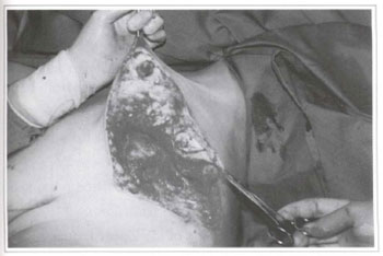
Fig. 5a - Reconstruction of the left breast with a lateral surgical flap of the upper pedicle.
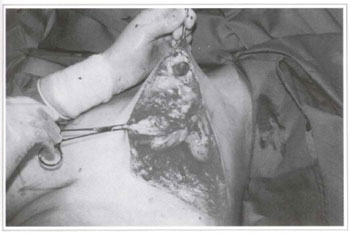
Fig. 5b - Reconstruction of the left breast with a lateral surgical flap of the upper pedicle.
Fig. 6 - Immediate postoperative period. Final suture. The right, breast was submitted to balance mammaplasty.
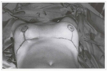
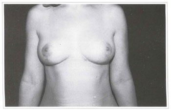
Fig. 7a - 1 year postoperative period; frontal view. Excellent healing of the surgical wound with satisfactory results.
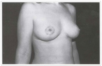
Fig. 7b - Postoperative period; right lateral view.
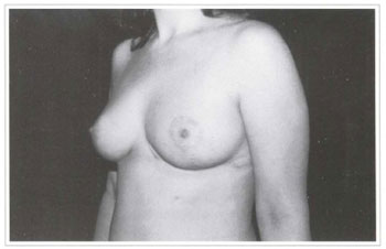
Fig. 7c - Postoperative period; left lateral view.


