


Case Report - Year 2024 - Volume 39 -
Anterolateral Thigh Flap for the Treatment of Sequelae of Ludwig's Angina: Case Report
Retalho anterolateral da coxa para o tratamento de sequelas da angina de Ludwig: Relato de caso
Olga Lanusa Leite Veloso1 ; Diego Arley Gomes da Silva2; Felipe Mazza de Lima3; Kariny Oliveira Silva4; Breno Bezerra Gomes de Pinho Pessoa1; José Maria Sampaio de Menezes4
ABSTRACT
Ludwig's angina is a polymicrobial infectious process, most often of odontogenic origin, which requires early diagnosis and treatment and is aggravated by systemic diseases. Surgical procedures may be necessary, such as debridement and the creation of flaps for reconstruction. The anterolateral thigh flap is a free flap with numerous advantages, such as a large donor area and minimal functional sequelae. The objective of the present article is to report a severe case of Ludwig's angina in which an anterolateral thigh flap was used to treat the sequelae.
Keywords: biological tissue flaps; Ludwig's angina; microsurgery; plastic surgery; surgical flaps
RESUMO
A angina de Ludwig é um processo infeccioso polimicrobiano, geralmente de origem odontogênica, que requer diagnóstico e tratamento precoces, e é agravada por doenças sistêmicas. Procedimentos cirúrgicos podem ser necessários, como desbridamento e criação de retalhos para reconstrução. O retalho anterolateral da coxa é um retalho livre com inúmeras vantagens, como grande área doadora e sequelas funcionais mínimas. O objetivo deste artigo é relatar um caso de angina de Ludwig grave em que um retalho anterolateral da coxa foi utilizado para tratar as sequelas.
Palavras-chave: angina de Ludwig; cirurgia plástica; microcirurgia; retalhos cirúrgicos; retalhos de tecido biológico
Introduction
Ludwig’s angina is a rapidly spreading polymicrobial infectious process that involves the submandibular, sublingual and submental spaces.1 In 90% of the cases, it is of odontogenic origin. The roots of the lower second and third molars extend inferiorly to the mylohyoid line, thus becoming the main route of infection dissemination to the submandibular space.2
Local or systemic diseases alter the body’s homeostasis, favoring the evolution and dissemination of the infectious process.3
The main symptoms are local pain, edema, and fever. Progression to the cervical region can cause limitations of cervical movement, sialorrhea, dysarthria, and dysphonia. Tachypnea, dyspnea, tachycardia, restlessness, and the need to maintain an upright posture suggest airway obstruction.
The initial treatment is clinical, based on broad-spectrum antibiotic therapy. Additional procedures such as drainage of collections and debridement of devitalized tissue may be necessary. If the infection is of odontogenic origin, exodontia of the affected tooth is essential for a successful treatment.4
The anterolateral thigh flap has a wide range of indications in the literature for the reconstruction of large losses of substance in the head and neck, trunk, and extremities of limbs.5 Its versatility is due to its long vascular pedicle, the possibility of removing a large island of skin, the different types of tissues used in a flap, and the low morbidity in the donor area.6
The objective of the current report is to present the application of the anterolateral thigh free flap in cervical reconstruction after major surgical debridement due to Ludwig’s angina.
Case Report
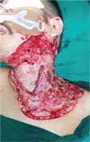
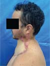
A 27-year-old man, with type-1 diabetes mellitus, admitted to Instituto Doutor José Frota, in the city of Fortaleza, State of Ceará, Northeastern Brazil, 12 days after beginning endodontic treatment in dental element 47, with Ludwig’s angina associated with necrotizing fasciitis in the bilateral submandibular region and upper thorax. The patient underwent 3 consecutive surgical procedures for abscess drainage, debridement of devitalized tissues, and extraction of tooth element 47 (►Fig. 1). Pleural involvement and deep mediastinal progression were ruled out by computed tomography (CT) scan.

Thirteen days postoperatively, the patient presented with sudden dyspnea associated with asymmetric edema of the lower limbs. A venous Doppler ultrasound of the lower limbs found deep vein thrombosis in the right lower limb. The patient referred to the intensive care unit (ICU) with a hypothesis of pulmonary thromboembolism, which was later confirmed, evolving with several conditions of infectious origin during hospital stay, such as urinary tract infection and pneumonia, among others.
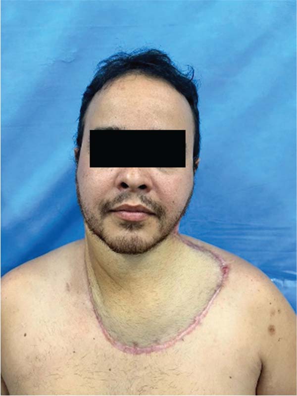
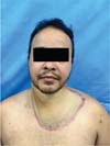
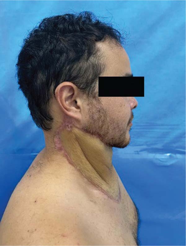
After clinical improvement, he was referred to plastic surgery, where he continued to use silver-based dressings and polyhexamethylene biquanide gauze. On day 57 of hospitalization, a free anterolateral flap was created from the left thigh to reconstruct the cervical region, through anastomosis of the right transverse cervical artery and vein, with a branch of the lateral circumflex femoral artery. The flap was fixed only at the edges of the wound, without the need for additional anchoring. The donor area on the left thigh was closed primarily and presented no complications.
Two additional surgical procedures were necessary, the first for debridement of the flap edge, and the second for grafting the remaining open areas.
During hospital stay, the patient used polymyxin B and teicoplanin for 14 days, followed by fluconazole for 10 days and meropenem for another 7 days.
The patient was discharged 104 days after admission. Flap refinement by liposuction was performed 244 days after surgery (►Figs. 2-4).


Discussion
Ludwig’s angina is a rapidly developing cellulitis, which involves the floor of the mouth and the submandibular space, and it is potentially fatal.7
Approximately 70% to 90% of cases are related to odontogenic infection, mainly in the lower second and third molars, since their roots are located below the mylohyoid line. In the eventual progression of the dental infection, the subsequent perforation of the cortical mandible bone in contact with the tongue will lead to the progression of the process to submandibular, sublingual, and submental spaces.4
Other causes include lacerations in the floor of the mouth, infected oral neoplasms, lymphadenitis, pharyngitis, foreign bodies in the floor of the mouth, compound mandibular fractures, salivary gland infections, tonsil abscesses, otitis media, and even the use of injectable drugs in the large cervical vessels.8
The microbiota that causes Ludwig’s angina is polymicrobial, of oral origin. The most frequently isolated pathogens are Streptococcus viridans and Staphylococcus aureus, but anaerobes such as Bacteroides, Peptostreptococcus, and Peptococcus are also represent frequent causes of this infection.9
The recommended empiric antimicrobial therapy is crystalline penicillin G associated or not with an aminoglycoside and metronidazole. For patients allergic to penicillin, clindamycin can be an alternative.10 Definitive therapy should be based on antibiograms. In the case described here, antibiotic therapy was advised by the Hospital Infection Control Committee.
In patients who develop the infection, there are generally associated local etiological factors, such as the presence of systemic diseases, such as diabetes mellitus, malnutrition, liver diseases, immunosuppressive diseases, and organ transplantation.11 In our clinical case, the patient was diagnosed with type-1 diabetes mellitus.
Among the various complications of Ludwig’s angina, we can highlight airway obstruction, mediastinitis, sepsis and septic shock, empyema, necrotizing fasciitis, pericardial effusion, osteomyelitis, subphrenic abscess, aspiration pneumonia, and pleural effusion.12 In the present case, the patient presented with necrotizing fasciitis, requiring major surgical debridement, causing an extensive area of substance loss in the cervical and upper chest region, with indication of reconstruction with a microsurgical flap due to the unavailability of viable local tissue.
As therapeutic options, partial skin grafts, local flaps such as the deltopectoral and trapezius muscle flaps, as well as free flaps can be used. The use of dermal matrix could also have been used.
Skin grafts evolve with contraction of the grafted region, and they are not a good option for areas with a lot of mobility, such as the cervical region. In the case of local flaps, such as the deltopectoral and trapezius muscle, they would have to be bilateral for full coverage of the defect, with greater morbidity of the donor area, as it could not be closed primarily and causing greater aesthetic damage. The use of dermal matrices is expensive and unavailable in the service, making the free flap the best option for the case.
The first description of the anterolateral thigh flap was made in 1984 by Song et al., as a flap based on septocutaneous perforators of the lateral circumflex femoral artery,12 which sends perforators through the septum or the vastus lateralis muscle to an extensive area of skin.13
Currently, the anterolateral thigh flap is gaining ground in microsurgery due to its versatility.14 Its long vascular pedicle can reach 8 cm to 16 cm in length, a large island of skin measuring approximately 25 cm x 18 cm can be perfused by just one perforator.7 This provides an extensive donor area,7 and enables a subsequent primary closure if the donor area is smaller than 9 cm.13
The possibility of carrying a flap with different types of tissue associated with it, such as adjacent muscles,13 also represents an advantage of this flap, as well as having low morbidity in the donor area and few functional sequelae.6 Other advantages related to this technique, such as the possibility of maintaining the patient’s decubitus position during the surgical procedure and the fact that this flap can be removed thinly, with a thickness of up to 4 mm, made it the best option for the case described. Furthermore, this flap can be sensitized by including the anterior branch of the lateral cutaneous nerve of the thigh.6,13
The disadvantages of anterolateral thigh flap include the large number of hair follicles, especially in male patients, the relative difficulty in dissecting myocutaneous perforators, and the need to refine the flap in patients with greater subcutaneous thickness in the anterior region of the thigh.13 In the case described here, there was a greater presence of hair follicles in the cervical region, as well as a need for future refinement of the flap for better adaptation and cervical mobility.
Conclusion
Ludwig’s angina is a serious, rapidly progressing, and potentially lethal infection that must be diagnosed and treated promptly by a multidisciplinary team.
In this clinical case, the anterolateral thigh flap represented an excellent option for covering the cervical defect, with acceptable functional and aesthetic results, minimal involvement of the donor area, and without sequelae.
REFERENCES
1. Goldberg HM, Topazian RG. Odontogenic infections and deep fascial space infections of dental origin. In: Topazian RG, Goldberg MH, Hupp JR, eds. Oral and Maxillofacial Infections. São Paulo, Brazil: Santos; 1997:124-136
2. Tschiassny K. Ludwig’s angina: an anatomic study of the role of the lower molar teeth in its pathogenesis. Arch Otolaryngol 1943; 38(05):485-496
3. Saifeldeen K, Evans R. Ludwig’s angina. Emerg Med J 2004;21(02): 242-243
4. Barakate MS, Jensen MJ, Hemli JM, Graham AR. Ludwig’s angina: report of a case and review of management issues. Ann Otol Rhinol Laryngol 2001;110(5 Pt 1):453-456
5. Koshima I, Fukuda H, Yamamoto H, Moriguchi T, Soeda S, Ohta S. Free anterolateral thigh flaps for reconstruction of head and neck defects. Plast Reconstr Surg 1993;92(03):421-428, discussion 429-430
6. Kimata Y, Uchiyama K, Ebihara S, Nakatsuka T, Harii K. Anatomic variations and technical problems of the anterolateral thigh flap: a report of 74 cases. Plast Reconstr Surg 1998;102(05): 1517-1523
7. Tavares SS, Mendes ML, Vasconcellos WA, Vasconcellos JR. Angina de Ludwig: revisão de literatura e relato de caso. Rev Cir Traumatol Buco-Maxilo-Fac 2009
8. Soares LP, Volpato LE, Ferrari CH, et al. Angina de Ludwig associada à presença de corpo estranho em região sublingual. Rev Fac Odontol UPF 2004;9(02):x
9. Parker E, Mortimore G. Ludwig’s angina: a multidisciplinary concern. Br J Nurs 2019;28(09):547-551
10. Fogaça PF, Cardoso CL, Magalhães MH, et al. Angina de Ludwig: uma infecção grave. Rev Port Estomatol Cir Maxilofac 2006;47 (03):156-157
11. Gulinelli JL, Correa NP, Mendes RM, et al. Angina de Ludwig. ROBRAC 2007
12. Miller CR, Von Crowns K, Willoughby V. Fatal Ludwig’s angina: cases of lethal spread of odontogenic infection. Acad Forensic Pathol 2018;8(01):150-169
13. Schulz JI. Retalho livre anterolateral da coxa para reconstrução de extremidades. Arq Catarin Med 2007;36(01):x
14. Xiong L, Gazyakan E, Kremer T, et al. Free flaps for reconstruction of soft tissue defects in lower extremity: A meta-analysis on microsurgical outcome and safety. Microsurgery 2016;36(06): 511-524
1. Division of Plastic Surgery, Instituto Doutor José Frota, Fortaleza, CE, Brazil
2. Division of Thoracic Surgery, Instituto do Coração (InCor), Hospital das Clínicas,
Faculdade de Medicina, Universidade de São Paulo (HCFMUSP), São Paulo, SP, Brazil
3. Universidade Federal do Ceará, Fortaleza, CE, Brazil
4. Division of Oral and Maxillofacial Surgery, Instituto Doutor José Frota, Fortaleza,
CE, Brazil
Address for correspondence Olga Lanusa Leite Veloso, Serviço de Cirurgia Plástica, Instituto Doutor José Frota, Fortaleza, CE, Brazil (e-mail: olgaveloso701@gmail.com).
Article received: March 04, 2024.
Article accepted: September 29, 2024.
Conflict of Interests
The authors have no conflict of interests to declare.
 Read in Portuguese
 Read in English
 PDF PT
 Print
Print
 Send this article by email
 How to Cite
 Mendeley
 Pocket