ISSN Online: 2177-1235 | ISSN Print: 1983-5175

Previous Article Next Article

Articles - Year2001 - Volume16 - Issue 3

ABSTRACT

A byplanar flap, composed by cutaneous and muscular flaps with common musculocutaneous pedicle, is proposed to an adequate resection of the cutaneous and muscle excess in lower blepharoplasty. The byplanar flap allows to resect different quantities of the skin and muscle, in accordance to the palpebral deformity. It provides an appropriate vascular support, either for the cutaneous and for the muscular flap, through its common pedicle. The byplanar flap in association with the lateral canthopexy promotes the recovery of the lower eyelid muscular sling with the lateral canthus and lower eyelid support. The resection and fixation of the muscular portion of the biplanar flap elevate the skin and muscle flaps together, providing the adequate correction ofthe excess of both tissues, decreasing the occurrence of scleral show or ectropion.

Keywords: Biplanar flap; lower blepharoplasty; canthopexy

RESUMO

Neste trabalho é discutido um retalho biplanar, formado por um retalho cutâneo e outro muscular, com pedículo comum musculocutâneo, para adequada ressecção do excesso de pele e de músculo nas blefaroplastias inferiores. O retalho biplanar permite retirar diferentes quantidades de pele e de músculo, de acordo com a participação de cada um deles na deformidade palpebral, assegurando um adequado aporte vascular, através de seu pedículo comum musculocutâneo. A cantopexia muscular junto ao canto lateral do olho recupera a cinta muscular da pálpebra inferior, promovendo maior sustentação ao canto lateral do olho e à pálpebra inferior. A fixação do retalho muscular que compõe o retalho biplanar eleva os tecidos cutâneo e muscular em um único bloco, permitindo a correção adequada dos excedentes de cada um desses tecidos, com menor risco de esclera aparente ou ectrópio.

Palavras-chave: Retalho biplanar; blefaroplastia inferior; cantopexia


INTRODUCTION

The palpebral contour involves eyelid skin, orbicularis oculi muscle and intraorbital fat. The balance among these anatomical elements is responsible for the youthful contour of the eyelid. During the aging process occurs progressive loss of the skin elasticity accomplished by muscular flaccidity with redundance of both tissues. The cutaneous changes are expressed by wrinkles and folds along the eyelid. The muscular flaccidity allows the herniation of the intraorbital fat as well as the laxity ofthe ciliary margin. Scleral show, ectropion or lagophthalmos are related to diverse degrees of muscular flaccidity. These alterations either separately or associated, are responsible for the loss of youthful eyelid contour. The adequate correction of skin and muscle excess is performed in accordance with the participation of each one of these elements in the palpebral deformity(1). Variation in the ratio of the proper elasticity provides a difference of the skin and muscle excess, difficulting its correction. According to Mustarde(2), the difficulty to define the exact amount of muscle and skin to be resected can produce eyelid secondary deformities. Several procedures are described to correct the eyelid deformities. Cutaneous flaps are indicated to correct the skin flaccidity, expressed by wrinkles and folds around the lateral canthus(3). The correction of the muscular hypertrophy at the ciliary margin is performed by resection of the hypertrophied portion(4). Lateral canthopexy can be associated to lower blepharoplasty to guaranty the eyelid support(5). The transconjuntival approach is employed to correct fat bags when excess of skin is not present(6). The musculocutaneous flap is indicated to cutaneous and severe muscular flaccidity(7) . Klatsky(8), proposes the association of cutaneous and muscle flaps to correct each anatomical deformity.

A biplanar flap, composed by cutaneous and muscular flaps with common musculocutaneous pedicle, is proposed to an adequate resection of the cutaneous and muscle excess in lower blepharoplasty.


METHOD

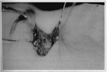
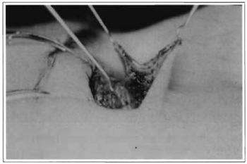
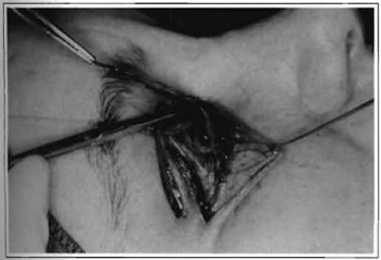
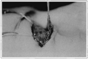
The lower blepharoplasty was performed with an incision along the eyelid border and extended 15 mm at the lateral canthus. It is located 2 mm below the ciliary margin. The incision transfixed the musculocutaneous plan toward the orbital septum with a 450 angle in relationship the skin surface, leaving a thin muscular layer at the ciliary border. A musculocutaneous flap was dissected from the incision to the orbital rim, exposing the fat bags (Fig. 1). The orbital septum was opened over the fat herniation and the fat excess was adequately fixed and removed. The orbital septum was not closed. Following the fat correction, the skin was dissected from the orbicularis muscle and from the border of the flap to the half of its entire length (Fig. 2). This maneuver creates a biplanar flap composed by two flaps, one cutaneous and the other muscular with a common pedicle. The muscular portion of the muscular flap was pulled up to achieve a moderate stretching along its length. Excess of muscle is resected remaining a triangular extension in its lateral portion at the lateral canthus (Figs. 3 and 4). Lateral canthopexy was performed by fixing the triangular extension of the lateral portion of the muscle flap on the periosteum of the lateral orbital rim throughout a tunnel created at lateral raphe of orbiculares oculi muscle. The muscular flap was sutured in the muscular layer at the ciliary margin, bringing the skin flap together. The muscular fixation raised the biplanar flap, defining the cutaneous excess. The excess was adequately resected, decreasing the risk of scleral show or ectropion.


Fig. 1 - Musculocutaneous flap dissected from the orbital septum and from the subciliary incision to the inferior orbital rim.


Fig. 2 -The skin is dissected from the orbicularis muscle and from the margin of the musculocutaneous flap to the half of its length, creating a biplanar flap with common pedicle


Fig. 3 -The excess of muscle is resected along the length of the muscular portion of the biplanar flap.


Fig. 4 -A triangular extension remains at the lateral portion of the muscular flap to be fixed at the periosteum of the lateral orbital rim.


Fig. 5a -Skin tumor next to the lateral canthus.


Fig. 5b -Biplanar flap associated to a lateral canthopexy allow its removal extending the resection 3 mm outside of the margin of the tumor.


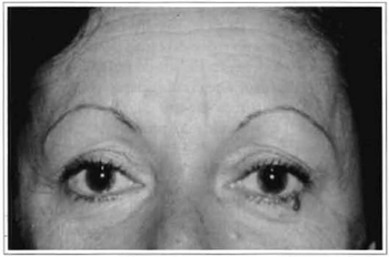
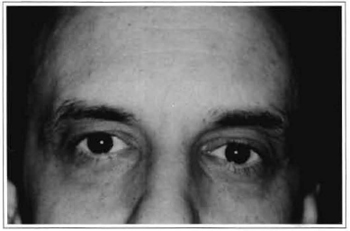
Fig. 6a -Severe lower eyelid fat herniation caused by muscle laxity.


Fig. 6b - 1 year postoperative recovery of lower eyelid contour.


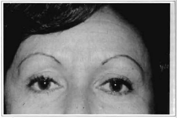
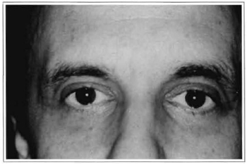
Fig. 7a -Lower eyelid asymmetry characterized by moderate fat herniation (right eyelid) and skin excess accomplished by mild fat herniation (left eyelid).


Fig 7b -Recovery of eyelids contour after lower blepharoplasty with a biplanar flap.


RESULTS

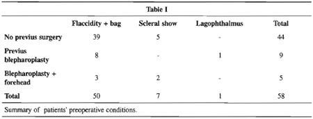
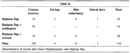
Fifty-eight patients, 46 females and 12 males, aged from 34 to 61 years, requiring improvement of their lower eyelids underwent a biplanar lower lid blepharoplasty. Blepharocalasis with moderate muscle flaccidity and large fat bags were observed in 50 patients. Seven patients presented bilateral marginal laxity with mild scleral show. A single patient presened a moderate unilateral lagophthalmos due to a prior muscle cutaneous blepharoplasty. Eleven patients have had a prior aesthetic procedure, 8 were submitted to a single blepharoplasty and 3 had associated a forehead surgery to the blepharoplasty. Table I, summarizes the preoperative conditions. Lateral canthopexy was associated to the biplanar blepharoplasty in 37 patients, forehead rhytidoplasty in 10 and 11 patients were submitted only to the biplanar blepharoplasty. No significant lower eyelid differences occurred in the patients submitted to the biplanar blepharoplasty. The recovery of "arcus marginalis" and lower lid contour were obtained in 103 eyelids of 51 patients, with a better support for the ciliar margin and lower lid muscular sling. The fat bags remained in 4 eyelids of 2 patients analyzed. Cutaneous redundance after biplanar blepharoplasty occurred in 8 eyelids of 4 patients and scleral show in only one eyelid, corresponding to the patient with preoperative lagophthalmos. The amount of periorbital fat removed from each side was proportional to the fat herniation preoperatively. Recurrence of the fat bags was achieved in 4 eyelids of 3 patients. Table II summarizes the results after lower blepharoplasty with a biplanar flap. The results were analyzed through photographs preoperatively, at 6 months and at 1 year postoperatively. The results obtained at 6 month postoperatively were maintained at 1 year after the surgery.


DISCUSSION

The recovery of the youthful palpebral contour is the objective ofthe blepharoplasty. It is obtained through the correction of each one of the anatomical deformities present on the lower eyelid. Several techniques were proposed to recovery the youthful contour of the lower eyelid, however, not even adequate to correct the quantitative disagreement of skin and muscle flaccidity(9). As described the biplanar flap allows to resect different quantities of the skin and muscle, in accordance with the palpebral deformity. It provides an appropriate vascular support, either for the cutaneous or for the muscular flap, through its conunon pedicle. Also, the biplanar flap in association with the lateral canthopexy promotes the recovery of the lower eyelid muscular sling with the lateral canthus and lower eyelid support. The resection and fixation of the muscular portion of the biplanar flap elevate the skin and muscle flaps together, providing the adequate correction of the excess of both tissues. The muscle fixation at the lateral canthus and at the muscular layer at the ciliary margin avoids the lower eyelid falling, decreasing the occurrence of scleral show or ectropion. In addition, it promotes the necessary stretching of the ciliary margin. The herniation of the intraorbital fat is appropriately corrected through the musculocutaneous approach. According to Netscher(10), there are no statistics differences between the transconjuntival and the subciliary transcutaneous incision, if skin excess is not presented. Other situation requires a subciliary transcutaneous incision.


REFERENCES

1. ABRAMO AC, VIOLA Jc. Lower blepharoplasty: Patially joined skin and muscle flaps. Aerth. Plast. Surg. 1993;17: 283-6.

2. MUSTARDE JC. Problems and pitfalls in blepharoplasry. Aesth. Plast. Surg. 1978; 1: 34954.

3. ADAMSON PA, STRECKER HD. Transcutaneous lower blepharoplasty. Facial Plast. Surg. 1996; 12: 171-83.

4. LOEB K. Necessity for partial resection of orbicularis oculi muscle in blepharoplasties in source young patients. Plart. Reconstr. Surg. 1977; 60: 176-8.

5. JELKS GW, GLAT PM, JELKS EB, LONGAKER MT. The inferior retimacular lateral cantoplasty: a new technique. Plast. Reconstr. Surg. 1997; 100:1262-70.

6. ZAREM HA, RESNICK JL. Expanded applications for transconjuntival lower lid blepharoplasry. Plart. Reconstr. Surg. 1999; 103:1041-3.

7. HINDERER UT. Correction of the weakness of lower eyelid and lateral canthus. Clin. Plart. Surg. 1993; 20:331-49.

8. KLATSKY AS, MANSON PN. Separate and muscle flaps in lower lid blepharoplasty. Plast. Reconstr. Surg. 1981; 67:151-6.

9. ADAMSON JE, McCRAW JB, CARRAWAY JH. Use muscle flap in lower-lid blepharoplasty. Plast. Reconstr. Surg. 1979; 63:359-63.

10. NETSHER DT, PATRINELY JR, PELTIER M, POLSEN C, THORBY J. Transconjuntival versus transcutaneous lower eyelid blepharoplasty: a prospective study. Plast. Reconstr. Surg. 1995; 96:1053-60.


I - Master and Doctor Degree in Plastic Surgery by the Escola Paulista de Medicina, Universidade Federal de São Paulo - UNIFESP Chairman of ACA - Grupo Integrado de Assistência em Cirurgia Plástica.
II - Assistant physician of ACA -Grupo Integrado de Assistência em Cirurgia Plástica.
III - Apprenticeship of ACA - Grupo Integrado de Assistência em Cirurgia Plástica.

Address for correspondence:
Antonio Carlos Abramo, MD
R. Afonso de Freitas, 641
04006-052 -São Paulo -SP Brazil

Hospital da Beneficência Portuguesa de São Paulo at the "ACA -Grupo Integrado de Assistência em Cirurgia Plástica", accredited to teaching plastic surgery by SBCP and AMB


Previous Article Back to Top Next Article

Indexers

Licença Creative Commons All scientific articles published at www.rbcp.org.br are licensed under a Creative Commons license