ISSN Online: 2177-1235 | ISSN Print: 1983-5175

Previous Article Next Article

Articles - Year1998 - Volume13 - Issue 3

ABSTRACT

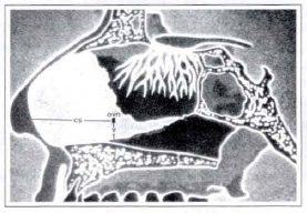
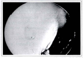
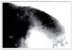
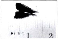
On this report the latest findings about the anatomical and funtional characteristics of the "vomeronasal organ" are evaluated. The high incidence of identification of the "vomeronasal organ" in nornal iudividuals indicates that the vomeronasal system is a universal feature of the adult human nasal cavity. Evaluation of the neuronal connections between this organ and the central nervous system shows that the "vomeronasal organ" is a functional chemosensory system with the ability to transduce signals that modulate certain autonomic parameters. Surgeons during nasal operations must consider the presence of the "vomeronasal organ" and its clinical significance.

Keywords: rhinoseptoplasty; vomeronasal organ; pheromones.

RESUMO

Neste trabalho, avaliam-se os últimos achados sobre as caraterísticas anatômicas e funcionais do "órgão vômero nasal" humano. A freqüente incidência da identificação do "órgão vômero nasal" em pessoas normais indica que o sistema vômeronasal é uma estrutura anatômica constante da cavidade nasal. A avaliação de conexões neuronais entre este órgão e o sistema nervoso central mostra que o "órgão vômero nasal" é um sistema quimio-sensorial funcional com habilidade de modular alguns parâmetros autonômicos. A presença do "órgão vômero nasal" e seu significado clínico devem ser considerados pelos cirurgiões nas rinoseptoplastias.

Palavras-chave: rinoseptoplastia; órgão vômero nasal; feromônios


INTRODUCTION

The nasal surgery is a frequent procedure in the practice of plastic surgery, where the final result must show a natural aspect and preserved function(l, 5). Nasal neurofisiology studies showed the importance of the "vomeronasal organ" for the humans being. This olphative organ was described initialy in animals(6), and posteriorly in humans(ll). The "vomeronasal organ" is described as a blind-ended tube, 2 to 7 mm long, located in the inferior and posterior portion septum, near to the vomer. Its opening is faced to the nasal cavity and located 2 cm from the caudal septum extremity(13).

Johnson et al(7) found the "vomeronasal organ" in 39 % of the cadavers analized. Garcia-Velasco and Mondragon(4) observed the presence of the "vomeronasal organ" in both nasal cavities in 91 % of 1000 patients analyzed. A pseudostratified columnar epithelium with 3 morphologically different types of cells without any similarity with other tissue of the body was described through histological analysis(1O). A prominent "vomeronasal organ" was observed in human fetus, presenting a direct connection with the central nervous system, with a chemosensorial system for detection of pheromones(12). Monti-Bloch et al.(9), in a double-blind study, compared the receptive potential of the "vomeronasal organ" to that of the olphative epithelium, employing different substances similar to the human pheromones named vomeropherins. Early studies demonstrated that picodoses(10, 11, 12) of vomeropherins applied directly on the "vomeronasal organ" are effective, with a quick and specific action on the central nervous system(2).

The objective of this paper is to determine the exact position of the "vomeronasal organ" in relation to the cartilaginous septum of the nose in order to preserve it during rhinoseptoplasty surgery.


MATERIAL & METHODS

Nasal dissections were done in 15 cadavers aged from 23 to 57 years old, with average of 41.8 year-old, being 9 male and 6 female. Table I shows the material used to locate the "vomeronasal organ". A complete cutaneous mucosa incision was made in me middle line of the nasal dorsum extended to the superior lip (Fig. 1). The lateral walls of the nose were laterally displaced, leaving the cartilaginous septum exposed (Fig. 2), allowing the identification of the opening of the "vomeronasal organ" on me septal mucosa (Fig. 3). Measures were taken to define the exact location of me "vomeronasal organ" in the cartilaginous septum using a millimetric ruler. Through a straight line was measured me distance from me caudal portion of me cartilaginous septum towards me anterior margin of the vomeronasal orifice, named CS (Fig. 4). The distance from the vomer to me inferior margin of the vomeronasal orifice was also used to determine the "vomeronasal organ" location and named V (Fig. 4).

The diameter of the orifice of the "vomeronasal organ" was also used as an evaluation parameter, measured in millimeters and identified by D (Fig. 5). In two cases me cartilaginous Septum and me mucosa were removed and me "vomeronasal organ" was identified by luminous transparency (Fig. 6a). After marking the location of me "vomeronasal organ" it was dissected from the mucosa of the cartilaginous septum (Fig. 6b), me "vomeronasal organ" orifice identified (Fig. 6c) and the material sent to hystological analysis.


Fig. 1- Incision made in the middle line of the nasal dorsum and upper lip.


Fig. 2 - The lateral walls of the nose laterally moved exposing the cartilaginous septum.


Fig. 3 - Localization of the "vomeronasal organ". The arrow shows the vomeronasal orifice.


Fig. 4 - Schematic representation of the nasal septum, showing the parameters used to determine the position of the "vomeronasal organ".


Fig. 5 - Image of the mucosa of the cartilaginous septum, showing the vomeronasal orifice.


Fig. 6a - Nasal septum mucosa dissected and analized by luminous transparency, showing the "vomeronasal organ".


Fig. 6b . "Vomeronasal organ" dissected from the septum mucosa, divided in half. '0' indicates the vomeronasal orifice.


Fig. 6c - Dissected cartilaginous septum. The arrow shows the precise localization of the "vomeronasal organ".


RESULTS

Fifteen cadavers were analyzed, with 2 present traumatic perforation in the cartilaginous septum, at the postero inferior portion, next to the vomer, not allowing the identification of the "vomeronasal organ".

From the remained 13 cadavers, 7 male and 6 female, the "vomeronasal organ" was identified by direct vision in 7 cases, corresponding to 53.84% of the analyzed cadavers as presented in Table II. Measures were taken to determine the exact position of the "vomeronasal organ", with average of 17.5 mm corresponding to the distance from the caudal portion of the cartilaginous septum to the anterior margin of the orifice of the vomeronasal organ" (CS). An average of 0.95 mm for the distance from the vomer to the inferior margin of the orifice of the "vomeronasal organ" (V), and an average of 1.3 mm for the diameter of the vomeronasal orifice (D), as presented in Table III, was found. The hystological analysis of the two dissected pieces showed a glandular structure sustained by loose conjunctive tissue lined by a pseudostratified columnar epithelium with three distinct cell types: lightly-stained cells that extend from the basement membrane to the free surface of the epithelium; columnar cells with densely-stained cytoplasm; and below these light and dark cells are basal cells, small cells located at the basement membrane (Fig. 7). The location of the "vomeronasal organ" did not suffer variation with age, sex or color of the skin of the analyzed cadavers.


Fig. 7 - Histological analysis of the "vomeronasal organ". Showing pseudoestratified tissue formed by light cells (cc), dark cells (co) and basal cells (cb), sorrounded by sustaining connective tissue (tc).


DISCUSSION

García-Velasco and Mondragon(4) identified the presence of the "vomeronasal organ" by direct vision in 91% of the human beings analized. Johnson et al.(7), in the same way, observed the "vomeronasal organ" in 39% of the cadavers evaluated. The disagreement in the amount of the "vomeronasal organ", identified by the authors, can be explained by the retraction of the mucosa in post-mortem, responsible for the oclusion of the orifice of the "Vomeronasal organ", making difficult its identification by direct vision. The different diameters presented in the orifice of the "vomeronasal organ" ranging from 0.2 mm to 2 mm, also affect the identification of the "vomeronasal organ", as reported by Moran et al.(10). In the present report, the "vomeronasal organ" was identified in 53.84% of the cadavers evaluated. This result disagrees from those described by García-Velasco and Mondragon(4), that identified the "vomeronasal organ" in human beings. The functional and activity of the "vomeronasal organ" in humans, as well as its connection with the central nervous system, is widely recognized in the literature(9). Its chemosensorial system, characterized by columnar pseudostratified epithelium, was recognized in this paper by the hystological findings of the two "vomeronasal organs" sent to analysis. Based on this property of the "vomeronasal organ", its preservation becomes necessary in the nasal surgery. Septal trauma and secondary distortions of the septal cartilage are responsible for the nasal deformities, being necessary to associate septoplasty to the aesthetic nasal surgery. The precise location of the "vomeronasal organ" is of great importance for the several septoplasty techniques, as advocated by Freer(3) and Killian(8). In this procedure the incision of the mucosa is described in the anterior portion of the cartilaginous septum, without its exact location, increasing the possibility of injury of the "vomeronasal organ". With the knowlegde of the exact location of the "vomeronasal organ", it is possible to perform the incision for septoplasty, preserving the area occupied by the "vomeronasal organ".


CONCLUSION

The importance of the "vomeronasal organ" as a chemosensorial system, connected to the central nervous system, requires its exact location at the mucosa of the cartilaginous septum, in order to preserve it in septal and aesthetic surgeries of the nose.


REFERENCES

1. ABRAMO AC, ABDO FLLHO D, GARCÍACASAS S. Extramucosal Rhinoplasty with Videoscopic Assistance. Aesth. Plast. Surg. 1998; 22:25-28.

2. BERLINER DL. Steroidal Substances Active in the Human Vomeronasal Organ Affect Hypothalamic Funtion. J. Steroid Biochem. Malec. Biol. 1996; 58:1.

3. FREER O. The Correction of Deflections of Nasal Septum with a Minimum of Traumatism. J.Amer. Med. Ass. 1992;38:636.

4. GARCÍA-VELASCO J, MONDRAGON M. The Incidence of the Vomeronasal Organ in 1000 Human Subjects and Its Possible Clinical Significance. J. Steroid. Biochm. Molec. Biol. 1991; 39:561-563.

5. GARCÍA-VELASCO J, GARCÍA-CASAS S. Nose Surgery and the Vomeronasal Organ. Aesth. Plast. Surg. 1995; 19:451-454.

6. JACOBSON L. Description Anatomique d'un Organe Observe Dans les Mammiferes. Ann. Mus. Hist. Nat. 1811; 412-424.

7. JOHNSON A, JOSEPHSON R, HAWKE M. Clinical and Histological Evidence for the Presence of the Vomeronasal (Jacobson's) Organ in Adult Humans. J. Otolar. 1985; 14:71-79.

8. KILLIAN G. Submucose Fensterresektion der nasenscheidewand. Arch. Lnryngol. Rhinol. 1904; 16:362.

9. MONT1-BLOCH L, JENNINGS-WHITE C, DOLBERG DS, BERLINER DL. The Human Vomeronasal System. Psyconeuroendocrinology. 1994; 19:673-686.

10. MORAN DT, JAFEK BW, ROWLEY JC III. The Vomeronasal (Jacobson's) Organ in Man: Ultraestructure and Frequency of occurrence. J. Steroid Biochem. Molec. 1991; 39:545-552.

11. POT1QUET M. Le Canal de Jacobson. Rev. Laryng. 1891; 737-753.

12. STENSAAS LJ, LAVKER RM, MONTIBLOCH L, GROSSER BI, BERLINER DL. Ultrastructure of the Human Vomeronasal Organ. J. Steroid Biochem. Mol. Biol. 1991; 39:553-560.

13. TESTUT L. Tratado de Anaromia Humana, Barcelona. Salvat Ed. 1912; 3:469.


I - Resident Doctors at ACA-Grupo Integrado de Assistencia em Cirurgia Plástica.
II - Head Professor of ACA-Grupo Integrado de Assistencia em Cirurgia Plástica.

Address for correspondence:
Rua Afonso de Freitas, 641
04006-052 - São Paulo - SP - Brazil

ACA - Grupo Integrado de Assistencia em Cirurgia Plástica.


Previous Article Back to Top Next Article

Indexers

Licença Creative Commons All scientific articles published at www.rbcp.org.br are licensed under a Creative Commons license