


Case Report - Year 2025 - Volume 40Issue 1
The Versatility of the Lateral Supramalleolar Flap in Covering Lower Limb Injuries: A Case Series
A versatilidade do retalho supramaleolar lateral para a cobertura de lesões nomembro inferior: Série de casos
David Christian Rodrigues Pereira1, ; Breno Bezerra Gomes de Pinho Pessoa1; Rebeca de Mesquita Oliveira Hamdan1; Sarah Cruz de Santana2; Giselle Ferreira de Souza2; Lucas Macedo Aurélio Paiva2; Victor Luis Almeida Pires de Castro2; Leticia Timbó Martins Ferreira2
ABSTRACT
The lateral supramalleolar flap is based on the perforating branch of the peroneal artery. This fasciocutaneous flap covers up to 8x6 cm on the dorsal foot and ankle. It receives blood supply from the ankle anastomotic arcade, and it is underutilized in pediatric patients for distal third leg injuries. The present retrospective study assessed 15 patients with complex lower limb wounds treated with the lateral supramalleolar flap at Instituto José Frota. All cases resulted from motorcycle accidents and showed good outcomes with minimal complications, proving the flap's effectiveness in foot and ankle injuries. Patients' ages ranged from 7 to 50 (mean: 28.5) years. Injuries were on the dorsum of the foot (46.6%), forefoot (26.6%), calcaneus (13.3%), lateral foot (13.3%), distal third of the leg (20%), and ankle (13.3%). Amean follow-up of 15 months showed stable coverage in all patients. The average number of preoperative debridements was 1.93 (range: 1-4). We used the standard fasciocutaneous flap dissection technique. The lateral supramalleolar flap is a reliable, simple, and safe option for lower limb injuries, with consistent outcomes and easy reproducibility.
Keywords: amputation, traumatic; lateral ligament, ankle; leg; lower extremity; surgical flaps; wounds and injuries
RESUMO
O retalho supramaleolar lateral, baseado na artéria peroneal, é um retalho fasciocutâneo utilizado para cobrir até 86 cm da região dorsal do pé e tornozelo. Recebe irrigação sanguínea da arcada anastomótica do tornozelo e é subutilizado em pacientes pediátricos para lesões no terço distal da perna. Este estudo retrospectivo avaliou 15 pacientes com feridas complexas em membros inferiores, tratados com esse retalho no Instituto José Frota. Todos os casos foram causados por acidentes de motocicleta e apresentaram bons resultados com complicações mínimas, comprovando a eficácia do retalho em lesões no pé e tornozelo. A idade dos pacientes variou entre 7 e 50 (média: 28,5) anos. As lesões ocorreram no dorso do pé (46,6%), antepé (26,6%), calcâneo (13,3%), lateral do pé (13,3%), terço distal da perna (20%) e tornozelo (13,3%). Um acompanhamento médio de 15 meses mostrou boa evolução em todos os pacientes. O número médio de desbridamentos pré-operatórios foi de 1,93 (variação: 1-4). Utilizamos a técnica padrão de dissecção do retalho fasciocutâneo. O retalho supramaleolar lateral é uma opção confiável, simples e segura para lesões nos membros inferiores, com resultados consistentes e fácil reprodutibilidade..
Palavras-chave: amputação traumática; ferimentos e lesões; ligamentos laterais do tornozelo; ligamentos; perna; retalhos cirúrgicos
Introduction
The lateral supramalleolar flap, first described by Masquelet and Gilbert in the late 1980s, represents a crucial advancement in reconstructive surgery for complex distal extremity defects, especially in the foot and ankle.1 Initially developed as a fasciocutaneous flap based on the perforating branches of the peroneal artery, its use has evolved over the years, providing reliable coverage for defects in traditionally challenging areas.
This flap quickly gained prominence due to its vascular reliability, versatility, and relatively simple dissection, making it a suitable option for covering traumatic wounds and chronic conditions with a large repair area.1 In recent decades, the lateral supramalleolar flap has proven effective in providing durable and sensitive coverage in regions such as the dorsum of the foot and malleolar areas, with fewer complications compared with free microsurgical flaps.2
The anatomical basis of this flap-using perforating branches of the peroneal artery, which anastomose with the anterior tibial artery-allows flap elevation as a reverseflow pedicled flap, sparing critical blood flow and ensuring tissue viability.3 Clinical studies have shown that this flap can cover defects up to 8 x 6 cm, expanding its application from trauma to include oncologic resections, diabetic ulcers, and other complex wounds.3
Although widely used in adults, the literature on this flap in pediatric cases is scarce, with few documented studies. The underutilization of the lateral supramalleolar flap in pediatric reconstructive surgery may be an area for further research, especially exploring its usefulness for congenital or traumatic defects in children.4
Objective
The current study presents the experience of the Plastic Surgery Service at Instituto José Frota (IJF), in the city of Fortaleza, state of Ceará, Brazil, in treating lower limb injuries with the lateral supramalleolar flap from January to June 2024.
Materials and Methods
The present retrospective study included all patients admitted to the IJF Plastic Surgery Service who underwent lower limb reconstruction due to loss of skin coverage with a lateral supramalleolar flap from January to June 2024.
These patients were electively admitted after clinical/ surgical wound treatment by other specialties, such as orthopedics and general surgery. We assessed the following variables: gender, age, characteristics of the tissue loss, and presence of exposed bone. The study included patients treated at the IJF Plastic Surgery Service for lower limb wounds during the study period and who underwent reconstruction with the lateral supramalleolar flap. The exclusion criteria were hemodynamic instability and uncooperative patients.
We performed debridement of lower limb injuries using one or more methods to obtain a clean wound with no secretions or areas of macroscopic necrosis and less fibrotic tissue.
Participants were exempted from informed consent because this was a retrospective study. The Research Ethics Committee of the State Department of Health of Brazil’s Federal District approved this study under CAAE number 84101424.6.0000.5047 and opinion number 1.167.841.
Surgical Technique
As described by Masquelet and Gilbert, the lateral supramalleolar flap technique uses a distally based pedicle. The flap is based on the anastomosis of the arterial arcade around the ankle with the perforating branch of the fibular artery, which emerges from the interosseous membrane approximately 5 cm from the lateral malleolus. This anastomosis provides branches to the skin in this region. These branches connect with the plexus accompanying the superficial fibular nerve, constituting the septocutaneous territory of the anterior tibial artery.
Results
The study sample consisted of 15 patients with complex lower limb wounds treated with lateral supramalleolar flaps. The mean age of patients at the time of initial treatment was 28.5 (range: 7-50) years. Men represented 86.6% of the sample (►Table 1). The mean number of preoperative surgical debridements was 1.93 (range: 1-4).
| Gender | N | % |
|---|---|---|
| Male | 13 | 86.6 |
| Female | 2 | 13.3 |
| Age group (years) | ||
| 0-19 | 3 | 20 |
| 20-29 | 3 | 20 |
| 30-39 | 4 | 26.6 |
| > 39 | 5 | 33.3 |
| Lesion location | ||
| Dorsum of the foot | 7 | 46.6 |
| Forefoot | 4 | 26.6 |
| Calcaneum | 2 | 13.3 |
| Lateral foot | 2 | 13.3 |
| Distal third of the leg | 3 | 20 |
| Ankle | 2 | 13.3 |

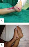
In all cases, the etiology of lower limb substance loss was motorcycle accidents. Substance loss was most common in the dorsum of the foot (46.6%), followed by the forefoot (26.6%), calcaneus (13.3%), lateral foot (13.3%), distal third of the leg (20%), and ankle (13.3%) (►Figs. 1-3). This case series had 6 fractures, and 7 patients presented with bone and/or tendon exposure (►Fig. 4).

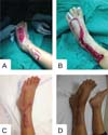
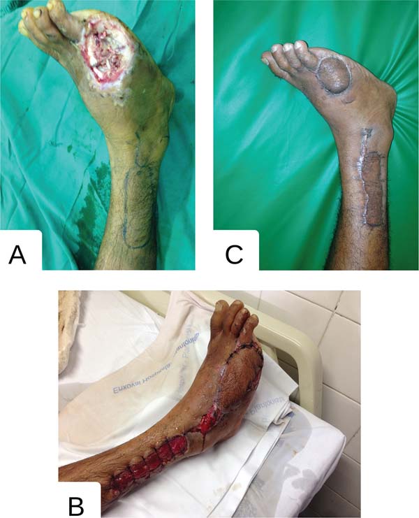
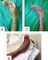
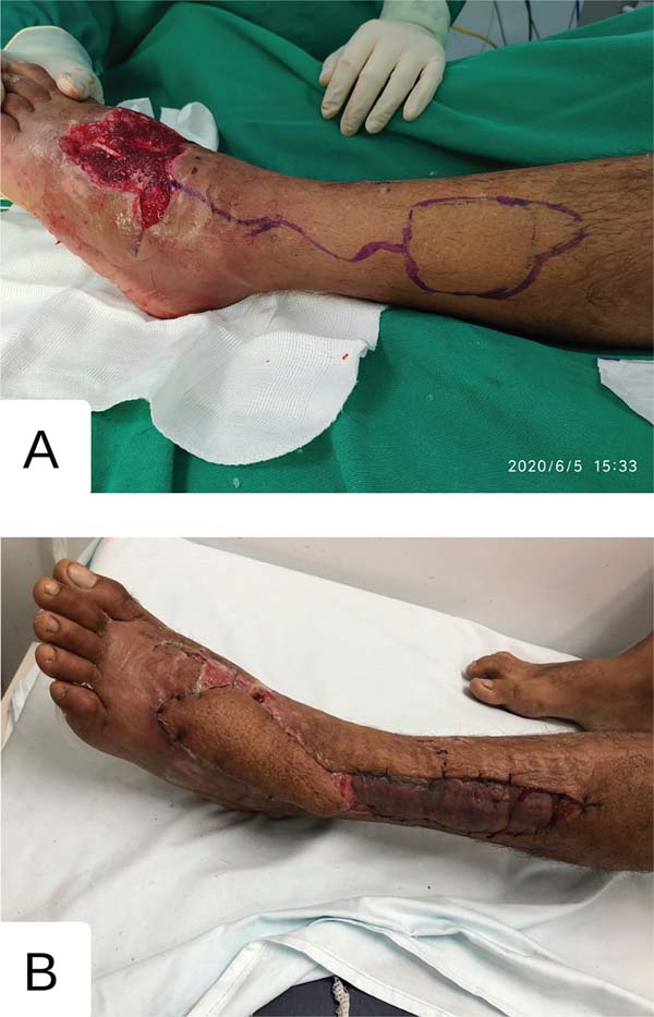


Surgical complications included four cases of epidermolysis, which required surgical or chemical debridement with dressings. In the remaining cases, the flap properly covered the lesion, leading to a good aesthetic and functional outcome.
Discussion
Injuries to the distal regions of the lower limbs can expose key anatomical areas and result in significant functional loss, compromising walking and other activities that depend on the proper functioning of this region. Most lower limb injuries resulted from motorcycle accidents; of the 15 surgeries performed in our study, all had this etiology. Therefore, the selection of a surgical flap to cover these injuries is crucial, considering both aesthetic and functional aspects.
For soft-tissue coverage in traumatic injuries to the distal portions of the lower limbs, such as the dorsum of the foot and ankle, microsurgical flaps are often the first option. With the improvement of surgical techniques, they have become increasingly popular, providing superior aesthetic and functional outcomes, even with smaller flap sizes. In the absence of an experienced microsurgical team or cases with microsurgical free-flap transplant failure, pedicled local flaps, such as fasciocutaneous flaps based on peroneal perforating arteries, are preferred.
Fasciocutaneous flaps from the lateral supramalleolar region are less common than those from the medial supramalleolar region.4 However, they constitute a versatile andeffectivealternativefordifficult-torepair lesions in the lower limbs. The reverse sural flap is another good option for reconstructing soft-tissue defects in the distal leg, ankle, and foot. Still, the lateral supramalleolar flap, which shares certain common factors with the reverse sural flap, remains underutilized. Both flaps are fasciocutaneous, can be isolated with a fascial pedicle, are based on perforating branches of the peroneal artery, contain a major cutaneous nerve, are distally based, have adequate pedicle length, offer a large skin surface area, and do not sacrifice any major artery of the corresponding limb.
The advantages of the lateral supramalleolar flap over the reverse sural flap include the possibility of performing surgery with the patient in the supine position and having an anterograde vascular supply when used in proximal defects. Conversely, the disadvantages of the lateral supramalleolar flap compared with the reverse sural flap include smaller volume, limited skin area, increased anesthesia area over the dorsum of the foot, and a higher incidence of venous congestion.
In 2011, Batista demonstrated the effectiveness of the lateral supramalleolar flap in covering lesions in various regions of the foot, distal leg, and ankle, in smalland medium-sized lesions.3 Furthermore, these flaps offer advantages such as easy reproducibility, simple dissection, a short learning curve, and not interfering with the creation of otherflaps. A case series fromConchaetal.5(2022) included 13 patients with traumatic injuries in several regions, such as the dorsum of the foot and ankle (lateral, medial, and anterior). A single patient developed complete necrosis due to the proximity of the donor site, where the advancement flap for the lateral ankle was created-the remaining cases presented with satisfactory flap fixation and good aesthetic outcomes. The authors concluded that this flap offers satisfactory versatility, being effective for patients of any age, although it is not the most suitable for the Achilles tendon region.
In our case series, we observed epidermolysis in four patients, which required surgical or chemical debridement, but no partial or complete necrosis. It is noteworthy that there was no documentation regarding the patients’ previous comorbidities, as all of them were admitted to the emergency department after motorcycle accidents. We followed up the patients for 180 days, and all showed good flap evolution, with satisfactory aesthetic and functional outcomes. The use of this flap requires professional expertise and adequate hospital infrastructure, given its complex soft-tissue reconstruction practices. Our experience and the evidence availableintheliterature leadus to believe that the lateral supramalleolar flap is an effective and relatively simple option for covering exposed fractures of the foot and ankle. This technique does not require advanced training in microsurgery and provides consistent results when performed by experienced plastic surgeons.
Conclusion
The lateral supramalleolar flap has proven to be an effective option for covering areas with exposed bone in the lower limbs. This technique offers versatility, ease of application, and a low complication rate, making it a valuable option in managing such injuries.
References
1. Masquelet AC, Gilbert A. Retalho supramaleolar lateral. In: __________. Atlas colorido de retalhos na reconstrução dos membros. Rio de Janeiro: Revinter; 1997:148-157
2. Saad FT, Almeida KGd, Almeida PYNGd, Silva TFd, Balbuena MB, Coutinho BBA, et al. Reconstrução de dorso do pé com retalho supramaleolar lateral de fluxo reverso em menor de 4 anos de idade. Rev Bras Cir Plást 2015;30(02):324-328. Doi: 10.5935/2177-1235. 2015RBCP0154
3. Batista JC. Retalho supramaleolar de fluxo reverso: aplicações clínicas. Rev Bras Cir Plást 2011;26(01):140-146. Doi: 10.1590/ S1983-51752011000100025
4. Quirino AC, Viegas KC. Fasciocutaneous flaps for covering foot and ankle injuries. Rev Bras Ortop 2014;49(02):183-188. Doi: 10.1016/ j.rboe.2014.03.004
5. Concha JM, Camaro PL, David A, Concha C. The lateral supramalleolar flap for the treatment of open foot and ankle fractures. Orthoplastic Surgery 2022;9:80-85. Doi: 10.1016/j.orthop. 2022.07.006
1. Plastic Surgery Service, Instituto Doutor José Frota (IJF), Fortaleza, CE, Brazil
2. Medicine Program, Universidade de Fortaleza, Fortaleza, CE, Brazil
Address for correspondence Dayvid Christian Rodrigues Pereira, Serviço de Cirurgia Plástica, Instituto Doutor José Frota (IJF), Fortaleza, CE, Brasil (e-mail: dayvid0309@hotmail.com sarahcruzsantana@gmail.com).
Artigo submetido: 12/02/2025.
Artigo aceito: 14/07/2025.
Conflict of Interests
The authors have no conflict of interests to declare.
 Read in Portuguese
 Read in English
 PDF PT
 Print
Print
 Send this article by email
 How to Cite
 Mendeley
 Pocket