


Case Report - Year 2024 - Volume 39 -
Pellon Technique for Chronic Wounds in the Frontal Region: A Case Report and Literature Review
Técnica de Pellon em ferida crônica na região frontal: Um relato de caso e revisão da literatura
Oona Tomiê Daronch1 ; Renata Fernanda Ramos Marcante1; Aristides Augusto Palliares1
ABSTRACT
Stem cells from adipose tissue can differentiate into fibroblasts, keratinocytes, and other cells; they can also secrete mediators with neoangiogenic and anti-inflammatory properties. Initially described for burn treatment, some authors suggested stem cells for treating complex, evolving wounds with 3 cm in length. After two fat grafting sessions, our patient presented granulation tissue in the frontal region. The properties of stem cells fromadipose tissue allow their use in burn treatment and chronic wounds, as in our case, which presented bone exposure in a prime region with few adjacent donor areas and a high failure potential for skin flaps.
Keywords: regenerative medicine; wounds and injuries; adipose tissue; wound healing; graft survival
RESUMO
Células-tronco derivadas do tecido adiposo podem se diferenciar em fibroblastos, queratinócitos e outras células; eles também podem secretar mediadores com propriedades neoangiogênicas e anti-inflamatórias. Inicialmente descrita no tratamento de queimaduras, ao longo do tempo também começaram a ser empregadas no tratamento de feridas complexas O presente caso relata o uso da gordura para o tratamento de uma ferida crônica com exposição óssea traumática na região frontal, com três anos de evolução e 3 cm de extensão. Após duas sessões de lipoenxertia, a paciente apresentava granulação do tecido da região frontal. As propriedades de células-tronco do tecido adiposo podem ser aproveitadas não apenas no tratamento das queimaduras,mas tambémemferidas crônicas, como o caso do presente trabalho, no qual a exposição óssea era em área nobre, com poucas áreas doadores adjacentes e grande possibilidade de insucesso no caso de retalhos cutâneos.
Palavras-chave: medicina regenerativa; ferimentos e lesões; gordura subcutânea; cicatrização; aloenxertos
Introduction
The properties of fat stem cells have been known for a long time. Further studies revealed that several growth factors participate in this function, including tumor necrosis factoralpha (TNF-α), interleukin-6 (IL-6), transforming growth factor-beta (TGF-ß), adiponectin, resistin, leptin, angiotensinogen, vascular endothelial growth factor (VEGF), and plasminogen activator inhibitor-1 (PAI-1).1 Stem cells from adipose tissue can differentiate into fibroblasts, keratinocytes, and other cells; they can also secrete mediators with neoangiogenic and anti-inflammatory properties.2 Initially described for burn treatment,1 some authors suggested their use in treating complex wounds. This case reports the use of fat to treat a chronic wound with traumatic bone exposure in the frontal region.
Objectives
The primary objective of this study was to report a case of treatment with fat grafting over the open area of a patient with an extensive chronic wound and exposed bone in the frontal region. The secondary objective was to conduct a brief literature review on this treatment type for complex wounds.
Case Report
VS, a female, 30-year-old, married, diabetic, homemaker patient, presented a trauma at the frontal region after hitting a wall 7 months ago due to an episode of hypoglycemia. She reported that there was no sign of injury at the time of the trauma and that, one week later, the entire frontal region presented blackened skin, initially in the left frontal area and then in the left hemiface, with a necrotic appearance. She also reported the blackened skin ulcerated and began to secrete an odorless serous fluid, with no bleeding, 2 weeks after the trauma. The patient reported initially receiving inpatient treatment for 6 days for a two-stage lesion debridement and antibiotic therapy (the last treatment occurred in November 2020). Since then, the lesion has been stable, with exposed frontal bone, no fever or purulent secretion, and she complains of local pain. She used collagenase ointment on the lesion. The patient had significant aesthetic complaints and loss of frontal motor skills, in addition to loss of selfesteem due to being unable to look at herself in the mirror anymore.
As comorbidities, the patient had insulin-dependent type 2 diabetes mellitus treated with metformin and NPH insulin and under current adequate control.
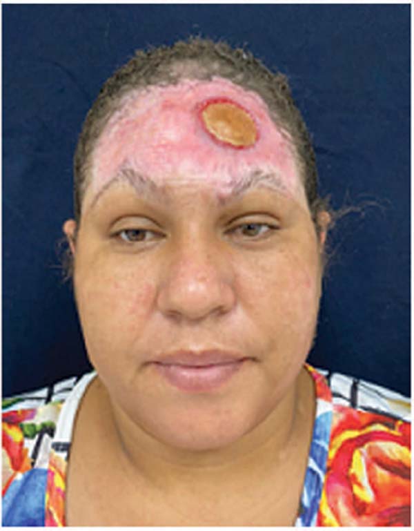
On physical examination, the patient presented fibrotic tissue throughout the extension of the frontal region, measuring 13 cm x 8 cm, from the eyebrow area to the upper and lateral hairline. In addition, there was a portion of the exposed cranial vault in the left frontal region, measuring 3 cm in diameter and presenting adherent fibrotic tissue with raised edges (►Fig. 1 ). There was a small bloody area in the right (middle third) frontal region, transversal, measuring 1 × 3 cm, with no inflammatory signs. The bone cortex was intact and had a yellowish appearance. The scar tissue in the right frontal region presented retraction, with rightsided eyebrow elevation (1 cm higher than the left eyebrow). There were no local secretions, inflammatory signs, or lack of mobility in the frontal region.
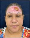
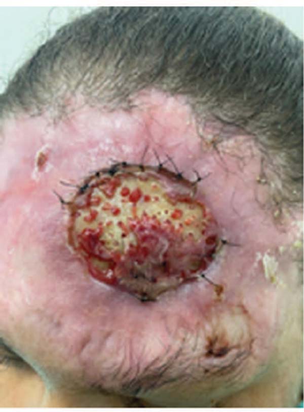
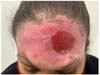
The patient underwent two fat grafting sessions using the Pellon technique (overlapping the raw area) with an average interval of 3 weeks between them. We injected 5 mL of fat in each session under local anesthesia with no fat centrifugation. We made small openings in the cortical bone until it bled to optimize the entry of mesenchymal fat cells into the bone. The patient received prophylactic antibiotics for 7 days in all fat grafting sessions, and her diabetes was under control. From the first to the second procedure, we noted a progressive reduction in the bone exposure area size (►Fig. 2). Four weeks after the last fat grafting session, granulation tissue completely filled the area (►Fig. 3). Subsequently, we performed a full-thickness skin graft to cover the granulation tissue. The donor area for the graft was the inguinal region.


Discussion
The popularity of autologous fat grafting increased significantly in the late 1980s when an abundance of fat from liposuction procedures allowed surgeons to experiment with its therapeutic potential.3 Adipocyte-derived stem cells were first isolated from liposuctioned tissue. Fat injection to further improve scars likely results in improvement by mesenchymal cells and numerous growth factors from liposuction coming from the skin and causing scar remodeling.2 Patients with subacute (over 3 weeks) burns with apparent progression to scarring and those with hypertrophic scarring after healing from a burn or other injury, such as trauma or keloids of any origin, are also candidates for fat grafting treatment. Repeated injections (up to four injections total) occur at intervals of 7 to 10 days for wounds or 6 to 8 weeks for scars.
Fat grafting as an adjuvant treatment in acute and subacute burns aims to take advantage of the benefits of fat: several metabolic and regenerative properties, increasing vascularization and enhancing tissue regeneration.2 These properties can treat burns and chronic wounds, as in our case report, in which the bone exposure was in a prime area with few adjacent donor areas and a high failure potential for skin flaps.
The gold standard treatment for wounds with exposed bone coverage with local or distant flaps. In our case report, the first option could be a microsurgical flap, such as a Chinese flap, given its thickness, which is adequate for frontal region covering. The disadvantages of this treatment are its higher morbidity and a higher chance of complications, especially considering the patient’s obesity and diabetes. In this case, fat grafting was sufficient to assure bone exposure coverage with low morbidity, the need for less complex procedures, and lower costs for the public health system. A study with 20 wound cases in different locations observed qualitative aesthetic and functional improvement.4 Most importantly, pain reduction or complete resolution and scar elasticity increase were objectively assessable in all cases. Fat can also cover prime areas in other sites, including those with exposed bone and ligaments in the calcaneus.5
A recent study on fat grafting to treat facial wounds showed its benefits in local scars. Although there are multiple graft donor sites, no data currently favors one over the others. Facial fat grafting occurs after fat collection and processing.6 Several face areas can undergo autologous fat grafting treatment, including the glabella, frontal region, temporal fossa depressions, labiomental fold, lips, pre-jowl grooves, and earlobes. Lipofilling can successfully treat scars in these areas with different degrees of volume deficit.6 Thus, fat grafting can treat prime areas with good outcomes, as in our study. An option to optimize fat grafting outcome is the use of vacuum pressure therapy.6 Grafted fat mobilization against the wound surface under negative pressure seems to transform into an autologous biological matrix with large amounts of mesenchymal cells and adipocytes.6 The wound requires debridement and an adequate bed for fat placement, as in our case report.
The most common method of fat graft harvesting is based on the Coleman technique,7 which collects liposuction samples from the donor site and processes them before injection into the recipient site. The standard technique is called lipotransfer, but there are several variations. A recent systematic review including 10 articles3 observed that most studies used a lipotransfer technique, and one administered unprocessed liposuction material with no centrifugation. In the latter study, 88% of the wounds healed completely, and 12% healed partially within 4 months. The mean wound healing time was 68 days (range, 40-107), with an average reduction in wound surface area of 90%.8 In our study, we collected abdominal fat using the conventional lipotransfer per the Coleman technique, with no centrifugation, and the bone exposure area showed complete granulation after two fat grafting sessions.
Other relevant issues for discussion, with a probable positive impact on wound evolution and granulation tissue formation, are the adequate control of the patient’s underlying disease (diabetes), the use of antibiotics, and wound debridement before fat graft placement.
REFERENCES
1. Pellon MA. Molecular and microanatomic characteristics of fat and its application in the treatment of acute burns and sequelae. Cirugia Plastica Ibero-Latinoamericana 2020;46:53-62
2. Piccolo NS, Piccolo MS, de Paula Piccolo N, et al. Fat Grafting for Treatment of Facial Burns and Burn Scars. Clin Plast Surg 2020;47 (01):119-130
3. Malik D, Luck J, Smith OJ, Mosahebi A, Mosahebi A, Mosahebi A. A Systematic Review of Autologous Fat Grafting in the Treatment of Acute and Chronic Cutaneous Wounds. Plast Reconstr Surg Glob Open 2020;8(05):e2835
4. Maione L, Lisa A, Vinci V, Bandi V, Klinger F, Klinger M. Autologous fat graft in foot calcaneal postsurgical chronic ulcer. Injury 2019; 50(Suppl 4):S64-S67
5. Klinger M, Caviggioli F, Klinger FM, et al. Autologous fat graft in scar treatment. J Craniofac Surg 2013;24(05):1610-1615
6. Klinger M, Klinger F, Caviggioli F, et al. Fat Grafting for Treatment of Facial Scars. Clin Plast Surg 2020;47:131-138 PubMed
7. Souza GMC, Amorim CCB, Vallejo CEA, et al. Fat grafting associated with negative pressure wound therapy. Acta Cir Bras 2019;34 (09):e201900907
8. Luck J, Smith OJ, Malik D, Mosahebi A. Protocol for a systematic review of autologous fat grafting for wound healing. Syst Rev 2018;7(01):99
1. Plastic Surgery, Universidade Estadual Paulista
“Júlio de Mesquita Filho” (UNESP), Botucatu, São Paulo, SP, Brazil
Address for correspondence Oona Tomiê Daronch, Universidade Estadual Paulista “Júlio de Mesquita Filho” (UNESP), Cirurgia Plástica, Botucatu, SP, Brazil (e-mail: oona.daronch@yahoo.com.br; oona.daronch@gmail.com).
Article received: April 02, 2024.
Article accepted: November 16, 2024.
Conflict of Interests
The authors have no conflict of interests to declare.
 Read in Portuguese
 Read in English
 PDF PT
 Print
Print
 Send this article by email
 How to Cite
 Mendeley
 Pocket