


Review Article - Year 2022 - Volume 37 - Issue 3
Cleft hand classification and treatment: literature review
Classificação e tratamento da mão em fenda: revisão da literatura
André Gustavo Pontes de Carvalho Pires1,* ; Marcela dos Santos Martins1; João Carlos Nakamoto1; Marcos Marcatto Abreu1; Renato Novaes Oliveira1; Arthur Perez Ferreira Neto1
ABSTRACT
Cleft hand is a rare congenital deformity characterized by a longitudinal deficiency of the central rays of the hand, which may be associated with other malformations. Due to the wide spectrum of manifestations, the treatment is challenging. This study presents the most suitable classifications, surgical techniques, and follow-up adopted according to the clinical manifestation. The search was performed in the Web of Science, PubMed, Scopus, Cochrane and Embase databases, descriptors and terms related to the hand anomaly in a typical cleft. Thirty-two articles were included and analyzed regarding the classification of the anomaly, classification of the severity of expression, surgical techniques and studies adopted with information on the surgical intervention for a cohort of patients. Considering that studies about cleft hands could be directly affected by embryological, genetic and molecular biology discoveries, different classifications have been described, and several studies to complement existing surgical techniques have been found. Innovative studies are scarce. In addition to better quality research, standardization in the description of techniques and results could elucidate existing treatment options gaps.
Keywords: Congenital abnormalities; Hand; Finger joint; Congenital, Hereditary, and neonatal diseases and abnormalities; Hand deformities.
RESUMO
A mão em fenda é uma deformidade congênita rara caracterizada por uma deficiência longitudinal dos raios centrais da mão, podendo estar associada a outras malformações. Devido ao amplo espectro de manifestações, o tratamento de mãos em fenda é desafiador. Este estudo objetiva apresentar as classificações, técnicas cirúrgicas mais indicadas e seguimentos adotados conforme a manifestação clínica. Foi realizada uma pesquisa nos bancos de dados Web of Science, PubMed, Scopus, Cochrane e Embase, descritores e termos relacionados à anomalia mão em fenda típica. Trinta e dois artigos foram incluídos, sendo analisados quanto a classificação da anomalia, classificação da gravidade de expressão, técnicas cirúrgicas e estudos com informações da intervenção cirúrgica adotada para uma coorte de pacientes. Considerando que estudos sobre mão em fenda são diretamente afetados pelas descobertas embriológicas, genéticas e de biologia molecular, diferentes classificações foram descritas e diversos estudos de complementação de técnicas cirúrgicas já existentes foram encontrados. Estudos inovadores são escassos. A padronização na descrição das técnicas e resultados, além de pesquisas de melhor qualidade, poderiam elucidar lacunas ainda existentes em torno das opções de tratamento.
Palavras-chave: Anormalidades congênitas; Mãos; Articulações dos dedos; Doenças e anormalidades congênitas, hereditárias e neonatais; Deformidades da mão
INTRODUCTION
Congenital cleft hand (CCH) was originally classified as typical versus atypical cleft hand1. With the advancement of genetics and molecular biology, the atypical cleft hand was reclassified as a teratological sequence of symbrachydactyly2.
This anomaly is characterized by the “V” shape, which may be associated with the absence of one or more digits, and may be unilateral or bilateral, with or without the involvement of the feet3,4. Generally, it is an autosomal dominant inheritance, with variable penetrance and expressiveness4.
Resulting from a longitudinal deficiency of the central rays of the hand, CCH can range from a simple skin cleft of soft tissues to the suppression of all rays except the smallest digit5. Based on the three axes of hand and upper limb development, CCH is currently classified as hand plate malformations - abnormal axis differentiation (patterning/late limb differentiation)6.
Manske & Halikis and Sharma and Sharma stand out among the most used classifications. The first is based on the involvement of the first commissure7. The second provides a complete hand description and assigns a numerical value to each element, with the subsequent recommendation of the indicated surgical procedure8.
Indications for surgical treatment range from space deficiency in the first commissure, absence of the thumb, progressive deformity to severe flexion contractures of one or more fingers4,9. However, this topic remains controversial and challenging, especially due to the patient’s adaptation to the deformity and acceptable functionality of the limb10.
OBJECTIVE
This literature review aims to present the classifications, the most relevant surgical techniques reported in the literature and the results obtained from the studies included.
METHODS
Databases and research
The bibliographic search was carried out between April and October 2020 in journals indexed in the Web of Science, PubMed, Scopus, Cochrane and Embase databases. The search terms used were a combination of “Typical Cleft Hand,” “Cleft Hand,” “Ectrodactyly,” “Central hand,” “Central ray deficiency,” “EEC syndrome,” “Cleft hand,” “Cleft-hand” , “Cleft- Hand Malformation”, “Lobster claw”, “Fingers/ abnormalities”, “Muscle, Skeletal/abnormalities”, “Hand Deformities, Congenital/pathology”, “Trigger Finger Disorder/congenital”, “SHFM”, “Collateral Ligaments/surgery”, “Hand Deformities, Congenital/ surgery”, “Metacarpophalangeal Joint/surgery”, “Tendon Transfer”, “Surgical Flaps”, “Suture Techniques”, “Syndactyly/surgery”, “Reconstructive Surgical Procedures”, “Hand deformities, Congenital/ surgery”, “Fingers/surgery”, “Congenital/surgery”, “Treatment”, and other related terms. All records returned by the search were imported into Mendeley’s bibliographic management software, and duplicate publications were removed. We also identified relevant articles through bibliographic linking with eligible articles.
Selection of studies
The included studies were related to CCH and may have the following approaches: classification of the anomaly, classification of severity of expression, surgical techniques, intervention or cohort of patients undergoing surgical treatment. The search did not limit language or study design. For intervention analysis, in order to observe current practical trends, the search was restricted to studies published between 2000 and 2020.
Studies that analyzed patients with cleft hands resulting from trauma sequelae or syndromic association, review articles or secondary analyses and publications that were incomplete or did not provide sufficient data for one of the outcomes of interest were excluded. Patient cohort data from studies approaching surgical techniques were not used for interventional analyses.
Data extraction
Two independent investigators reviewed the search results to select eligible studies using pre-established inclusion and exclusion criteria. Disagreeing decisions were discussed with a third reviewer. Data were extracted using a form according to predefined variables for each analysis. In order to summarize the findings in the literature, we chose to include a topic unifying surgical techniques and classification of severity of expression according to suggestive reports observed in the literature.
RESULTS
Selection of studies
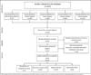
Five hundred twenty-seven studies were identified by searching the descriptors in the databases. Three hundred sixty-nine articles were excluded due to duplicity or by title, abstract and/or keywords. After applying the determined inclusion and exclusion criteria, 36 were considered potentially eligible; four were discarded based on clinical outcome, lack of data or inadequate study design, resulting in 32 studies for analysis of results. In Figure 1, the flowchart of the search for studies in the chosen databases is represented.

Analyses of included studies
The included studies were divided into four groups: (1) congenital anomaly classification (n=8); (2) expression severity rating (n=10); (3) surgical techniques (n=12); and (4) intervention analysis (n=4). Of these, only two studies met the inclusion criteria in two concomitant groups; therefore, they were counted as unique inclusions11,12. Therefore, the main features of the 32 included studies stratified into groups are presented in subsequent topics.
CCH functional classification
The classification system for congenital limb malformations was developed by Swanson et al.13, based on the grouping of anomalies according to the part affected during development.
This system, accepted by the American Society for Surgery of the Hand (ASSH), the International Federation of Societies for Surgery of the Hand (IFSSH) and the International Society for Prosthetics and Orthotics (ISPO), is called the IFSSH classification14. Subsequently, the Japanese Society for Surgery of the Hand (JSSH) suggested modifications to include two groups: “Abnormal lightning induction” and “Unclassifiable cases “15,16.
A new classification for congenital anomalies of the upper extremity, known as the OMT classification and considered an alternative to the Swanson, Barsky and Entin classification17,18, was presented by Oberg et al.19 in 2010. Since its publication, the OMT classification has been vigorously evaluated by several authors for its usefulness and reliability, and recently, in 2020, an update was published6.
Therefore, according to the current classification, CCH is classified as IB1IV: I- Malformation; B- Hand plate: abnormal axis differentiation (late limb standardization/ differentiation); 1- Proximaldistal Axis; IV: Cleft hand (cleft foot/hand malformation)6.
Congenital differences can also be classified according to their severity of expression, which can help in the functional determination and treatment orientation. Due to the unpredictability and peculiarity of the phenotypic presentations of this anomaly, a large number of classification systems have been proposed, which may be based on the number of defective rays11,20,21, teratological mechanism of aplasia and synostosis22, first commissure contracture7,12, the complexity of associated anomalies23 and radiological morphology and cleft position - medioulnar and ulnar24.
More current classifications tend to present a greater complexity of information. Valenti et al.25 proposed a classification based on six groups, each with a therapeutic strategy based on describing all clinical and radiographic abnormalities observed. In line with this, Sharma & Sharma8 described a new comprehensive functional classification considering all morphological determinants of the anomaly, such as absent digits, associated anomalies, cleft location and thumb functional status, calling it DAST8.
Among these, the most widely used clinical classification is that of Manske & Halikis7, which is based on the condition of the first commissure, with type I being normal (normal web); type II (narrow web) with moderate (IIA) or severe (IIB) narrowing; type III (syndactylized web) fused first commissure, syndactyly between thumb and forefinger; type IV (merged web) the first commissure included in the cleft, there is no index finger and the thumb is unstable; and type V (absent web) with the absence of the commissure due to the absence of the thumb and forefinger.
The most recent functional classification with multivariate analysis, advocated by Sharma & Sharma8, can be presented as follows:
Type I: described as having a normal first commissure, characterized by having the first commissure not reduced, with a slight cleft and no abnormal bone;
Type IIA: described as having a slightly narrowed first commissure, characterized by having a slightly reduced first commissure and abnormal bone;
Type IIB: described as having a severely narrow first commissure, characterized by a severely reduced first commissure, abnormal bony syndactyly;
Type III: described as having thumb/ index syndactyly without first commissure, characterized by fused first commissure, syndactyly between thumb and second finger and abnormal bone;
Type IV: described as having the first commissure included in the cleft, characterized by the suppression of the second finger and syndactyly of the ulnar digit;
Type V: described as having an absent first commissure, characterized by the absence of the thumb.
As a general rule, a sum of scores greater than 4 or individual scores in any morphological determinant of the anomaly greater than 2 indicates a potentially more complex deformity with less possibility of satisfactory functional and aesthetic results8.
Surgical technique
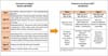
Recommended surgical procedures for treating central deficiency include cleft closure, reduction of the intermetacarpal space, the release of syndactyly, and excision of polydactyl or transverse bone elements when present. Different techniques can reduce intermetacarpal space and interventions secondary to cleft closure, and the most frequently reported in the literature, along with indications, advantages and disadvantages, as reported by the study when available, are listed in Figure 2.

Surgical technique according to expression severity classification
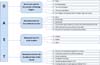
Grading systems are essential to facilitate communication and guide surgical reconstruction. Therefore, we present the behaviors most commonly reported by the included studies, stratified according to the classification by Manske & Halikis7.
In order to expose the advances in knowledge of this anomaly, we present in Figure 3 the Manske & Halikis7 classification and the recommendations for procedures suggested by some authors of the new classification system for hands with typical clefts using the DAST8 classification.

Intervention analysis
Surgical information from cohort studies of patients undergoing procedures for the treatment of cleft hands is presented in Chart 1.
| Author, Year | Technique | Advantages disadvantages |
|---|---|---|
| Barsky, 19641 |
Uses a retail place, pedicle foursquare, in diamond shape to recreate the commissure after the cleft is narrowed. Indication: Cleft hand. |
Benefits: Aesthetic improvement. Disadvantages: Insufficient functional concern; without reconstruction of the first commissure. |
| Snow and Littler, 196726 |
The cleft is elevated like a palmar flap, with a small radial flap preserved by recreating the commissure; the first commissure space is freed, which may require splitting the dorsal interosseous and surrounding fascia; the second metacarpal is transposed and attached to the remainder of the third metacarpal base; Fixation is obtained with axial and transverse wires, and the palmar flap is transferred, recreating the new commissure between the second and fourth ray. Indication: Third metacarpal segment present. |
Benefits: Functional; Gain cosmetic. Disadvantages: Risk of distal flap necrosis due to its high length-to-base ratio; Traction of the adductor and dorsal interosseous muscles may cause some radial angulation in the local translocation, incompletely correcting the central cleft. |
| Miura and Komada, 197927 |
Index transposition in a central position and palmar and dorsal redesign as separate flaps to create the first commissure. Cleft incised from side to side. The index finger is raised in its neurovascular bundles and transposed by osteosynthesis with the third metacarpal or by angulation osteotomy. Indication: Cleft hand with an adducted thumb. |
Advantage: Small flaps of random transposition of the dorsal and palmar skin. Disadvantage: Incidence in necrosis distal and contracture secondary |
| Ueba, 198128 |
Use of transverse flaps from any edge of the cleft and transposition of the index digit; Reconstruction of the intermetacarpal ligament by a free tendon. Indication: Total absence of the third metacarpal. |
Advantage: Improved aesthetics without changing the function of the hand. Disadvantage aesthetic of transferring the palmar to dorsal skin and dorsal to palmar skin. |
| Buck-Gramcko, 198529 |
Cleft narrowing, syndactyly separation, crossbones removal, correction of joint flexion contractures, rotation or wedge osteotomies for axial deviations and ulnar translocation of the index digit. Indication: Deep intermetacarpalpal ligament reconstruction. |
Advantages: Cosmetically acceptable without translocation. Disadvantages: Inadequate correction of the central space. |
| Ogino, 199011 |
The index and ring fingers reconstruct the deep metacarpaltransverse ligament using flexor sheaths (part of the A1 or A2 pulleys). Indication: Total absence of the third metacarpal. |
Advantages: Possibility of spontaneous correction of the deformity in flexion of the ring finger. Disadvantages: - |
| Upton, 200412 |
Wide incision that extends from the ulnar side of the cleft around the malpositioned index digit to the thumb; may include index transposition, metacarpal and/or phalangeal osteotomies, joint releases, phalangeal osteotomies, adductor pollicis muscle preservation, first dorsal interosseous muscle release, syndactyly separation(s), and thumb duplication correction. Indication: Typical cleft hand. |
Advantage: Provides clear identification of all anatomical structures of the palm. Disadvantage: Grasp and precision maneuvers remain poor despite considerable functionality. |
| Foucher, Loréa, Hovius, Pivato, Medina, 200630 |
Translocation in the radial direction of the ulnar finger(s) by intracarpal osteotomy; When necessary, a synostosis metacarpal can be performed in the same procedure. Indication: Type IIA of the Manske & Halikis classification7. |
Advantage: No functional loss; Good alignment of the second metacarpal with the radius. Disadvantage: Mobility between hamate and capitate is physiologically limited in all biomechanical studies. |
| Oberlin, Korchi, Belkheyar, Touam, MacQuillan, 200931 |
Reverse policing: The incision wraps around the second digit in the middle, extends over the dorsal edge of the cleft, and ends on the radial side of the third digit, where the second commissure space should be created. The index metacarpal is released (extraperiosteally) and translocated into the space of the absent third ray. After internal bone fixation, the flap, with its volar cutaneous pedicle, is transposed to reconstruct the first space of the mesh. Indication: Type II of the Manske & Halikis classification7 |
Advantages: Preservation of the dorsal venous network; no need for grafting; It does not harm the normal musculature of the thumb. Disadvantages: Possibility of ectopic bone deformation; Index finger misalignment; Divergence of the metacarpals if reconstruction of the transverse metacarpal ligament is insufficient. |
| Upton, Taghinia, 20109 |
Simple circumferential incision around the index radius; straightline incisions inside the cleft; and thumb extensions; Elevation of the dorsal and palmar flaps to provide exposure of the metacarpal portion of the hand; Reconstruction of the intermetacarpal ligament employing simple sutures, circumferential sutures around adjacent metacarpals, or by joining adjacent A1 pulleys. The new space of the first commissure is closed by a small flap based on the radial side of the radius of the annulus with a dorsal-palmar slope of 45°. Indication: Type II and Type III of the Manske & Halikis7 classification. |
Advantages: Preservation of the adductor pollicis muscle for a functional pinch. Disadvantages: Instability of the thumb in the joint metacarpophalangeal; Progressive camptodactyly of fingers adjacent to central cleft; Narrowing of the first commissure; Excessive length and radial deviation of the transposed index radius; Risk of recurrence of commissure narrowing due to scarring along the edge. |
| Christen, Dautel, 201310 |
Dorsal skin incision; Autologous tendon graft (preferably the palmar longus, otherwise the plantaris or a toe extensor); Positioning the tendon in a figure of eight around the base of the proximal phalanx and corresponding metacarpal neck. Indication: Hyperlaxity of the joint(s) metacarpophalangeal adjacent to the cleft. |
Advantages: Prevents the occurrence of excessive progressive narrowing; Disadvantages: Over-tightening of the graft will result in a limited range of motion in flexion and extension, whereas under-tensioning will lead to persistent laxity. |
| Yasin, Amin, Mahmoud, Abdel-Ghani, 202032 |
Use cleft skin as a bipedicled flap for reconstruction and widening of the first commissure’s narrow space of the first commissure. The skin of the cleft maintains its fixations on the dorsal and palmar surfaces of the hand, and the index digit is passed (tunneled) under it so that the skin of the cleft will occupy the space of the U commissure. Indication: Type IIB and Type III of the Manske & Halikis classification7. |
Advantages: Ensures the maintenance of a good blood supply to the skin of the cleft; a rounded edge of the U commissure without scars. Disadvantages: In a deep fissure, the skin retains its shape after reconstructing the first commissure. |

DISCUSSION
Many cases of CCH do not require surgical treatment10,33. Therefore, the follow-up to be adopted demands delicate management when outlining the indications for intervention and the surgical plan must specifically address the unique abnormality of the patient34. Since several authors report the psychological consequences of CCH caused by social stigmas, members of an interdisciplinary team may be requested to advise families with this condition35.
The main indication for correcting the deformity is restoring function as close to normal and as early as possible, with the esthetic correction being a natural result of the intervention8. As described in the literature, surgeons must assess the spacing of the first commissure, the presence or absence of metacarpals in the central rays, transverse bones, syndactyly that may be present and, especially, the vascular supply to the hand4. If the cleft hand is due to vascular insufficiency, as in cases of associated syndromes, or if there are other concomitant deformities, a stepwise surgical correction is suggested34.
It has been observed that understanding of the etiology of congenital limb anomalies is restricted and continues to progress, and we confirm this in the recent modification of cleft hand subcategorization in the OMT classification update published in 20206.
A large number of classifications according to the severity of CCH expression were found, with Manske & Halikis7 being the most frequently used, being cited in 100% of the studies included for analysis of surgical intervention32,36,37,38,39. However, the most complete and current classification with recommendations for surgical procedures and prognosis is that of Sharma & Sharma8.
It is important to highlight that some studies were redundant, describing modifications of the skin incision or refining techniques already consolidated, with the main objective of correcting the first commissure. If the first commissure is normal, so-called ‘simple cleft closure’ is suggested, with the reconstruction of soft tissue, ligaments, tendons and bones. The incision can be closed using different methods; the skin is usually removed from the lateral side of the adjacent digits to form a longitudinal scar. Often, a flap from the ulnar half of the cleft is used to increase the depth of the first commissure, creating a wider, more functional space between the index finger and thumb. Authors emphasize that detecting the neurovascular pedicle and careful division of the cleft are important steps in identifying additional tendons.
According to studies included in the intervention analyses, if the second and fourth digits remain divergent, an osteotomy of the base of one of the metacarpal bones can be considered. In cases of transverse bones, total or partial removal is suggested, leaving parts of it in continuity with the metacarpal joints to avoid damage to collateral ligaments, instability or stiffness40. Reconstruction of the deep, transverse metacarpal ligament can be performed using flexor sheaths (part of the A1 or A2 pulleys) of the index and annular digits, dorsal base rotation graft11, tendon/ fascia grafts36, or even absorbable stitches and K-wires.
For complex cases with syndactyly of the first commissure, options are presented according to the severity of the narrowing. If the syndactyly is proximal and loose, it can be released with an appropriate flap (z-plasty, combined z-plasty, or rotation-transposition flap) in association with cleft closure, as described.
For severe narrowing, attention should be paid to the distal bifurcation of the neurovascular bundle, not only between the thumb and index but also between the ring and little digit. If microvascular procedures cannot solve this problem, authors warn of the possibility of necrosis41,42. Therefore, the transposition of the second ray to the third metacarpal bone (if any) may be indicated. In advanced cases of partial or complete aplasia, transfer of toes or bone graft covered with periosteum is suggested42. Conservative treatment is reserved for the most severe cases, in which there is usually a great functional adaptation and in patients with severe neuropsychomotor developmental deficit, making adequate rehabilitation impossible43,44.
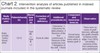
It was observed that practical application studies and long-term follow-up are limited. Only four studies that presented details of surgical intervention for a cohort of patients undergoing cleft hand treatment were identified, as shown in Chart 2. Of these, the most frequent distribution of cleft hands was type II. No type IV or V patients were described, a similar observation described by Manske & Halikis7, who reported that they are the rarest subtypes and are difficult to manage, a factor that would justify the absence of studies containing these subtypes.
| Study (Author, year) | Patients/hands (n) | Mansk type and (n) | Correction of central deficiency | Additional procedures (n) | Component and transverse bone (n) | Observation |
|---|---|---|---|---|---|---|
| Rider, Grindel, Tonkin, Wood, 200045 | 12/12 |
Type IIB: (-) Type III: (-) |
Snow & Littler26 | Osteotomy: 3 Osteotomy for delta phalanx: 2 Revision of the first commissure: 3 Revision of the syndactyly scar: 1 Religation of the cleft: 1 None: 2 | Dorsal base rotation graft11, bone suture or tendon graft: 8 | There were no cases of graft necrosis, although two grafts showed ischemia at the edge; Four (36%) secondary revisions of the first commissure were performed. |
| Goldfarb, Chia, Manske, 200836 | 12/16 |
Type I: 5 Type IIA: 7 Type IIB: 1 Type III: 3 |
Cleft reconstruction using soft tissue and/or bone procedures.* | Osteotomy: 3 Revision of the first commissure: 2 Proximal interphalangeal joint extensions: 3 Extensor mechanism imbrication: 2 Extensor indicis proprius transfer: 1. | Dorsal base rotation graft11 or tendon/ fascia grafts37 wrapped around the adjacent metacarpal neck: 8. | Flexion contracture of the proximal interphalangeal joint of the ring finger with a mean of 31° was the most notable clinical finding. The metacarpal divergence angle significantly improved from 33° to 12°, and the phalangeal divergence angle improved. significantly from 38° to 12°. |
| Aleem, Wall, Manske, Calhoun, Goldfarb, 201438 | 18/23 |
Type I: 5 Type IIA: 9 Type IIB: 5 Type III: 4 |
Standard cleft closure; Soft tissue reconstruction alone or combined with bone transposition of the index ray; and deepening of the first commissure space.* | Corrective osteotomies, tendon realignment, and soft tissue capsular tightening: 11 | Excised with intrinsic muscles inserted: 11 | The presence of transverse bone in the cleft hand was not associated with worse outcomes after cleft reconstruction. The use of the forceps cleft was more dependent on the status of the index digit and preoperative thumbindex space than on the presence of a transverse bone. |
| Beck, Chang, Jones, 201539 | 1/1 | Type IIB | Miura & Komada27 | It was not necessary. | Transverse osteotomy at the base of the index metacarpal; Ulnar translocation and fixation at the base of the metacarpal of the middle finger with K wire: 1 | All incisions and flaps healed mostly without any evidence of skin necrosis. Bone union was present 6 weeks after surgery. |
In recent studies, the Snow procedure is often related to its limitations, such as low viability of the palmar flap, technical difficulties, and records of complications resulting from necrosis. However, Rider et al.45, when studying the technique, observed a low flap necrosis rate, but the revision was necessary for one third of the patients. Despite this finding, there is a relevant use of the technique by Miura & Komada27, which is justified in the simpler design and less risk of flap necrosis while producing similar functional and cosmetic results39. A study with longterm results of the technique by Miura & Komada27 demonstrated excellent patient satisfaction in function and esthetics39. The same occurrence was reported in the literature for interventions performed using the Upton technique12.
In general, patients with a cleft hand due to vascular deformity are at high risk of skin loss and poor perfusion of the surgical site after surgery, especially if the procedure is not staged properly. In addition, finger stiffness remains the most common postoperative complaint, despite improving functional results34. Because of this, we emphasize that the median cleft of the hand is a complex but rare malformation that requires individualized management based on the severity of expression.
CONCLUSION
Studies on FCM are directly affected by discoveries in embryological, genetic and molecular biology. During the last few years, advances in these fields have led to restructuring the classification system and understanding different presentations. Regarding treatments, pioneering techniques include cleft closure and reconstruction of the first commissure. The main complications described were problems with necrosis of the distal flap and stiffness. Several studies on updating these techniques were found. In addition to better quality research, standardization in the description of techniques and results could elucidate existing treatment options gaps.
REFERENCES
1. Barsky AJ. Cleft Hand: Classification, incidence, and treatment. review of the literature and report of nineteen cases. J Bone Joint Surg Am. 1964;46:1707-20.
2. Manske PR. Symbrachydactyly instead of atypical cleft hand. Plast Reconstr Surg. 1993;91(1):196.
3. Falcochio DF, Da Costa AC, Durigan CPI, Nascimento VDG, Santili C, Chakkour I. Epidemiological and Clinical Aspects of Cleft Hand: Case Series From a Tertiary Public Hospital in São Paulo, Brazil. Hand (N Y). 2019;14(6):814-8.
4. Wolfe SW, Pederson WC, Hotchkiss RN, Kozin SH, Cohen MS. Green’s operative hand surgery: the pediatric hand E-book. Philadelphia: Elsevier Health Sciences; 2010.
5. Tonkin MA, Tolerton SK, Quick TJ, Harvey I, Lawson RD, Smith NC, et al. Classification of congenital anomalies of the hand and upper limb: development and assessment of a new system. J Hand Surg Am. 2013;38(9):1845-53.
6. Goldfarb CA, Ezaki M, Wall LB, Lam WL, Oberg KC. The Oberg-Manske-Tonkin (OMT) Classification of Congenital Upper Extremities: Update for 2020. J Hand Surg Am. 2020;45(6):542-7.
7. Manske PR, Halikis MN. Surgical classification of central deficiency according to the thumb web. J Hand Surg Am. 1995;20(4):687-97.
8. Sharma A, Sharma N. A comprehensive functional classification of cleft hand: The DAST concept. Indian J Plast Surg. 2017;50(3):244-50.
9. Upton J, Taghinia AH. Correction of the Typical Cleft Hand. J Hand Surg Am. 2010;35(3):480-5.
10. Christen T, Dautel G. Metacarpophalangeal ligamentoplasty in typical cleft hand. Tech Hand Up Extrem Surg. 2013;17(2):120-2.
11. Ogino T. Cleft hand. Hand Clin. 1990;6(4):661-71.
12. Upton J. Simplicity and treatment of the typical cleft hand. Handchir Mikrochir Plast Chir. 2004;36(2-3):152-60.
13. Swanson AB, Barsky AJ, Entin MA. Classification of limb malformations on the basis of embryological failures. Surg Clin North Am. 1968;48(5):1169-79.
14. Swanson AB. A classification for congenital limb malformations. J Hand Surg Am. 1976;1(1):8-22.
15. De Smet L; IFSSH. International Federation for Societies for Surgery of the Hand JSSH. Japanese Society for Surgery of the Hand. Classification for congenital anomalies of the hand: the IFSSH classification and the JSSH modification. Genet Couns. 2002;13(3):331-8.
16. Iba K, Horii E, Ogino T, Kazuki K, Kashiwa K; Congenital Hand Committee of Japanese Society for Surgery of the Hand. The Classification of Swanson for Congenital Anomalies of Upper Limb Modified by the Japanese Society for Surgery of the Hand (JSSH). Hand Surg. 2015;20(2):237-50.
17. Ezaki M, Baek GH, Horii E, Hovius S. IFSSH Scientific Committee on Congenital Conditions. J Hand Surg Eur Vol. 2014;39(6):676-8.
18. Tonkin MA, Oberg KC. The OMT Classification of Congenital Anomalies of the Hand and Upper Limb. Hand Surg. 2015;20(3):336-42.
19. Oberg KC, Feenstra JM, Manske PR, Tonkin MA. Developmental biology and classification of congenital anomalies of the hand and upper extremity. J Hand Surg Am. 2010;35(12):2066-76.
20. Tsuge K, Watari S. Surgical treatment of cleft hand and its associated deformities. Bull Hosp Jt Dis Orthop Inst. 1984;44(2):532-41.
21. Nutt JN 3rd, Flatt AE. Congenital central hand deficit. J Hand Surg Am. 1981;6(1):48-60.
22. Blauth W, Falliner A. Morphology and classification of cleft hands. Handchir Mikrochir Plast Chir. 1986;18(3):161-95.
23. Glicenstein J, Guero S, Haddad R. Fentes médianes de la main. Classification et indications thérapeutiques a propos de 29 cas [Median clefts of the hand. Classification and therapeutic indications apropos of 29 cases]. Ann Chir Main Memb Super. 1995;14(6):253. French.
24. Falliner A. The cleft hand. Proposal of a classification based on 279 cleft hands. Handchir Mikrochir Plast Chir. 2004;36(1):47-54.
25. Valenti P, Lozano Gonzales E, Vergara Amador E, Cogswell LK. Cleft hand: a review of 33 cases and new ideas about classification. Chir Main. 2008;27 Suppl 1:S121-8. DOI: 10.1016/j.main.2008.07.015
26. Snow JW, Littler JW. Surgical treatment of cleft hand. In: Transactions of the International Society of Plastic and Reconstructive Surgery 4th Congress. Rome: Excerpta Medica Foundation; 1967.
27. Miura T, Komada T. Simple method for reconstruction of the cleft hand with an adducted thumb. Plast Reconstr Surg. 1979;64(1):65-7.
28. Ueba Y. Plastic surgery for the cleft hand. J Hand Surg Am. 1981;6(6):557-60.
29. Buck-Gramcko D. Cleft hands: classification and treatment. Hand Clin. 1985;1(3):467-73.
30. Foucher G, Loréa P, Hovius S, Pivato G, Medina J. Radial shift of the ulnar fingers: a new technique for special cases of longitudinal central deficiency. J Hand Surg Br. 2006;31(2):156-61.
31. Oberlin C, Korchi A, Belkheyar Z, Touam C, Macquillan A. Digitalization of the second finger in type 2 central longitudinal deficiencies (clefting) of the hand. Tech Hand Up Extrem Surg. 2009;13(2):110-2.
32. Yasin E, Amin H, Mahmoud M, Abdel-Ghani H. Using Skin of the Cleft as Bipedicle flap for Release of the First Web Space in Congenital Central Deficiency. J Hand Surg Am. 2020;45(7):665. e1-665.e7.
33. Kozin SH, Zlotolow DA. Common Pediatric Congenital Conditions of the Hand. Plast Reconstr Surg. 2015;136(2):241e-57e.
34. Davis DD, Kane SM. Cleft Hand. Treasure Island (FL): StatPearls Publishing; 2020.
35. Flatt AE. The Care of Congenital Hand Anomalies. St. Louis: Quality Medical Publishing; 1994. p. 292-316.
36. Goldfarb CA, Chia B, Manske PR. Central ray deficiency: subjective and objective outcome of cleft reconstruction. J Hand Surg Am. 2008;33(9):1579-88.
37. Tada K. Central ray deficiency of the hand. Operative treatment and results. Int Orthop. 1984;8(3):229-33.
38. Aleem AW, Wall LB, Manske MC, Calhoun V, Goldfarb CA. The transverse bone in cleft hand: a case cohort analysis of outcome after surgical reconstruction. J Hand Surg Am. 2014;39(2):226-36.
39. Beck JD, Chang B, Jones NF. Over 20-year follow-up of Miura reconstruction of cleft hand. Hand (N Y). 2015;10(2):319-22.
40. Wood VE. The treatment of crossbones of the hand. Handchir Mikrochir Plast Chir. 2004;36(2-3):161-5.
41. Buck-Gramcko D. Microsurgery in congenital malformations of the hand. In: Brunelli G, ed. Textbook of Microsurgery. Milano: Masson; 1988. p. 933-40.
42. Buck-Gramcko D. Congenital malformations of the hand and forearm. Chir Main. 2002;21(2):70-101.
43. Kay SP, Platt A. Congenital disorders: cleft hand. In: Berger RA, Weiss APC, eds. Hand surgery. Philadelphia: Lippincott Williams & Williams; 2004. p. 1465-75.
44. França Bisneto EN. Deformidades congênitas dos membros superiores: parte I: falhas de formação. Rev Bras Ortop. 2012;47(5):545-52.
45. Rider MA, Grindel SI, Tonkin MA, Wood VE. An experience of the Snow-Littler procedure. J Hand Surg Am. 2000;25(4):376-81.
1. Universidade Estadual de Campinas, Departamento de Ortopedia e Traumatologia, Campinas,
SP, Brazil.
AGPCP Data analysis and/or interpretation, Statistical analysis, Data Collection, Conceptualization, Conception and design of the study, Resource Management, Project Management, Investigation, Methodology, Carrying out operations and/or experiments, Writing - Preparation of the original
MSM Data analysis and/or interpretation, Methodology, Writing - Preparation of the original, Writing - Review and Editing
JCN Final Manuscript Approval, Project Management, Writing - Review and Editing, Supervision, Preview
MMA Final Manuscript Approval, Project Management, Supervision
RNO Conceptualization, Conception and design of the study, Research
APFN Study conception and design, Writing - Review and Editing, Software, Validation
Corresponding author: Marcela dos Santos Martins Cidade Universitária Zeferino Vaz - Barão Geraldo, Campinas, SP, Brazil Zip Code: 13083-970 E-mail: marcela.m.1509@gmail.com
Article received: May 16, 2021.
Article accepted: December 13, 2021.
Conflicts of interest: none.
Institution: Universidade Estadual de Campinas, Hospital de Clínicas, Campinas, SP, Brazil.
 Read in Portuguese
 Read in English
 PDF PT
 Print
Print
 Send this article by email
 How to Cite
 Mendeley
 Pocket