
Case Report - Year 2021 - Volume 36 - Issue 1
Scalp reconstruction with expanded flap
Reconstrução de couro cabeludo com retalho expandido
ABSTRACT
Introduction: The presence of extensive scalp defects is
a major reconstructive challenge for the plastic surgeon.
These defects have a vast etiology, such as traumatic, thermal
or electrical burns, benign and malignant or congenital
tumor resections, radiotherapy treatments sequelae, and
infections. Noting that injuries such as scalping and burns
(thermal or electrical), generate significant repercussions
such as severe tissue loss, chronic osteomyelitis or minor
sequelae such as scar alopecia. This study aims to report
a case of late scalp reconstruction with a tissue expander
and posterior advancement flap, due to cicatricial alopecia,
in an 11-year-old female, victim of scalding by hot water in
the right frontotemporal region.
Methods: It was performed
a retrospective analysis of the patient's medical record.
The present work follows the standards of the Helsinki
ethics committee.
Conclusion: The scalp tissue expansion
technique by stages and subsequent scalp advancement flap
performing proved to be effective in restoring the patient's
hair structure and hairline with minimal local distortion,
restoring the scalp's shape and aesthetics of the patient.
Keywords: Hemangioendothelioma; Hemangioma; Plastic surgery; Surgical pathology; Neoplasms of vascular tissue
RESUMO
Introdução: A presença de defeitos extensos em couro cabeludo apresenta-se como um grande desafio reconstrutor para o cirurgião plástico. Estes defeitos têm vasta etiologia como: traumática, queimaduras térmicas ou elétricas, ressecções tumorais benignas e malignas ou congênitas, sequelas de tratamentos radioterápicos e infecções. Destacando-se lesões como o escalpelamento e queimaduras (térmicas ou elétricas), geram repercussões significativas como a perda de tecido grave, osteomielite crônica ou sequelas menores como uma alopecia cicatricial. O objetivo deste trabalho é relatar um caso de reconstrução de couro cabeludo em fase tardia com expansor tecidual e posterior retalho de avanço, devido à alopecia cicatricial, em paciente feminina de 11 anos vítima de escaldamento por água quente em região frontotemporal direita.
Métodos: Foi realizada análise retrospectiva de prontuário da paciente em questão. O presente trabalho segue os padrões do comitê de ética de Helsinque.
Conclusão: A técnica de expansão tecidual de couro cabeludo por estágios e posterior confecção de retalho de avanço de escalpo demonstrou ser eficaz de restaurar a estrutura pilosa e linha da implantação capilar da paciente com mínima distorção local, restituindo a forma e a estética do couro cabeludo da paciente.
Palavras-chave: Queimaduras; Dispositivos para expansão de tecidos; Cirurgia plástica; Procedimentos cirúrgicos
INTRODUCTION
The presence of extensive scalp defects presents is a major reconstructive challenge for the plastic surgeon1,2. These defects have a vast etiology, such as traumatic, thermal, or electrical burns, benign and malignant or congenital tumor resections, radiotherapy treatments sequelae, and infections. Deformities can range from small defects, which can be closed primarily, to extensive defects, which require tissue expansion or even the transfer of a free flap for closure.
Noting that injuries such as scalping and burns (thermal or electrical), generate significant repercussions such as severe tissue loss, chronic osteomyelitis or minor sequelae such as scar alopecia.
In a young female patient, cicatricial alopecia is a very stigmatizing condition in her social life, and may give the patient the experience of intense psychological and social suffering throughout the treatment and throughout her life. It causes significant damage to self-esteem, identity, body perception, humor, sociability, and global affective relationships3.
A successful reconstruction plan requires in-depth knowledge of the relevant anatomy, careful analysis of the defect, and consideration of various reconstruction options. Each reconstruction plan must be carefully adjusted to meet the patient’s specific needs and the characteristics of the associated wound¹.
The plastic surgeon can decide between numerous techniques with varying degrees of complexity, such as skin grafts, tissue expanders, local or free flaps, among others1,4-7. In the case of late scalp reconstruction for scarring alopecia, the use of tissue expander with scalp advancement flap is an excellent alternative to restore the hair area in alopecia topography.
Developed in 1976 by Radovan et al. and improved by Manders from 1980, tissue expanders’ placement made it possible to treat regions of alopecia through the expansion and advancement of the adjacent hairy scalp regions. The tissues, once expanded, are repositioned in the form of rotation or advancement flaps to cover the defect region8,9,10
This study aims to report a case of late scalp reconstruction with a tissue expander and posterior advancement flap due to scar alopecia in an 11-year-old female, victim of scalding by hot water in the right frontotemporal region.
CASE REPORT
Female patient, white, 11 years old, without comorbidities, a victim of thermal burn by scalding with boiling water, thermal injury of deep second degree with approximately 2% of burned body surface, affecting the right frontal-temporal region. During the patient’s hospitalization, a dressing with 1% silver sulfadiazine was applied without local grafting. The patient in question evolved with good local epithelialization and scar alopecia due to scalding injury.
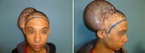
At the age of 13, the patient comes to the doctor’s office with the desire to improve the area of scar alopecia (Figure 1). In the first surgery, a 200cc (ml) semi-lunar silicone tissue expander was implanted under the scalp in the subgaleal plane (width 14.6cm x height 7.6cm x projection 4.3cm) for tissue expansion with 40ml of sf 0.9% already placed in this surgical act.
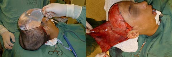
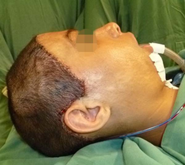
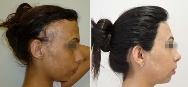
In the second week after surgery, 20 ml was instilled weekly for ten weeks until the volume of 240 ml of the expander was reached (Figure 2). After maximum expansion, the patient underwent a new surgical procedure after ten weeks in which the expander was removed, and an advancement flap was made for the region with scarring alopecia (Figure 3). A trichophytic incision was made in the scalp to place the flap on the topography of the hair’s cutlet and contour in the frontal-parietal region. The patient evolved well in the early and late postoperative periods without complications (Figures 4 and 5).
The current methodology was the retrospective analysis of the patient’s medical record. This paper follows the standards of the Helsinki ethics committee and CEP approval.
DISCUSSION
As in the case report in question, the patient desired to have hair in an area of scar alopecia (chronic defect due to thermal scalding burns) in the right parietal and temporal region, it was decided to use gradual tissue expansion with subsequent confection of advancement flap seen to the desired area for local capillary restoration.
The use of tissue expansion is a powerful resource because it allows the surgeon to replace tissue with a similar one. The technique increases the amount of tissue available locally, preserves sensitivity, and maintains hair follicles and attached structures. Defects of up to 50% of the scalp can be reconstructed with minimal distortion of the hairline1.
Before inserting a tissue expander, care must be taken to mark the vascular territories on the scalp. Expander placement is not random 1. In this case, the occipital vessels’ vascularization was preserved, and the contralateral vessels were used to make the flap (supratrochlear, supraorbital, superficial temporal, and posterior auricular).
The main indications for tissue expansion in the scalp are chronic injuries, such as scar alopecia, consistent with our patient’s condition. We can also highlight some contraindications to the method, such as acute traumatic injuries or active infectious processes, due to the risk of contamination of the expander and, consequently, its extrusion and loss of result. It is also contraindicated in children under three years of age, as there is the immaturity of the skullcap, which may, during expansion, cause deformities in the bone structure by an external pressure mechanism8.
Tissue expansion can be performed intraoperatively or in stages. In the intraoperative period, 3 to 4 cycles of inflation and disinflation of the expander are performed 3 to 5 minutes after placing the device, then, it is removed, and the wound is closed primarily.
A device is placed in the subcutaneous or subgaleal position in the staged technique and connected to a one-way valve. Expansion begins two weeks after placement. The device is expanded weekly or biweekly. The expansion must be continued until the expanded flap is 20% larger than the size of the defect (so that the skull’s curvature and the primary contracture of the flap during insertion are taken into account) 1. This technique was used in the surgical procedure with subsequent removal of the expanding device and making an advancement flap to cover the area of scar alopecia.
There were no early or late surgical complications in this case. The patient evolved well in the tissue expansion procedure, with weekly returns to the doctor’s office. After three months, a new surgical procedure was performed to remove the scalp expander, and the advancement flap was made.
CONCLUSION
The scalp scaling tissue expansion technique and subsequent scalp advancement flap preparation proved to be effective in restoring the patient’s hair structure and hairline with minimal local distortion, restoring the shape and aesthetics of the patient’s scalp.
REFERENCES
1. Neligan PC. Cirurgia plástica: princípios. 3a ed. Rio de Janeiro: Elsevier; 2015. v. 1.
2. Cunha CB, Sacramento RMM, Maia BP, Marinho RP, Ferreira HL, Goldenberg DC, et al. Perfil epidemiológico de pacientes vítimas de escalpelamento tratados na Fundação Santa Casa de Misericórdia do Pará. Rev Bras Cir Plást. 2012 Mar;27(1):3-8. DOI: http://dx.doi.org/10.1590/S1983-51752012000100003
3. Ribeiro NS. Necessidade e dilemas das famílias vítimas de escalpelamento atendidas na FSCMP: desafios para o serviço social [dissertação]. Belém: Universidade Federal do Pará; 2009.
4. Tutela JP, Banta JC, Boyd TG, Kelishadi SS, Chowdhry S, Little JA. Scalp reconstruction: a review of the literature and a unique case of total craniectomy in an adult with osteomyelitis of the skull. Eplasty. 2014 Jul;14:e27.
5. Makboul M, Abdel-Rahim M. Simple flaps for reconstruction of pediatric scalp defects after electrical burn. Chin J Traumatol. 2013;16(4):204-6.
6. Souza CD. Reconstrução de grandes defeitos de couro cabeludo e fronte em oncologia: tática pessoal e experiência - análise de 25 casos. Rev Bras Cir Plást. 2012 Jun;27(2):227-37. DOI: http://dx.doi.org/10.1590/S1983-51752012000200011
7. Zayakova Y, Stanev A, Mihailov H, Pashaliev N. Application of local axial flaps to scalp reconstruction. Arch Plast Surg. 2013 Sep;40(5):564-9. DOI: http://dx.doi.org/10.5999/aps.2013.40.5.564
8. Mélega JM. Cirurgia plástica: reconstrução de couro cabeludo e calota craniana. In: Mélega JM, ed. Cirurgia plástica: fundamentos e arte. Rio de Janeiro: MEDSI; 2012. p. 195-208.
9. Radovan C. Breast reconstruction after mastectomy using the temporary expander. Plast Reconstr Surg. 1982;69(2):195-208.
10. Manders EK, Schenden MJ, Furrey JA, et al: Soft-tissue expansion: Concepts and complications. Plast Reconstr Surg 1984; 74:493-505.
1. Service Prof. Dr. Ricardo Baroudi, Plastic
Surgery, Campinas, SP, Brazil.
2. Pontifical Catholic University of Campinas,
Faculty of Medicine, Campinas, SP, Brazil.
DNK Analysis and/or data interpretation, Conception and design study, Conceptualization, Data Curation, Final manuscript approval, Formal Analysis, Investigation, Methodology, Project Administration, Resources, Visualization, Writing - Original Draft Preparation, Writing - Review & Editing
ACN Supervision, Visualization, Writing - Review & Editing
LCS Visualization, Writing - Review & Editing
JGS Conceptualization, Supervision
Corresponding author: Daniel Nowicki Kaam Avenida Benjamin Constant, 1971, Apto 1603, Cambuí, Campinas, SP, Brazil. Zip Code: 13025-005 E-mail: danielnkaam@gmail.com
Article received: October 14, 2019.
Article accepted: February 22, 2020.
Conflicts of interest: none


 Read in Portuguese
 Read in English
 PDF PT
 Print
Print
 Send this article by email
 How to Cite
 Mendeley
 Pocket
 Twitter