
Fig. 1 - Preoperative frontal view 2 hours after the trauma.

Fig. 2 - Preoperative profile view showing laceration of the left ear.

Fig. 3 - Preoperative view of the avulsioned segment in saline solution after trichotomy and cleaning.

Fig. 4 - Preoperative frontal view of the avulsioned segment where the eyebrows can be visualized.

Fig. 5 - Transoperative view of the microanastomosis performed in the arteria and left superficial temporal vein.
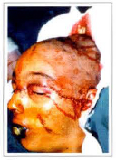
Fig. 6 - Transoperative view of the sutures after vascular reimplantation.

Fig. 7 - 30 days postoperative frontal view. Painless complete integration of the avulsioned segment.

Fig. 8 - 30 days postoperative right profile view.
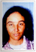
Fig. 9 - Postoperative view after 6 years and 10 months. Frontal view showing hair growth.

Fig. 10 - Postoperative view after 6 years and 10 months. Left profile view, showing face and ear scar sequelae.

Fig. 11 - 30 days postoperative rear view.

Fig. 12 - Postoperative view after 6 years and 10 months showing hair length, due ro religious beliefs.


