ISSN Online: 2177-1235 | ISSN Print: 1983-5175

Previous Article Next Article

Original Article - Year2013 - Volume28 - Issue 3

ABSTRACT

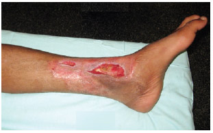
BACKGROUND: Fractures of the distal third of the leg, ankle, and foot frequently present with exposed bone, requiring specialized teams to accomplish skin coverage. These teams are often not readily available, which can prevent the standard treatment of extensive debridement, fracture fixation, and use of flaps, which is termed the "fix and flap" method.
METHODS: The author proposes a 2-stage treatment method for such cases: negative pressure is imposed during debridement and bone exposure by the orthopedic team, followed by the elective preparation of free flaps for definitive coverage. Five patients were treated with a total of 6 free flaps, including 1 latissimus dorsi muscle and 5 of anterolateral thigh flaps. There was 1 total flap loss. Thus, the success rate was 83.34%.
RESULTS: The patients had good outcomes with limb salvage, preserved function, and no osteomyelitis.
CONCLUSIONS: Negative-pressure wound therapy is an option for the emergency treatment of open fractures of the lower limb, allowing the survival of elective free flaps without resulting in impairment as a final outcome.

Keywords: Leg bones. Leg injuries. Tissue transplantation. Surgical flaps.

RESUMO

INTRODUÇÃO: Traumas do terço distal da perna, do tornozelo e do pé cursam frequentemente com exposição óssea, demandando equipes especializadas para realizar a cobertura cutânea definitiva. Muitas vezes, essas equipes não estão prontamente disponíveis, o que pode impedir o tratamento padrão, que consiste em amplo desbridamento, fixação das fraturas e utilização de retalhos (fix and flap).
MÉTODO: Neste trabalho, o autor propõe tratamento em duas etapas, sendo a primeira a instituição de terapia por pressão negativa quando do desbridamento e exposição óssea pela equipe da ortopedia, seguida pela realização de retalhos livres para cobertura definitiva, de forma eletiva. Foram tratados 5 pacientes, com realização de 6 retalhos livres, sendo 1 do músculo grande dorsal e outros 5 da face ântero-lateral da coxa. Houve perda total de um retalho, com índice de sucesso de 83,34%.
RESULTADOS: Os pacientes apresentaram boa evolução, com salvamento do membro, função preservada, e sem osteomielite.
CONCLUSÕES: A terapia por pressão negativa é uma opção no tratamento de urgência das exposições ósseas do membro inferior, permitindo a realização de retalhos livres de forma eletiva, sem prejuízo no resultado final para o paciente.

Palavras-chave: Ossos da perna. Traumatismos da perna. Transplante de tecidos. Retalhos cirúrgicos.


INTRODUCTION

Trauma of the lower limbs is a special challenge for plastic surgeons. Because of their anatomical features, the distal third of the leg, ankle, and foot regions are more susceptible to bone exposure after loss of soft tissue. Furthermore, local flap options are more limited than in other body areas. The standard treatment for bone exposures in these areas is extensive debridement, fracture fixation, and flap coverage within 72 hours, which is known as "fix and flap" 1,2. Despite the good results with this method reported in the literature, the following factors limit the wide applicability of the technique 3:
  • Local wound conditions: it is sometimes difficult to determine which tissue is viable and must be debrided, warranting consecutive cleaning until the time of definitive treatment.
  • Clinical condition of the patient: the combination of debridement, fracture fixation, and flap creation demand a prolonged surgical time. Some multiple trauma patients may not have adequate stability or clinical condition for large surgical procedures.
  • Plastic surgery teams with high availability: the creation of free flaps demands specialized staff and equipment, which is not always possible within the proposed 72-hour window.
  • Accordingly, this report presents the author's experience working as an elective plastic surgeon at a secondary referral hospital for the treatment of orthopedic cases by using negative-pressure wound therapy, which is an increasingly widespread modality and use for a temporary coverage in an attempt to resolve some of the abovementioned limiting factors.


    METHODS

    Orthopedic and nursing teams were previously trained to perform the dressing for negative-pressure therapy. In cases of debridement performed by the orthopedic team that would result in the exposure of bone in the distal region of the lower limb with no possibility of coverage with adjacent soft tissues, negative-pressure therapy was performed in the operating room and changed every 72 hours by nursing staff. Evaluation by a plastic surgeon was performed at the first subsequent elective schedule.

    During the 7 months of the study, 9 patients underwent debridement and negative-pressure therapy. Among them, 2 were treated with regional flaps of the gastrocnemius muscle, 2 received skin graft coverage after adequate granulation, and 5 were treated with free flaps, which will be described herein.

    Negative-pressure wound therapy, also known as vacuum dressing, was performed as follows:
  • Bone covered with rayon
  • Placement of polyurethane foam covering the entire wound
  • Complete sealing of the dressing with waterproof plastic film
  • Introduction of a plastic drain tube for continuous suction
  • Application of negative-pressure through a vacuum pump
  • After evaluation by the plastic surgeon, the options were expansion of debridement, maintaining negative-pressure therapy, or surgical planning for definitive treatment with free flaps.

    Case Descriptions

    Case 1 was a 15-year-old male patient who was a victim of a Bothrops snakebite. The patient was referred to a specialist service, where he received Bothrops anti-venom; he developed edema of the left ankle and foot as well as skin necrosis and purulent drainage after 10 days. He was referred for treatment at the hospital where this study was conducted. Three debridements were carried out by surgical and orthopedic teams. In the last debridement, the extensor retinaculum and tendon of the anterior tibialis muscle were removed, leading to exposure of the bones and ankle joint. There was also an accidental injury of the dorsalis pedis artery, which was ligated. Negative-pressure therapy was subsequently initiated and maintained for 13 days until local edema was reduced and it was certain no progression of tissue necrosis would occur. The patient then underwent treatment with a free flap of the ipsilateral dorsal large muscle with a skin island monitoring flap. Because of the dorsalis pedis artery injury and wound location, terminoterminal anastomosis in the anterior tibial vessels was performed with separate sutures of 9-0 and 10-0 nylon for the arterial and single venous anastomoses, respectively. After one week, the viability of the flap was confirmed, and partial skin graft on the latissimus dorsi muscle was subsequently performed. The patient was discharged one week after grafting and is currently receiving outpatient treatment. Coverage is stable without evidence of osteomyelitis. Ambulation is possible with mild restriction because of the loss of function of the anterior tibial muscle, which is partially offset by the adjacent extensor muscles. Figures 1 to 5 illustrate this case.


    Figure 1 - Case 1: debridement with exposure of ankle bones and joints.


    Figure 2 - Case 1: appearance after negative-pressure therapy.


    Figure 3 - Case 1: appearance 1 week after latissimus dorsi free flap.


    Figure 4 - Case 1: appearance after latissimus dorsi partial skin graft.


    Figure 5 - Case 1: patient with good ambulation and no infections in ambulatory monitoring.


    Case 2 was a 40-year-old male patient who was victim of a motorcycle accident. He had left distal tibial fracture initially treated by fixation with plates and screws. Five days after fracture fixation, he developed necrosis of the adjacent skin. He underwent debridement and negative-pressure therapy, which resulted in good granulation and closure by secondary intention. Therefore, he was discharged. Five months later, he returned to the hospital with drainage of pus at the wound site. After surgical debridement, fracture healing was observed, the plate and screws were removed, and he started treatment with vacuum therapy. After 18 days of negative-pressure therapy, there was good granulation but exposure of the tibia persisted in the callus region. Coverage with free fasciocutaneous flap from the anterolateral region of the contralateral thigh (ALT Flap) was subsequently performed with termino-terminal anastomosis of the posterior tibial vessels with separate 9-0 and 10-0 nylon sutures in the arterial and venous anastomoses, respectively. As the flap remained congested, a second venous anastomosis was performed in a concomitant vein. The patient recovered well and was discharged after a week. In outpatient treatment, coverage remains stable even after a new automobile accident and ambulation is normal. No signs of osteomyelitis were present after the free flap was completed. Figures 6 and 7 illustrate this case.


    Figure 6 - Case 2: persistent exposure of the tibia after negative-pressure therapy.


    Figure 7 - Case 2: free flap (ALT Flap) at 5 months.


    Case 3 was a 22-year-old male patient who was hit by a truck. He presented with broken bones in the right ankle and loss of substance in the dorsum of the foot and knee. Debridement was performed by the orthopedic team, exposing the ankle bones and extensor tendons. Negative-pressure therapy was initiated and maintained for 20 days. Debridement was subsequently expanded with removal of the extensor hallucis longus tendon and coverage of the dorsal aspect of the foot and ankle with an ipsilateral ALT flap. A termino-terminal anastomosis was performed using anterior tibial vessels, with separate 9-0 and 10-0 nylon sutures in the arterial and single venous anastomoses, respectively. The patient recovered after 12 hours. However, he developed venous congestion and was taken to the operating room, where venous thrombosis caused by excessive stretching of the flap was detected. After fixation in the most suitable position, thrombus removal, and recreation of the venous anastomosis, evolution was favorable with a loss of 5 mm from the distal edge of the flap. The remaining areas were grafted after a week. The patient was discharged one week after grafting. Three months after free flap creation, he has normal ambulation and no signs of osteomyelitis. Figures 8 to 11 illustrate this case.


    Figure 8 - Case 3: appearance after debridement with exposure of ankle extensor tendons and bones.


    Figure 9 - Case 3: appearance after negative-pressure therapy.


    Figure 10 - Case 3: loss of 5 mm from the distal edge of the ALT flap.


    Figure 11 - Case 3: appearance after treatment with free flap and grafting.


    Case 4 was a 17-year-old male patient who fell from standing height. He presented with closed fracture of the left tibial pilon. An external fixation device was initially placed in the emergency room. After 10 days, open reduction internal fixation was performed with plates and screws, followed by primary closure of the incisions. He developed necrosis of skin and subcutaneous tissue in the medial malleolus region after 5 days with exposure of the synthetic material and fracture site. He underwent debridement and received negative-pressure therapy for 9 days. After this period, as the exposure of synthetic material persisted, contralateral ALT flap coverage was indicated with termino-terminal anastomosis in the posterior tibial vessels using separate 9-0 and 10-0 nylon arterial and single venous sutures, respectively. During monitoring of the flap, pallor as well as loss of turgor and temperature were detected after 12 hours. Therefore, the patient underwent surgical re-exploration. After unsuccessful rescue maneuvers, we opted to create another flap - this time from the anterolateral surface of the ipsilateral thigh. There was partial flap loss after one week, which was treated conservatively on an outpatient basis through debridement with a scalpel and papain. After 3 months of outpatient treatment, there was good healing without signs of osteomyelitis. The orthopedic team recommended restricted ambulation for this patient. He had preserved movement and sensitivity and performed physical therapy to gain strength and recover proprioception. Figures 12 to 15 illustrate this case.


    Figure 12 - Case 4: appearance after debridement, tibial exposure, and bone synthesis.


    Figure 13 - Case 4: appearance after negative-pressure therapy and persisting exposure of osteosynthesis material.


    Figure 14 - Case 4: partial necrosis of the free flap edges.


    Figure 15 - Case 4: appearance after ambulatory debridement and healing.


    Case 5 was a 45-year-old male patient who suffered a fall from a ladder. He presented with a distal closed fracture of the left tibia. The patient was initially treated with an external fixation device in the emergency room. After one week, the external device was exchanged for internal fixation with plates and screws, and iliac crest bone grafting for comminuted bone fracture was performed. Five days after open reduction internal fixation, the patient developed skin necrosis and adjacent pus drainage. During debridement, bone graft loss was observed and was treated by placement of an orthopedic cement spacer with antibiotics, followed by grafting of skin and subcutaneous tissue, and then negativepressure therapy. After 11 days of negative-pressure therapy, the orthopedic cement was replaced and a contralateral ALT flap was placed with termino-terminal anastomosis in the posterior tibial vessels and separate 9-0 and 10-0 nylon sutures for the arterial and singe venous anastomoses, respectively. The patient recovered well and was discharged after 7 days. He received outpatient treatment for 2 months, during which, he presented with stable coverage without evidence of osteomyelitis. He is awaiting definitive treatment, according to the treatment plan of the orthopedic team. Figures 16 to 18 illustrate this case.


    Figure 16 - Case 5: appearance after debridement of skin and subcutaneous tissue, and removal of infected bone graft.


    Figure 17 - Case 5: appearance after negative-pressure therapy and use of orthopedic cement spacer with antibiotics.


    Figure 18 - Case 5: appearance after ALT free flap.


    RESULTS

    A total of 5 patients were treated, with the completion of 6 free flaps. One total flap loss was attributed to microthrombosis, identified by the no-reflow phenomenon. Thus, the success rate of the flaps was 83.34%. All patients ultimately achieved stable coverage. As of writing, no patient has presented with osteomyelitis. Two patients have still not completely recovered ambulation and are awaiting definitive orthopedic treatment; however, both have good motor response in physiotherapy.


    DISCUSSION

    Coverage of exposed bone of the distal third of the leg, ankle, and foot poses unique challenges to plastic surgeons. As these regions are anatomically unfavorable for the creation of appropriately sized locoregional flaps, it is often necessary to use free flaps, which require specialized equipment and trained staff. Inability to maintain specialized staff always available sometimes restricts the use of the standard technique fix and flap technique described in the literature, which advocates extensive debridement of unviable tissue, fracture fixation, and stable coverage during surgery or up to 72 hours during the acute phase of bone exposure 1-3.

    Previous studies demonstrate the treatment failure rate measured according to the occurrence of osteomyelitis or non-healing is larger when the covering flap is applied in the subacute phase of the wound between 72 hours and 6 weeks after bone exposure 2,4. The advent of negative-pressure therapy may prolong the optimal treatment phase, allowing a narrower, staged debridement as well as allowing the surgery to be performed by the plastic surgery team on an elective basis 3,5,6. Furthermore, negative-pressure has been suggested to reduce the initial complexity of the cases, decreasing the size of the flap required for coverage or even enabling the use of grafts or healing by second intention6,7.

    At the hospital where this study was conducted, the author acts as a single hired plastic surgeon with an orthopedic team comprising 15 doctors attending to cases brought by rescue teams or referred from smaller hospitals as needed. The treatment protocol presented herein was successfully introduced in order to balance demand with the availability of surgical time. The implementation of the negative-pressure dressing was taught to orthopedic and vascular surgery teams, who often perform debridement of the lower limbs. The nursing team was also able to conduct exchanges at bedside. Thus, to ascertain the need for skin coverage by the plastic surgery team, negative-pressure therapy is set and the schedule for the free flap is programmed electively when needed.

    In addition to the abovementioned benefits, the technique improves safety with respect to the vascularity of the affected region, which allows most cases to be treated with fasciocutaneous flaps; this reduced patient morbidity, because there was no loss of muscle function associated with flaps traditionally used for these reconstructions (i.e., latissimus dorsi and rectus abdominis).

    Despite the small number of cases, the present results are encouraging, because the patients have preserved limbs and function, stable coverage, and bone integrity without infections.

    Finally, the combination of negative-pressure therapy in the acute phase with final coverage in the subacute phase offers a treatment option for less-equipped centers. Because of its easy preparation, the vacuum dressing can be performed by any surgical or nursing staff depending only on simple training. Patients can subsequently wait for the availability of specialized staff or venues for definitive treatment, possibly without endangering the final results.


    CONCLUSIONS

    The exposed bone of the lower limb entails complex treatment that requires specialized plastic surgical and orthopedic teams. Treatment in the acute phase requires high availability of professionals, which is not always possible. The use of negative-pressure therapy extends the ideal phase for definitive treatment without risk to the patient. In some cases, this combination treatment even appears to be superior to the traditional fix and flap as it allows a more parsimonious debridement, greater assurance of the viability of the remaining tissue, and less morbid flaps or even simpler treatments such as grafts or closure by second intention. The use of this treatment opens possibilities for limb salvage for smaller hospitals and more remote locations by enabling patient transfer to specialized centers after initial debridement.


    REFERENCES

    1. Byrd HS, Spicer TE, Cierney G 3rd. Management of open tibial fractures. Plast Reconstr Surg. 1985;76(5):719-30.

    2. Gopal S, Majumder S, Batchelor AG, Knight SL, De Boer P, Smith RM. Fix and flap: the radical orthopaedic and plastic treatment of severe open fractures of the tibia. J Bone Joint Surg Br. 2000;82(7):959-66.

    3. Rinker B, Amspacher JC, Wilson PC, Vasconez HC. Subatmospheric pressure dressing as a bridge to free tissue transfer in the treatment of open tibia fractures. Plast Reconstr Surg. 2008;121(5):1664-73.

    4. Choe EI, Kasabian AK, Kolker AR, Karp NS, Zhang L, Bass LS, et al. Thrombocytosis after major lower extremity trauma: mechanism and possible role in free flap failure. Ann Plast Surg. 1996;36(5):489-94.

    5. Stannard JP, Singanamala N, Volgas DA. Fix and flap in the era of vacuum suction devices: what do we know in terms of evidence based medicine? Injury. 2010;41(8):780-6.

    6. Krug E, Berg L, Lee C, Hudson D, Birke-Sorensen H, Depoorter M, et al.; International Expert Panel on Negative Pressure Wound Therapy [NPWT-EP]. Evidence-based recommendations for the use of negative pressure wound therapy in traumatic wounds and reconstructive surgery: steps towards an international consensus. Injury. 2011;(42 Suppl 1):S1-12.

    7. DeFranzo AJ, Argenta LC, Marks MW, Molnar JA, David LR, Webb LX, et al. The use of vacuum-assisted closure therapy for the treatment of lower-extremity wounds with exposed bone. Plast Reconstr Surg. 2001;108(5):1184-91.


    Physician, plastic surgeon, Hospital Universitário, Universidade de São Paulo, full member of the Brazilian Society of Plastic Surgery, São Paulo, SP, Brazil

    Correspondence to:
    Gustavo Flosi Stocchero
    Rua Abílio Soares, 1.337 - Paraíso
    São Paulo, SP, Brazil - CEP 04005-005
    E-mail: gustavo@vivermelhor.com.br

    Submitted to SGP (Sistema de Gestão de Publicações/Manager Publications System) of RBCP (Revista Brasileira de Cirurgia Plástica/Brazilian Journal of Plastic Surgery).
    Article received: 26/7/2012
    Article accepted: 15/12/2013

    Work performed at Hospital Universitário, Universidade de São Paulo, São Paulo, SP, Brazil.


    Previous Article Back to Top Next Article

    Indexers

    Licença Creative Commons All scientific articles published at www.rbcp.org.br are licensed under a Creative Commons license