
Case Report - Year 2024 - Volume 39 -
Circumferential congenital scar on the lower limb due to amniotic band syndrome addressed with Wplasty
Cicatriz congênita circunferencial em membro inferior devido à síndrome da banda amniótica abordada com dablioplastia
ABSTRACT
Amniotic band syndrome (ABS) is defined as a condition in which chorioamniotic constriction occurs in fetal parts, causing congenital scars. Most commonly, it affects the upper and lower limbs and can cause growth restriction in the affected region. It can also result in intrauterine amputations, lymphedema, congenital clubfoot, syndactyly, and death. The stratification of the condition is given by the Patterson scale, which takes into account the ring formation and its consequences for the affected part. The objective of this work is to report the case of a female patient following an uneventful pregnancy who suffered involvement of the left lower limb by ABS. The scar caused growth restriction, requiring surgical repair treatment. We opted for the Wplasty approach, so that, in the immediate and late postoperative period, the scar retraction was properly corrected, without signs of dehiscence or inflammation in the region.
Keywords: Congenital abnormalities; Cicatrix; Child; Case reports; Plastic surgery procedures.
RESUMO
Síndrome da banda amniótica (SBA) é definida por uma condição em que ocorre uma constrição corioamniótica em partes fetais, causando cicatrizes congênitas. Mais comumente, acomete os membros superiores e inferiores, podendo causar restrição do crescimento da região afetada. Pode, ainda, resultar em amputações intrauterinas, linfedema, pé torto congênito, sindactilias e morte. A estratificação da condição é dada pela escala de Patterson, que leva em consideração a formação anelar e sua consequência para a parte acometida. O objetivo deste trabalho é relatar o caso de uma paciente feminina advinda de gestação com intercorrências que sofreu acometimento do membro inferior esquerdo pela SBA. A cicatriz causava restrição de crescimento, sendo preciso proceder ao tratamento cirúrgico de reparo. Optou-se pela abordagem de dablioplastia, de modo que, no pós-operatório imediato e tardio, a retração cicatricial foi devidamente corrigida, sem sinais de deiscência ou de inflamação da região.
Palavras-chave: Anormalidades congênitas; Cicatriz; Criança; Relatos de casos; Procedimentos de cirurgia plástica.
INTRODUCTION
Amniotic band syndrome (ABS) comprises a wide range of congenital changes attributed to the constriction of fetal parts in fibrous chorioamniotic rings. Its incidence is estimated between 1:1200 and 1:15000 live births1.
To clarify the etiology of this syndrome, several theories have emerged. Initially, in 1930, Streeter presented the endogenous theory, in which defects in the germplasm with diffuse vascular rupture and chorioamniotic morphogenetic alterations result in the formation of fibrotic bands and abnormalities of fetal development. In 1968, Torpin2 proposed the exogenous theory, in which the rupture of the amnion before the 12th week, caused by exogenous factors, would provide direct contact between the fetus and the chorionic surface, with consequent oligohydramnios following extravasation of the amniotic fluid and easy protrusion through the rupture. Thus, favoring the formation of amniotic fibrous bands in the ruptured portion, which compromises the development of the structures involved distal to the band3, 4.
Both theories explain the presence of malformations of the limbs, abdominal wall, internal organs, and craniofacial5. Constrictions can result in extensive symptoms, from lymphedema, intrauterine amputations, syndactyly, congenital clubfoot, and death1. There are no known factors that have statistically proven associations with ABS. Some conditions that are related, but are not statistically significant, include primiparous women, age under 25 years, collagenopathies, trauma resulting from attempted abortions, abnormal glycemic indices, and prematurity6, 7.
Patterson listed the diagnostic criteria based on the symptoms in order of increasing severity as a way of stratifying the symptoms as follows: 1) simple constriction rings with a normal end distal to the ring; 2) ring with distal atrophy and lymphedema; 3) ring with syndactyly on the affected extremities; 4) ring that causes amputation1, 8.
OBJECTIVE
Because of the above, this work aims to report a case of a congenital circumferential scar on the lower limb due to amniotic band syndrome in a 2-year-old female patient, detailing the therapy used, in addition to carrying out a literature review. detailing the surgical approach to this syndrome, thus helping clinical management and the prognosis of future cases related to ABS.
METHOD
This is a documentary and retrospective study, carried out through an active search of the medical records of a patient diagnosed with ABS at the Walter Cantídio University Hospital (HUWC), in the city of Fortaleza, Ceará, Brazil. The hospital is a unit that provides highly complex health care, in addition to being a reference for the training of human resources and the development of research in the health area. Provides support for the tertiary care network in the state of Ceará, serving patients from the Unified Health System (SUS). Relevant information was collected by evaluating the patient’s medical records.
Regarding ethical aspects, the present project, as it is research with human beings, was submitted for evaluation by the HUWC Research Ethics Committee following Resolution 466/12 of the National Health Council (CNS). All established ethical precepts will be respected concerning ensuring the legitimacy of information, preserving anonymity, privacy, and confidentiality of information taken from the patient’s medical record.
CASE REPORT
A female patient, 2 years old, from a twin pregnancy with feto-fetal transfusion syndrome discovered in the 17th week of pregnancy. After a week, an intrauterine fetoscopy was performed to correct the syndrome; however, the second fetus died on this occasion. Born by premature cesarean section (26 weeks of gestation), with fetal distress and weighing 635g (small for gestational age - SGA).
She was intubated shortly after birth due to the immaturity of her respiratory system, requiring three doses of surfactant - the first at 40 minutes of life. In this context, she remained hospitalized in the Intensive Care Unit (ICU) for 93 days, 57 days of which were on mechanical ventilation. During hospitalization, she developed necrotizing enterocolitis, stage IIIb, requiring exploratory laparotomy with the creation of an ileostomy.
Furthermore, during his hospital stay, he also presented: early and late neonatal infections, fungal infection, bacterial pneumonia, and grade I peri-intraventricular hemorrhage (IVPH), in addition to a convulsive episode that ended with the use of phenobarbital. She was discharged from the hospital 3 months and 28 days after her birth, clinically stable.
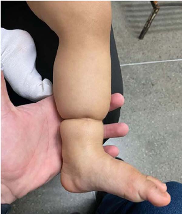
At 7 months of age, she was diagnosed with an amniotic band scar on her left lower limb, precisely in the ipsilateral calf region, which caused significant constriction of the limb, compromising its physiological growth and development (Figure 1). Therefore, she was recommended surgical correction and was referred to the plastic surgery service.
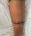
The scar retraction correction procedure was performed in the affected region, using Wplasty with the patient under general anesthesia, on August 8, 2022 (Figure 2). Anesthetic infiltration with epinephrine was performed, followed by infusion and detachment of the retracted area. Incisions continued to relax the fascia, as well as rigorous hemostatic inspection. The plans were closed.
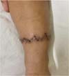
In the immediate postoperative period (PO), the patient developed an intact surgical wound, without tension on the limb and with the region distal to the wound well perfused (Figure 3). She was discharged from the hospital on the 1st day of PO, without complaints, and was advised to return to the outpatient clinic after three days.
At the time of return, the surgical wound was without inflammatory signs, without dehiscence, and with well-coadapted edges that were not tensioned (Figure 4). The second evaluation, on the 18th postoperative day, found an ideal appearance of the wound, as well as preserved normal tension. The stitches were removed and continued on an outpatient basis (Figure 5).
DISCUSSION
As it is a rare congenital condition, most of the literature on amniotic band syndrome (ABS) still consists of case reports, with less diversity in the theoretical approach to this condition9, 10.
Regarding recognized therapies, the possibility of surgical treatment strongly depends on the topography and functionality of the affected organ. The presence of noticeable deformity with or without lymphedema indicates surgical intervention. The approach can be performed between 3 months and 2 years of age unless there is the possibility of significant neurovascular involvement or lymphedema. This does not contraindicate performing the release later, but the results concerning limb growth are enhanced when the intervention is early11, 12.
Lower limb constriction associated with clubfoot is the most prevalent deformity in Brazil, followed by upper limb involvement9, 11. However, epidemiology differs concerning international articles, which define the upper limbs as the most affected area, with emphasis on the fingers7, 13.
Among the anatomofunctional deformities that may be included in the list of consequences of ABS, Drury & Rayan13 listed: partial limb constriction, complete constriction band, intrauterine amputation, fenestrated acrosyndactyly, partial syndactyly, bone growth at the site of intrauterine amputation, deficient interdigital space, lymphedema, remaining digits, ectopic implantation of amputated fingers in another part of the body, proximal interphalangeal joint contracture, nerve compression, complete nerve rupture, among others.
From this perspective, indications range from bridle resection, with deep dissection and release of the neurovascular bundle, flap reconstruction using Z-plasty, Wplasty, and even, in the most serious cases, amputation of affected limbs. When the upper or lower limb is affected in isolation, the release of the retracted structures with subsequent Z-plasty, single or multiple, has a high potential for a good prognosis, with single procedures having fewer reports of complications.
Furthermore, the bibliography points out that the Wplasty approach, despite not having a significant number in practice, is seen as a safe technique with satisfactory postoperative results. In cases of lower limbs associated with foot involvement, surgical treatment in two sequential stages is recommended9.
In this case, the patient benefited from Wplasty therapy in just one surgical procedure, since the constriction was isolated to the left calf, and, despite there being growth delay, there was no tortuosity or deformity in the feet. According to the literature analysis, the one-stage surgical approach was not responsible for an increase in ischemic complications or the development of venous congestion12.
However, the discussion regarding the circumferential constrictive band approach in one or more surgical stages remains fruitful. The issue discussed involves the personalization of treatment, necessary given the large number of possibilities for deformities resulting from the amniotic band. The importance of surgical planning is highlighted to obtain the best possible result within the child’s aesthetic and functional aspects7.
It must be analyzed which structures are affected, whether there is lymphedema, and the impact on the growth of the limb, among other factors. The approach in two or more surgical stages may be preferable in cases of association of multiple deformities (which would make it difficult to approach on just one occasion), blood or lymphatic circulation disorders in the distal segment, need to accommodate growth, and in patients who tend to worse healing (as more overt approaches can carry greater risks in this regard)11, 12, 14.
In any of these cases, mobilization of a fatty tissue flap may improve the aesthetics of the repair in the area of the constriction. After excision of the skin around the band, there may be an hourglass-shaped deformity of the affected limb, so that, even after resolution of the constriction, the patient remains with aesthetic impairment. One way to minimize this issue is to excise excess adipose tissue from surrounding areas11.
It was decided to make an incision and detachment of the retraction area, followed by relaxing the local fascia through another incision, ending with closure in layers. Postoperatively, the patient had no complaints or bleeding. At ectoscopy, the wound was well closed, without tension or areas of dehiscence, and with adequate perfusion. She was discharged from the hospital and scheduled to return for outpatient follow-up.
CONCLUSION
As it is a disruptive malformation, amniotic band syndrome compromises the life of the fetus and alters its development, which can range from the amputation of one or more limbs to fetal death. Therefore, early diagnosis is extremely important so that the best approach can be chosen, which can be helped by applying the Patterson classification8, and the main point must be the preservation of the functionality of the affected limbs, aiming to improve the patient’s prognosis.
It should be noted that establishing a good doctor-patient relationship with parents is extremely important to reassure them both about the pregnancy and the child’s development. Furthermore, there must be a multidisciplinary treatment, intending to obtain better long-term results, whether functional or aesthetic.
REFERENCES
1. Guillen Botaya E, Pino Almero L, Molini Menchón MO, Gonzalez Alonso V Pérez-Montejano M, Minguez Rey MF Tratamiento de las malformaciones en extremidades en el síndrome de bridas amnióticas: a propósito de un caso. Arch Argent Pediatr. 2020;118(5):e486-90.
2. Torpin R. Fetal malformations caused by amnion rupture during gestation. Arch Intern Med. 1968;122(2):191.
3. Nogueira FCS, Cruz RB, Machado LIP Ramos BLF, Madureira Júnior JL, Pinto RZA. Síndrome da banda amniótica: relato de caso. Rev Bras Ortop. 2011;46(Suppl 4):56-62.
4. Chatterjee S, Rao KSM, Nadkarni A. Amniotic band syndrome associated with limited dorsal myeloschisis: a case report of an unusual case and review of the literature. Childs Nerv Syst. 2021;37(2):707-13.
5. He T, Xu H, Sui P Wang X, Sun Y. Amniotic constriction band syndrome resulting in amputation caused by septate uterus: a case report. J Int Med Res. 2020;48(9):300060520949755.
6. Costa EN, Alves MP, Fraga CEC, Silva Júnior JAT, Daher O. Síndrome das bandas de constrição congênita. Estudo de 16 casos. Rev Bras Ortop. 1996;31(4):341-6.
7. Estanbouli MA, Anadani A, Albobah H, Dakkak T, Mokresh R, Etr A. Late management of amniotic bands syndrome with incomplete syndactyly: A case report of 4-year-old child. Int J Surg Case Rep. 2024;115:109277.
8. Patterson T. Congenital ring-constrictions. Br J Plast Surg. 1961;14:1-31.
9. Claro KTV Portinho CP? Ramirez JLH, Cubilla JJ, Collares MVM, Bampi R, et al. Síndrome de bandas amnióticas: relato de caso. Rev Bras Cir Plást. 2018;33(Suppl.1):148-9.
10. Falsaperla R, Arrabito M, Pavone P, Giacchi V, Timpanaro T, Adamoli P. Diagnostic Clue in a Neonate with Amniotic Band Sequence. Case Rep Pediatr. 2020;2020:8892492.
11. Upton J, Tan C. Correction of constriction rings. J Hand Surg Am. 1991;16(5):947-53.
12. Inglesby DC, Janssen PL, Graziano FD, Gopman JM, Rutland JW Taub PJ. Amniotic Band Syndrome: Head-to-Toe Manifestations and Clinical Management Guidelines. Plast Reconstr Surg. 2023;152(2):338e-46e.
13. Drury BT, Rayan GM. Amniotic Constriction Bands: Secondary Deformities and Their Treatments. Hand (N Y). 2019;14(3):346-51.
14. Chan AHW, Zeitlinger L, Little KJ. Multiple Continuous Y-to-V-Plasties for Excision and Reconstruction of Constriction Band Syndrome: Case Series and Description of Surgical Technique. Plast Reconstr Surg. 2022;149(4):774e-8e.
1. Hospital Universitário Walter Cantídio, Serviço de Cirurgia Plástica e Microcirurgia
Reconstrutiva, Fortaleza, CE, Brazil
2. Universidade de Fortaleza, Curso de Medicina, Fortaleza, CE, Brazil
Corresponding author: Isabela Franco Freire Av. Engenheiro Santana Júnior, 2937, apto 202, Cocó, Fortaleza, CE, Brazil. Zip Code: 60192-205. E-mail: isabelafrancofreire@edu.unifor.br


 Read in Portuguese
 Read in English
 PDF PT
 Print
Print
 Send this article by email
 How to Cite
 Mendeley
 Pocket
 Twitter