ISSN Online: 2177-1235 | ISSN Print: 1983-5175

Previous Article

Original Article - Year2011 - Volume26 - Issue 1

ABSTRACT

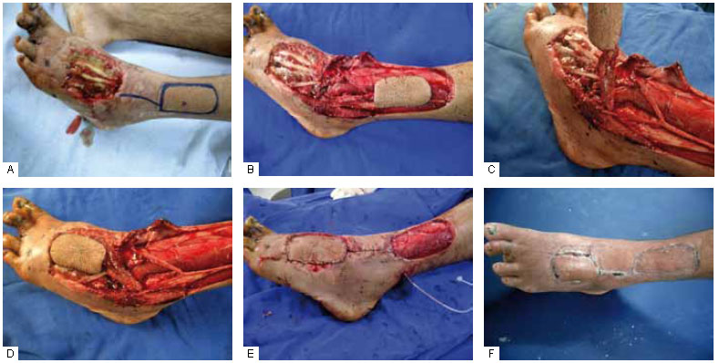
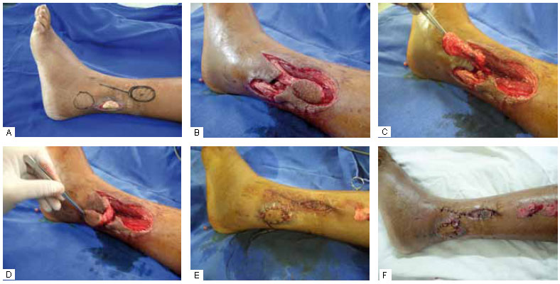
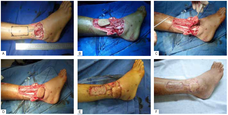
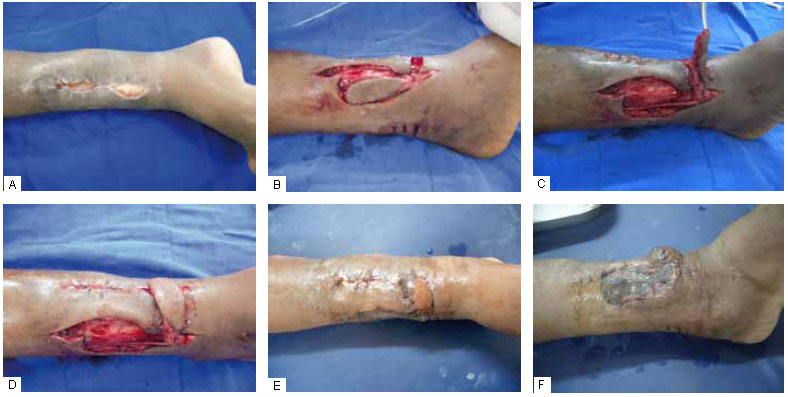
The distal regions of the lower limbs, including the lower third of the legs and feet, are very prone to soft tissue loss due to trauma and trophic lesions. The anterior part of the tibia with its slightly thick, vulnerable cover and large number of tendons, which cannot remain exposed, requires fast and complex skin coverage. The blood-flow direction, lack of muscle bellies for flaps, and inelastic skin for viable randomized flaps complicate this situation. In the past, intermediate-transfer flaps and cross-legged flaps were used for skin coverage, with drawbacks: they require more than one surgery, cause great discomfort to the patient, and are not always effective. Microsurgery is an option that greatly helps to cover lesions at this site; however, it requires a team with specialized training and equipment. Donor areas that are larger, may cause functional impairment, and use larger vascular trunks. Fasciocutaneous flaps with reverse flow may help to cover small and medium lesions at this location, with minor aesthetic and functional sequelae, and less surgical time. They are an effective surgical option because of their ease, safety, and versatility. The aim of this paper is to demonstrate the use of reverse-flow supramalleolar flaps in 4 different situations and discuss their advantages and applications.

Keywords: Surgical flaps. Leg injuries. Ankle injuries. Lower extremity/surgery.

RESUMO

As regiões distais do membro inferior, incluindo o terço inferior da perna e os pés, são áreas muito propensas a perda de partes moles por trauma e lesões tróficas. A posição anterior da tíbia, com sua cobertura pouco espessa e vulnerável, além da grande quantidade de tendões, que não podem permanecer expostos, exigem uma cobertura cutânea rápida e, ao mesmo tempo, complexa. A situação em relação à direção do fluxo sanguíneo, a escassez de ventres musculares para retalhos e pele pouco elástica para retalhos randomizados viáveis complicam esta situação. No passado, retalhos com transferência intermediária foram utilizados; assim como os de perna cruzada, "cross leg", ainda são, com desvantagens importantes: dependem de mais de um tempo cirúrgico, geram muito desconforto ao paciente e nem sempre são eficazes. A microcirurgia tornou-se uma opção que contribui muito para cobertura de lesões neste aspecto, porém, depende de equipe com treinamento e material especializado. As áreas doadoras são maiores, podem causar relativo déficit funcional e utilizam troncos vasculares maiores. Os retalhos fasciocutâneos de fluxo reverso podem contribuir para a cobertura de lesões pequenas e médias nesta localização, com sequelas funcionais e estéticas menores; e menor tempo cirúrgico. Constitui eficiente opção cirúrgica por sua facilidade, segurança e versatilidade. Neste estudo, temos o objetivo demonstrar o retalho supramaleolar de fluxo reverso utilizado em 4 casos, com aplicações em situações diferentes, com resultados satisfatórios de fácil execução e reprodução.

Palavras-chave: Retalhos cirúrgicos. Traumatismos da perna. Traumatismos do tornozelo. Extremidade inferior/cirurgia.


INTRODUCTION

Studies on the distribution of vessels and blood flow enabled advances in the treatment of skin lesions. With improvements in the procedures to save parts of the extremities, including orthopedic solutions for more complex trauma, the repair of structures that do show good wound coverage with simple skin grafts and simpler flaps was not viable1. This prompted the creation of intermediate transfer flaps. The impossibility of reconstruction with local flaps fostered the field of microsurgery with its applications.

Pontén2, in 1981, showed that fasciocutaneous flaps are an alternative to microsurgical flaps. Since then, several studies have demonstrated the continuous vascularization of these flaps and their safe clinical applications throughout the body3.

The distal position of some lesions in the limbs makes the application of direct-flow flaps difficult and quite impossible. The reverse-flow Chinese flap is a solution for upper-extremity lesions, as is the posterior interosseous artery flap with a pivotal point based on the wrist anastomoses. In the lower limb, the appendicular and distal positions of the leg, ankle, and instep third, and the dynamics of the region facilitate trauma. Lesions in these areas are still a challenge in plastic surgery. There are flaps similar to those of the upper limb, such as the anterior tibial artery flap4 and the sural flap. The supramalleolar flap can be considered analogous to the posterior interosseous artery flap.

Reverse blood flow is maintained and explained by the anastomoses around the ankle. The venous flow, a subject under discussion, is obtained by valvular insufficiency through the pressure gradient itself; communicating veins, capillaries, and collaterals; and denervation of the valves during pedicle release and elevation4,5. These factors also explain the temporary edema associated with reverse flaps.

The aim of this paper is to demonstrate the use of reverse-flow supramalleolar flaps in 4 different situations and discuss their advantages and facilities.


METHODS

Between May 2008 and March 2010, 4 reverse-flow supramalleolar flaps were applied to treat lesions in different areas of the lower limbs. The surgeries were not performed simultaneously with the orthopedic procedures. None of the patients presented clinical signs of peripheral arterial disease. The cases are summarized in Table 1.


Surgical Technique

The lateral supramalleolar flap technique, usually used with the distal base pedicle, has been well described by Masquelet and Gilbert5. It is based on the arterial arch anastomosis around the ankle with the perforating branch of the peroneal artery, which arises from the interosseous membrane about 5 cm from the lateral malleolus. This anastomosis sends branches to the skin and joins the plexus accompanying the superficial peroneal nerve, which constitutes the septocutaneous territory of the anterior tibial artery6.

Doppler ultrasound was not used for preoperative identification in any case. Instead, the possible location of the perforating artery was marked at the palpable depression area, located at the distal third of the space between the tibia and the fibula. This region was included in the flap, which was limited by the anterior tibial muscle tendon and the medial surface of the fibula, without exposing bone. The proximal portion of the flap was delimited up to the middle of the leg.

All the patients received spinal anesthesia. A pad was positioned under the ipsilateral buttock to facilitate internal rotation of the leg. The lesions were cleaned from necrotic tissues until viable tissue was reached. The marked flap was first dissected, without applying a tourniquet, in an anterior subfascial manner, preserving the septum, and subsequently from the proximal to the distal, preserving the superficial peroneal nerve, which was released from the rear aspect of the fascia. The septal vessels were proximally linked and the perforator was identified and linked when necessary. The septum was released from the fibula to allow flap transposition. For distal coverage, until the instep, the distal rotation point was the tarsal canal, via pedicular dissection through a retinacular incision.

The flap was transposed through a separate incision and sutured to the wound edges. It did not pass through the tunnel to reach the receptor site in order to prevent irrigation damage due to local characteristics. The dissection was not microvascular. The superficial peroneal nerve was buried beneath the long extensor muscles of the toes and the long fibular muscles. The flap donor area was covered with a partial skin graft. A pressure dressing was carefully applied over the graft so as not to compress the flap.

Figures 1 to 4 illustrate the cases.


Figure 1 - A: Extensor tendon exposure of the left foot. Delimitation of the flap edges and perforator in the palpable depression; B-D: Release of the flap with a pedicle at the level of the tarsal canal and elevation and transposition of the flap, preserving the superficial peroneal nerve; E: The positioned flap; F: The final result, showing coverage of the donor area with a partial skin graft.


Figure 2 - A: Marking the flap for covering the exposed peroneal tendon; the lower area demarcates the lateral malleolus; B-D: Release of the flap with a pedicle at the level of the tarsal canal and elevation and transposition of the flap, preserving the superficial peroneal nerve; E: Donor area coverage with a partial skin graft; F: Late result.


Figure 3 - A: Exposure of bone and synthesis material in the right lateral malleolar fracture and delimitation of the flap edges; B: Flap release; C: Flap elevation; D: Flap transposition, preserving the superficial peroneal nerve; E: The positioned flap and donor area coverage with a partial skin graft; F: Late result.


Figure 4 - A: Exposure of the anterior tibial tendon and tibia; B and C: Flap release and transposition; D: Transposed flap for anterior tibial tendon coverage and approach for tibial coverage; E: Edge epitheliosis of the flap due to adjacent graft compression; F: Graft application in the donor area at the same surgical time.


RESULTS

Effective and stable coverage of the lesions of the lower third of the leg, ankle, and instep was achieved. No patient developed dehiscence and/or postoperative infection. No necrosis was observed in any of the flaps, except for slight edge epitheliosis in case 4, due to probable compression of the graft area, which resolved by spontaneous epithelialization.

The surgical time ranged from 65 min to 135 min, as needed for distal dissection of the pedicle. All the flaps had temporary postoperative edema, which spontaneously resolved.


DISCUSSION

Skin loss of the lower extremity may expose bones, joints, and tendons. Muscular flaps have well-defined applications in cases of osteomyelitis, by providing effective blood supply to the affected areas, especially in the upper (gastrocnemius) and middle (soleus) thirds. However, more complex treatment is required for the most proximal portion of the distal third, such as a reverse hemisoleus flap, which is risky because of the need for type II irrigation. The latissimus dorsi and rectus abdominis microsurgical flaps, among others, are an option for covering the lower third of the leg and foot; however, they restrict function and worsen the aesthetic results, besides being technically more difficult.

The lower third of the leg, specifically, is an area with dynamic function (tendons and the ankle joint) and thinner flaps, with less fibrosis, and therefore more flexibility, allowing better functional and esthetic results in the donor and receptor areas7. Pontén2, who described fasciocutaneous flaps with greater vascularization by fascial inclusion, also explained standardization of these flaps and increased referral for coverage, particularly in the distal regions of the lower limbs8,9. In 1991, Hyakusoku described a propeller-shaped flap; this flap, which can be rotated 180º, is based on a perforator as its axis10. Jiga et al.11 showed the versatility of this type of flap with success, even in patients with peripheral obstructive arterial disease, such as coverage of ulcers after revascularization. The Gent consensus states that perforator flaps are skin and subcutaneous compounds, nourished by perforator vessels of the deep vascular system, crossing to the surface and more often passing through muscles and the intermuscular septum12.

Perforators arteries are randomly distributed in the leg, and some regions have more frequent ones. Whetzel et al.6 described 10 predictable fasciocutaneous territories based on intermuscular septa. Further, Schaverien and Saint-Cyr13 analyzed and described the predominant positions of these perforators, which can be found at 3 separate intervals of 5 cm in relation to the intermalleolar line (4-9, 13-18, and 21-26 cm in length, considering the leg's length from 34 to 41 cm). These studies are useful in planning flaps, mainly to cover the distal third of the lower limbs14. The delimitations may involve different forms of advancement and rotation through their pivotal point, enabling the preparation of a propeller-type flap with 180º rotation, reaching more distal areas through the expansion of the proximal limits, which is still a point of controversy15. The indications overlap those of the sural flap, except that it can include the muscular component to cover the heel area, providing more thickness16. The success rates show this to be an alternative to microsurgical flaps, which are more complex, require more surgical time and training, and have the risk of loss.

The echo Doppler enables quantitative and qualitative definition of vessels and allows planning of the type of flap, reducing complications. However, it is not essential10,17. Perforators consistency in these intervals is useful for planning options of flaps instead18. In the cases presented, the anatomy and palpation of the palpable depression point in the distal third between the tibia and the fibula was used for planning the flap.

The advantages of this type of flap are preserved arteries and muscles; no need for microanastomosis; quickness; and simulation of the receptor area in terms of texture, thickness, pigmentation, and flexibility13. The complications that may occur are the same for any type of flap. Technical errors in the preparation may lead to ischemia, with partial or total necrosis. The fascia is less resistant to infection than muscular flaps; therefore, it is important to consider the local conditions and directions for its performance19.


CONCLUSION

Microsurgical flaps are certainly a great option for the treatment of complex lesions, especially for the longer length of the lower limbs. However, there has been some concern about delivering more aesthetic outcomes in reconstructive surgeries and even microsurgical flaps with characteristics closer to the receptor area20.

Fasciocutaneous flaps have improved the possibility for adequate skin coverage of small and medium lesions in areas where this coverage is harder to achieve, such as in the distal regions of the leg, ankle, and foot21. Thinner flaps offer ideal characteristics: matching color, thickness, and texture; a constantly predictable and reproducible pedicle; acceptable donor area morbidity; avoidance of sacrificing the artery and muscle; and shorter and less complex surgical time (including the type of anesthesia)7,13, with risks similar to those of other flaps.

The fascia makes rotation harder and sometimes requires a subsequent delicate cut for increased release and less difficulty in rotation2. The flap extension exceeds the lesion size, causing skin ear formation, which is usually repaired subsequently, especially for lesions more distant from their base. This problem does not occur with rotation of the perforator flaps in asymmetric propeller-type flaps, because they have only the vessels as the rotation point. This allows rotation up to 180º, and a part of the donor area is covered by the smaller component of the propeller22.

The use of distal island pedicle flaps and supramalleolar plexus arteries pedicle flaps along with the release and sectioning of the septum proximally enable flap transposition without increasing its length (as noted even in propeller-type flaps), reaching the most distal areas without increasing the length of the donor area and improving aesthetic results9 without compromising important vessels and nerve lesions.

The supramalleolar flap demonstrates the ideal characteristics for covering distal lesions of the leg, ankle, and foot, of small and medium proportions. It is easily reproducible, with simple anatomical dissection and without interfering with the possibility of delimiting other flaps. Its application is an alternative in centers far from those with technical and laboratory availability for microsurgery.


REFERENCES

1. Ferreira LM, Andrews JM, Laredo Filho J. Retalho fasciocutâneo de base distal: estudo anatômico e aplicação clínica nas lesões do terço inferior da perna e tornozelo. Rev Bras Ortop. 1987;22(5):127-31.

2. Pontén B. The fasciocutaneous flap: its use in soft tissue defects of the lower leg. Br J Plast Surg. 1981;34(2):215-20.

3. Bloch RJ, Retalhos fasciais, fasciocutâneos e osteomiofasciocutâneos. Rio de Janeiro:Revinter;2002. 319p.

4. Wee JT. Reconstruction of the lower leg and foot with the reverse-pedicled anterior tibial flap: preliminary report of a new fasciocutaneous flap. Br J Plast Surg. 1986;39(3):327-37.

5. Masquelet AC, Gilbert A. Atlas colorido de retalhos na reconstrução dos membros. Rio de Janeiro:Revinter;1997. p.148-57.

6. Whetzel TP, Barnard MA, Stokes RB. Arterial fasciocutaneous vascular territories of the lower leg. Plast Reconstr Surg. 1997;100(5):1172-83.

7. Demirtas Y, Kelahmetoglu O, Cifci M, Tayfur V, Demir A, Guneren E. Comparison of free anterolateral thigh flaps and free muscle-musculocutaneous flaps in soft tissue reconstruction of lower extremity. Microsurgery. 2010;30(1):24-31.

8. Monteiro Jr AA, Morais J, Figueiredo, JCA, Amaral AB, Kalô FJ. Cirurgia reparadora do membro inferior. In: Mélega JM, ed. Cirurgia reparadora de tronco e membros. Cirurgia Plástica Fundamentos e Arte. Rio de Janeiro:Guanabara Koogan;2004. p.714.

9. Almeida OM, Monteiro AA Jr, Neves RI, Lemos RG, Braz JC, Brechtbuhl ER, et al. Distally based fasciocutaneous flap of the calf for cutaneous coverage of the lower leg and dorsum of the foot. Ann Plast Surg. 2000;44(4):367-73.

10. Pignatti M, Pasqualini M, Governa M, Bruti M, Rigotti G. Propeller flaps for leg reconstruction. J Plast Reconstr Aesthet Surg. 2008;61(7):777-83.

11. Jiga LP, Barac S, Taranu G, Blidisel A, Dornean V, Nistor A, et al. The versatility of propeller flaps for lower limb reconstruction in patients with peripheral arterial obstructive disease: initial experience. Ann Plast Surg. 2010;64(2):193-7.

12. Blondeel P, Van Landuyt K, Monstrey SJ, Hamdi M, Matton GE, Allen RJ, et al. The "Gent" consensus on perforator flap terminology: preliminary definitions. Plast J Surg. 2003;112(5):1378-83.

13. Schaverien M, Saint-Cyr M. Perforators of the lower leg: analysis of perforator locations and clinical application for pedicled perforator flaps. Plast Reconstr Surg. 2008;122(1):161-70.

14. Monteiro Júnior AA, Alonso N. Repair of cutaneous losses on the leg and foot with the distally based fasciocutaneous flap. Reparação de perdas cutâneas na perna e no pé com retalho fasciocutâneo de pedículo distal. Rev Soc Bras Cir Plást. 1991;6(1/2):33-40.

15. Rezende MR, Rabelo NT, Wei TH, Mattar Junior R, Paula EL, Zumiotti AV. Skin coverage of the middle-distal segment of the leg with a pedicled perforator flap. J Orthop Trauma. 2010;24(4):236-43.

16. Chang SM, Zhang K, Li HF, Huang YG, Zhou JQ, Yuan F, et al. Distally based sural fasciomyocutaneous flap: anatomic study and modified technique for complicated wounds of the lower third leg and weight bearing heel. Microsurgery. 2009;29(3):205-13.

17. Panagiotopoulos K, Soucacos PN, Korres DS, Petrocheilou G, Kalogeropoulos A, Panagiotopoulos E, et al. Anatomical study and colour Doppler assessment of the skin perforators of the anterior tibial artery and possible clinical applications. J Plast Reconstr Aesthet Surg. 2009;62(11):1524-9.

18. Tajsiç N, Winkel R, Hoffmann R, Husum H . Sural perforator flap for reconstructive surgery in the lower leg and the foot: a clinical study of 86 patients with post-traumatic osteomyelitis. J Plast Reconstr Aesthet Surg. 2009;62(12):1701-8.

19. Robertson KM, Woodberry KM, Flaps, fasciocutaneous flaps. http://emedicine.medscape.com/article/1284631-overview

20. Kim NG, Lee KS, Choi TH, Kim JS, Choi JH, Jang PY, et al. Aesthetic reconstruction of lower leg defects using a new anterolateral lower leg perforator flap. J Plast Reconstr Aesthet Surg. 2008;61(8):934-8.

21. Amarante J, Costa H, Reis J, Soares R. A new distally based fasciocutaneous flap of the leg. Br J Plast Surg. 1986;39(3):338-40.

22. Teo TC. Reconstrucción de la extremidad inferior con colgajos de perforantes locales. Cir Plást Iberlatinamer 2006;32(4).


Plastic Surgeon, Full member of the Brazilian Society of Plastic Surgery, Doctor at Santa Casa de Misericórdia de Passos - MG, Brazil; Former resident of the Plastic Surgery Department at Universidade Federal do Triângulo Mineiro - Uberaba, MG, Brazil.

Corresponding author:
Juliano Candido Batista
Rua Águas Formosas, 182 - Umuarama
Passos, MG, Brazil - CEP 37902-352
E-mail: juliano.candido@terra.com.br

Submitted to SGP (Sistema de Gestão de Publicações/Manager Publications System) of RBCP (Revista Brasileira de Cirurgia Plástica/Brazilian Journal of Plastic Surgery).
Received: November 7, 2010
Accepted: March 15, 2011

Work performed at Santa Casa de Misericórdia de Passos, Passos, MG, Brazil.
Article submitted for promotion to the status of full member of the BSPS.


Previous Article Back to Top

Indexers

Licença Creative Commons All scientific articles published at www.rbcp.org.br are licensed under a Creative Commons license