
Case Report - Year 2024 - Volume 39 -
Serial lipectomy and liposuction for management of Madelung syndrome: About a case
Lipectomia e lipoaspiração seriadas para manejo de síndrome de Madelung: A respeito de um caso
ABSTRACT
Madelung's disease is characterized by the symmetric and diffuse accumulation of adipose tissue with slow and progressive tumor growth. In this case report, a patient with this syndrome was treated at Hospital das Clínicas, in Recife, PE, to reduce the lipomatous content through a lipectomy. The patient presented large masses of elastic adipose tissue around the neck and supraclavicular region and the therapeutic proposal consisted of a two-stage surgery, with liposuction and anterior cervical lipectomy, followed by liposuction and posterior cervical lipectomy. Before surgery, a cardiological opinion was requested, which indicated an intermediate risk but did not contraindicate the procedure. Four days after surgery, the patient presented bruises and peripheral edema in the cervical surgical wound but was asymptomatic. Lipectomy and liposuction techniques are considered the most effective treatment methods for Madelung disease. However, the choice of surgery should be based on a comprehensive assessment of the severity of the disease, location of the mass, and patient expectations, as both techniques have advantages and disadvantages.
Keywords: Lipectomy; Lipoma; Neck; Plastic surgery procedures; Lipomatosis, multiple symmetrical.
RESUMO
A doença de Madelung é caracterizada pelo acúmulo simétrico e difuso de tecido adiposo com crescimento tumoral lento e progressivo. Neste relato de caso, um paciente portador dessa síndrome foi atendido no Hospital das Clínicas, em Recife, PE, com o objetivo de realizar uma redução do conteúdo lipomatoso através de lipectomia. O paciente apresentava volumosas massas de tecido adiposo elástico ao redor do pescoço e região supraclavicular e a proposta terapêutica consistiu em uma cirurgia em dois tempos, com lipoaspiração e lipectomia cervical anterior, seguida de lipoaspiração e lipectomia cervical posterior. Antes da cirurgia, foi solicitado um parecer cardiológico, que indicou um risco intermediário, mas não contraindicou o procedimento. Após 4 dias da cirurgia, o paciente apresentou equimoses e edema periférico na ferida operatória cervical, porém estava assintomático. As técnicas de lipectomia e lipoaspiração são consideradas os métodos de tratamento mais eficazes para a doença de Madelung. No entanto, a escolha da cirurgia deve ser baseada em uma avaliação abrangente da gravidade da doença, localização da massa e expectativas do paciente, uma vez que ambas as técnicas possuem vantagens e desvantagens.
Palavras-chave: Lipectomia; Lipoma; Pescoço; Procedimentos de cirurgia plástica; Lipomatose simétrica múltipla
INTRODUCTION
Madelung’s disease, known as multiple symmetric lipomatosis or Launois Bensause syndrome, was described by the Englishman Sir Benjamin Brodie for the first time in 1846 as a diffuse concentration of fat in the cervical region. In 1888, Otto Madelung published the first case series with 33 patients; from then on, the disease began to be discussed more. It is characterized by the diffuse symmetrical accumulation of adipose tissue, with slow tumor growth and progressive increase1.
The etiology of the syndrome remains uncertain, but it appears to be related to the chronic use of alcoholic beverages and alterations in the MFN2 genes, which interfere with the metabolism of leptin and adipokines, and the LIPE gene, which acts on key hormones for triglyceride metabolism2, 3. Computed tomography is the main method for diagnosis, preoperative staging, and postoperative monitoring of patients. The tomographic characteristic of the disease is the distribution of non-encapsulated and homogeneous lipomatous tissue, with imprecise limits and no cleavage plane with the adjacent subcutaneous tissue4.
Just as the etiology is unknown, definitive therapeutic measures have not yet been established, only palliative forms, such as dermolipectomy and liposuction, are used to improve the patient’s quality of life, since it is a disease that in most cases is asymptomatic, but with important aesthetic changes1.
The treatments performed seek to stabilize comorbidities and improve the aesthetic appearance, which negatively impacts the quality of life of patients, especially due to the recurrence of tumors5. Therefore, the objective of this study is to describe the case of a patient with Madelung syndrome, reporting the conduct and therapeutic management used to treat changes in the underlying disease.
CASE REPORT
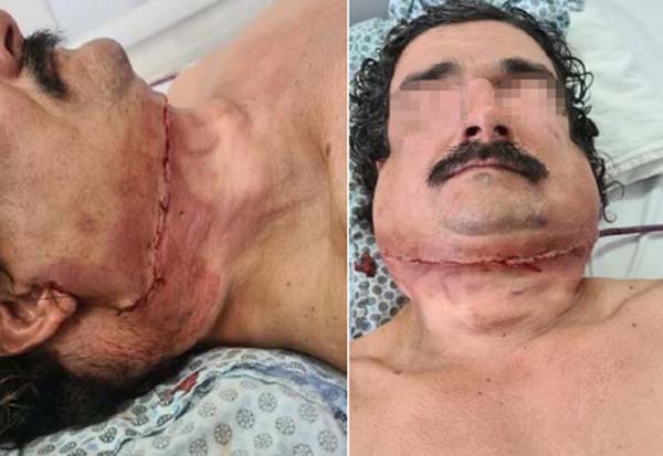
VS.G., 42 years old, male, was seen at the plastic surgery outpatient clinic of Hospital das Clínicas, in Recife, PE, in February 2022, with a diagnosis of Madelung’s disease. He reports stopping alcohol intake for 2 years, without comorbidities and use of medication. Furthermore, he underwent a biopsy in May of the previous year compatible with lipoma. On physical examination, large masses of elastic consistency were found around the neck and supraclavicular region (Figure 1).
The therapeutic proposal was liposuction with the removal of the tumor in three surgical stages. The requested cardiological opinion indicated intermediate risk but without contraindication to surgery. The 1st stage consisted of liposuction associated with left anterior cervical lipectomy (Figure 2). On the 4th postoperative day (POD) of the 1st surgical procedure, the patient was asymptomatic and presented with bruises and peripheral edema in the cervical surgical wound (SW) and drain output of less than 30ml. On the 26th POD, the patient reported hardening of the operated area, without other complaints.
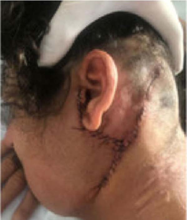
Six months after the first surgical stage, the 2nd surgical stage was performed, which consisted of a lipectomy on the left side of the neck and face and dorsal region (Figure 3). On the 5th POD, the patient was asymptomatic, but with a large amount of seroma in the drain, which resolved spontaneously by the 15th POD. Three months after the second surgical procedure, the patient presented discreetly hypertrophic and hyperchromic scars.
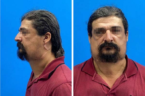
The 3rd surgical stage was scheduled 5 months after the 2nd stage, to perform liposuction with right cervical lipectomy (Figure 4). On the 1st POD, the patient denied pain, fever, and sensory and motor changes in the region ipsilateral to the procedure.
He also presented with the presence of edema and ecchymosis lateral to the scar and content of the serohematic drain <20ml, but progressing with a healed and good-looking OF. On the 12th POD, the patient presented with SW with well-coopted edges and no pain on mobilization but still presented ecchymosis lateral to the scar and slight edema on palpation but without evidence of infection. The final appearance of the late postoperative period of 4 months after the 3rd surgical stage can be seen in Figure 4.
DISCUSSION
In this study, the reported patient fits the classic profile of the syndrome: a 45-year-old man with an alcoholic past. Madelung’s disease is characterized by predominantly affecting middle-aged male patients with a history of alcoholism. However, cases in non-alcoholic women can occur. Furthermore, the main comorbidities associated with the disease are nephropathy, hepatopathy, and metabolic abnormalities6. The only comorbidity reported by the patient was a history of hepatic steatosis.
The disease does not usually present with painful symptoms. In this case, the patient’s main complaint was aesthetic as it affected areas of high exposure. Initially, Hugo and Conway classified diffuse symmetric lipomatosis as predominantly on the trunk and thighs; and predominantly cervical, as described by Madelung. A decade later, benign lipomatoses were classified into 3 clinical groups by Carlsen and Thomnsen: congenital diffuse lipomatosis (Type 1), symmetric diffuse lipomatosis (Type 2), which the reported patient falls into, and multiple lipomatosis (Type 3).
However, this classification is not quite accurate. More recent studies have already classified the syndrome into two distinct types: type I, more common in men and which generally manifests itself through a symmetrical distribution of superficial fat deposits, giving a “pseudo-athletic” appearance and possibly causing compression symptoms; and type II, which affects both men and women, presenting in a similar way to generalized obesity7.
The use of alcoholic beverages can influence or worsen the condition. In this case, the patient had already stopped drinking alcohol 2 years ago and did not return after surgery, which was beneficial for postoperative recovery. From a histological point of view, in Madelung’s disease, the adipocytes found in fatty masses are smaller, and there is a greater presence of fibrous and vascular tissues than normal7. Loss of large myelinated cells is also observed, although demyelination or axonal degeneration associated with chronic alcohol consumption does not occur. After surgery, the risk of recurrence is reduced by abstaining from alcohol. Some lifestyle changes that improve blood sugar levels and lipid control can prevent the growth of fat masses but do not reduce their size.
Some studies have demonstrated good long-term results in non-invasive techniques such as intralipotherapy, in which the mass is injected with phosphatidylcholine/deoxycholate. Intralipotherapy limits the growth of adipose masses but is not highly effective in reducing their volume8. Therefore, in this patient, surgery was the therapeutic technique of choice.
The combination of cervical lipectomy associated with liposuction demonstrated satisfactory results in the management of our patient. Among the techniques of choice, lipectomy is the most commonly used therapeutic option, as allows satisfactory exposure and complete removal, with good control of iatrogenic injuries to nearby structures, especially vessels and nerves8. Despite this, lipectomy is not technically easy, because lipomas are not encapsulated and easily infiltrate the surrounding tissue9.
A disadvantage of lipectomy is the increased rate of surgical complications, such as infections, hemorrhages, hematomas, lymphatic fistulas, and pathological scarring9,10. Our patient developed a hematoma and a postoperative seroma, but it resolved shortly afterward, leaving no sequelae. Despite having discreet unsightly scars, the patient reported a great improvement in quality of life after removal of the lipoma.
One study looked at 95 patients with Madelung syndrome treated with lipectomy and/or liposuction. Of those who underwent lipectomy alone (n=74), the relapse rate was 5%. Among those who performed both techniques (n=16), the rate was 12.5%. However, the sample population is different between groups and there are no randomized studies comparing combined and isolated techniques. Liposuction is less traumatic and has better aesthetic results than surgery. However, there is little clinical experience with liposuction in this group of patients, and it is an adjuvant therapy in many cases6. In this study, the combination of techniques proved to be favorable in the patient’s therapy, not resulting in recurrences to date.
CONCLUSION
The combination of cautious liposuction followed by serial lipectomy is beneficial, increasing the safety of the management of cervical structures and preserving the vascularization of the flaps. Furthermore, the combination of techniques allows for a higher quality aesthetic result, guaranteeing the patient a better quality of life.
REFERENCES
1. Liu Q Lyu H, Xu B, Lee JH. Madelung Disease Epidemiology and Clinical Characteristics: a Systemic Review. Aesthetic Plast Surg. 2021;45(3):977-86.
2. Capel E, Vatier C, Cervera P, Stojkovic T, Disse E, Cottereau AS, et al. MFN2-associated lipomatosis: Clinical spectrum and impact on adipose tissue. J Clin Lipidol. 2018;12(6):1420-35.
3. Rocha N, Bulger DA, Frontini A, Titheradge H, Gribsholt SB, Knox R, et al. Human biallelic MFN2 mutations induce mitochondrial dysfunction, upper body adipose hyperplasia, and suppression of leptin expression. Elife. 2017;6:e23813.
4. Perseguim AB, Aquino JLB, Pereira DAR, Rodrigues-Silva AMGM, De-Faria JCM. Madelung’s disease: combined surgical approach of lipectomy and liposuction. Rev Bras Cir Plást. 2022;37(1):105-10.
5. Enzi G, Busetto L, Ceschin E, Coin A, Digito M, Pigozzo S. Multiple symmetric lipomatosis: clinical aspects and outcome in a long-term longitudinal study. Int J Obes Relat Metab Disord. 2002;26(2):253-61.
6. Chen CY, Fang QQ, Wang XF Zhang MX, Zhao WY, Shi BH, et al. Madelung’s Disease: Lipectomy or Liposuction? Biomed Res Int. 2018;2018:3975974.
7. Ramos S, Pinheiro S, Diogo C, Cabral L, Cruzeiro C. Madelung disease: a not-so-rare disorder. Ann Plast Surg. 2010;64(1):122-4.
8. Orasmo CR, Ocanha JP Barraviera SR, Miot HA. Syndrome in question. An Bras Dermatol. 2014;89(3):525-6.
9. Bassetto F Scarpa C, De Stefano F Busetto L. Surgical treatment of multiple symmetric lipomatosis with ultrasound-assisted liposuction. Ann Plast Surg. 2014;73(5):559-62.
10. Verhelle NA, Nizet JL, Van den Hof B, Guelinckx IP Heymans O. Liposuction in benign symmetric lipomatosis: sense or senseless? Aesthetic Plast Surg. 2003;27(4):319-21.
1. Hospital das Clínicas da Universidade Federal de Pernambuco, Recife, PE, Brazil
2. Faculdade Pernambucana de Saúde, Recife, PE, Brazil
3. Universidade Federal de Pernambuco, Recife, PE, Brazil
Corresponding author: Pedro Henrique de Araújo Silva Avenida Prof. Moraes Rego, 1235, Recife, PE, Brazil. Zip Code: 50670-901, E-mail: henrique2_araujo@hotmail.com
Article received: September 8, 2023.
Article accepted: April 30, 2024.
Conflicts of interest: none.
Institution: Hospital das Clínicas da Universidade Federal de Pernambuco, Recife, PE, Brazil.


 Read in Portuguese
 Read in English
 PDF PT
 Print
Print
 Send this article by email
 How to Cite
 Mendeley
 Pocket
 Twitter