


Original Article - Year 2024 - Volume 39 -
Reconstruction of the distal third of the nose: case series and literature review
Reconstrução de terço distal do nariz: série de casos e revisão de literatura
Raissa Barakatt de Figueiredo1,* ; Eduardo Machado Mariano1; Wilson Cintra-Junior1; Hélio Kiyoto Maebayashi1; An Wan Ching1; José Antônio Cezaretti1
ABSTRACT
Introduction: The nose has great aesthetic and functional importance, with a high incidence of malignant lesions. There are several techniques for reconstructing the distal third of the nose, but there is no universal indication; will depend on the characteristics of the injury. Surgical options vary between skin grafts and local, regional, and microsurgical flaps. The objective is to present a series of cases of reconstruction of the distal third of the nose using different surgical techniques, discussing the peculiarities and the results obtained.
Method: This is a retrospective study carried out at the Hospital do Servidor Público Estadual de São Paulo (HSPE), evaluating a series of eight patients diagnosed with non-melanoma skin cancer located in the distal third of the nose and who underwent reconstruction by the team of Plastic Surgery.
Results: Satisfactory results were obtained for all patients undergoing distal nose reconstruction, using total skin graft techniques (n=1) and local flaps (n=7), such as the bilobed, nasolabial, and dorsal nose flap. nose, paramedian frontal, and nasolabial transposition.
Conclusion: Reconstruction of defects in the distal third of the nose is challenging and involves great technical variability. A careful assessment of the patient and the injury must be carried out, risks and benefits assessed and the decision shared with the patient.
Keywords: Nose neoplasms; Nose; Surgical flaps; Skin transplantation; Reconstructive surgical procedures
RESUMO
Introdução: O nariz apresenta grande importância estética e funcional, com alta incidência de lesões malignas. Existem várias técnicas de reconstrução do terço distal do nariz, não havendo uma indicação universal; irá depender das características da lesão. As opções cirúrgicas variam entre enxerto de pele, retalhos locais, regionais e microcirúrgicos. O objetivo é apresentar uma série de casos de reconstrução de terço distal do nariz com diferentes técnicas cirúrgicas, discutindo as peculiaridades e os resultados obtidos.
Método: Trata-se de estudo retrospectivo realizado no Hospital do Servidor Público Estadual de São Paulo (HSPE), avaliando uma série de oito pacientes com diagnóstico de câncer de pele não melanoma localizados em terço distal de nariz e que foram submetidos a reconstrução pela equipe de Cirurgia Plástica.
Resultados: Foram obtidos resultados satisfatórios para todos os pacientes submetidos a reconstrução distal do nariz, tendo sido utilizadas técnicas de enxerto de pele total (n=1) e retalhos locais (n=7), tais como o retalho bilobado, nasogeniano, dorsal do nariz, frontal paramediano, e transposição nasolabial.
Conclusão: A reconstrução de defeitos do terço distal do nariz é desafiadora e com grande variabilidade técnica. Deve-se realizar avaliação criteriosa do paciente e da lesão, avaliar riscos e benefícios e compartilhar a decisão com o paciente.
Palavras-chave: Neoplasias nasais; Nariz; Retalhos cirúrgicos; Transplante de pele; Procedimentos cirúrgicos reconstrutivos.
INTRODUCTION
The nose is a complex, unique, three-dimensional anatomical structure with great functional and aesthetic importance, located in the center of the face1-4. Its prominence on the face favors sun exposure and, consequently, presents a high incidence of skin cancer, mainly basal cell carcinoma (BCC), followed by squamous cell carcinoma (SCC), the most common cancer in the world population2,3,5,6.
Demands for nasal reconstruction due to malignant lesions are on the rise. The surgical technique is challenging, especially in the distal third of the nose, where the skin is thicker and adheres to the underlying cartilage, and there is a greater risk of distortion of the nasal margins7,8.
It is important to note that deeper injuries may compromise the cartilaginous framework and the nasal mucosa, requiring an even more complex reconstruction, aiming to maintain the functionality of the nose1. Another important aspect is the analysis of the anatomical units, first described by Millard, and the nasal aesthetic subunits, later described by Burget and Menick, to adjust the surgical sutures where the subunits meet, thus creating a natural contour and hiding the sutures in the natural creases of the skin5,7,9-11.
There are several techniques for reconstructing the distal third of the nose, but there is no universal indication3. The choice for each technique depends on the characteristics of the lesion, size, anatomical position, skin quality, patient comorbidities, and the surgeon’s experience1,7. Surgical options vary between skin grafts, and local, regional, and microsurgical flaps9.
OBJECTIVE
The objective of this work is to present a series of cases of reconstruction of the distal third of the nose using different surgical techniques, discussing the peculiarities and the results obtained.
METHOD
This is a retrospective study carried out in a single center, from March 2021 to March 2022. Eight patients diagnosed with non-melanoma skin cancer located in the distal third of the nose and who had reconstructions performed by the Surgery team were evaluated. Plastic surgery at the Hospital do Servidor Público Estadual de São Paulo (HSPE), in São Paulo, SP. After authorization from the Institutional Ethics Committee, the Free and Informed Consent Form was applied and the following data were collected from the patient’s records: sex, age, comorbidities, histological type of tumor, reconstruction technique used, complications in the postoperative period, aesthetic and functional result, remission of the injury.
The diagnosis of non-melanoma skin cancer was made by prior biopsy, and written consent was signed before the procedure. Surgical excision of the lesion was performed with lateral margins of 4 to 6 mm. Intraoperative frozen section examination was performed until malignancy-free margins were obtained. Except for reconstruction with a paramedian frontal flap, which required general anesthesia and hospitalization, all other reconstructions were performed with local anesthesia and on an outpatient basis.
Preoperative injuries and recent and late postoperative outcomes were documented by digital imaging.
RESULTS
Five women (62.5%) and 3 men (37.5%) were treated, with a mean age of 82.2 years. Seven patients were Caucasian, with a Fitzpatrick classification between 1 and 2. Five patients had comorbidities, such as isolated arterial hypertension or associated with type 2 diabetes mellitus, and one patient had, in addition to these, asthma and dyslipidemia. Other patient data are summarized in Table 1 and some cases will be described below.
| Case | Sex | Age years) | Size of the injury | Location of the injury | Histological type of pre-op/ post-op lesion | Reconstruction carried out | Compli-cation | Aesthetic/ functional result | Injury remis-sion |
|---|---|---|---|---|---|---|---|---|---|
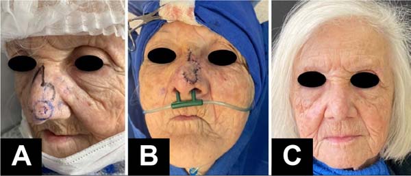
| 1 (Figure 1) | Feminine | 88 | 0.4cm | Left nasal tip | CEC “in situ”/ actinic keratosis | Bilobed flap | None | Satisfactory / Satisfac-tory | Yes |
| 2 | Masculine | 83 | 0.6cm | Left nasal wing | Nodular and micronodular BCC / nodular and superficial BCC | Auricular car-tilage graft and flap bilobate | None | Satisfactory / Satisfac-tory | Yes |
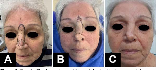
| 3 (Figure 2) | Feminine | 74 | 1.0cm | Transition between back and nasal tip | Nodular BCC / nodular and micronodular BCC | Nasal dorsum flap | None | Nasal tip deviation / Satisfactory | Yes |
| 4 | Feminine | 84 | 1.0cm | Right nasal wing | Nodular BCC/ micronodular and superficial BCC | Conch auricu-lar cartilage graft and naso-labial flap pedicled superiorly | None | Satisfactory /Satisfac-tory | Yes |
| 5 (Figure 3) | Masculine | 85 | 4.0cm | Nasal dor-sum and right and left lateral wall | Nodular and micronodular BCC /Nodular, micronodular, superficial and sclerodermiform BCC | Pedicled para-median frontal flap | None | Satisfactory / Satisfactory | Yes |
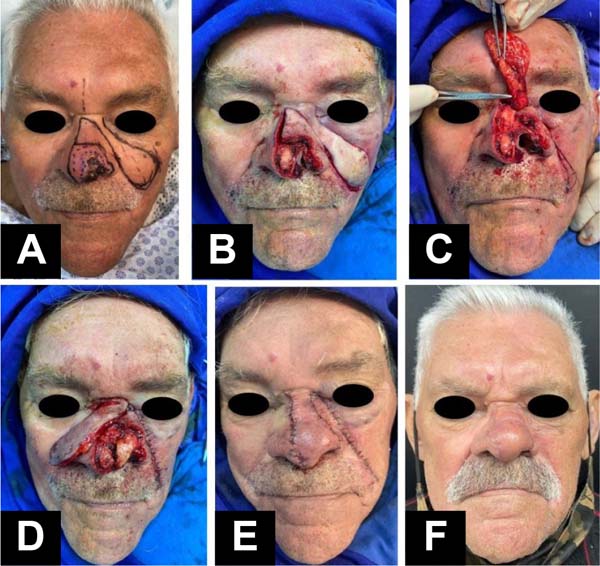
| 6 (Figure 4) | Masculine | 91 | 1.5cm | Left nasal tip | Nodular BCC/ nodular and infiltrative BCC | Tragal auricu-lar cartilage graft and ear flap transpositionnasolabial | None | Quite satisfactory/ Quite satisfactory | Yes |
| 7 | Female | 69 | 3.5cm | Nasal dor- sum |
Nodular, mi-cronodular and sclerodermiform BCC / Nodular and micronodular BCC | total skin graft | None | Regular / Satisfactory | Yes |
| 8 | Female | 84 | 1.5cm | Right nasal wing and nasal tip | Nodular, micronodular and superficial BCC / nodular and superficial BCC superficial | total skin graft | None | Regular / Regular | Yes |
Caption: BCC = basal cell carcinoma; SCC = squamous cell carcinoma


DISCUSSION
The oncological concept must be sovereign. The main objective is complete resection of the lesion, with evaluation of all margins before reconstruction. Mohs surgery, if available, is the gold standard for intraoperative margin assessment; Another form of evaluation is the frozen section examination, a method used in the patients in this study. If intraoperative evaluation is not possible, secondary intention healing, primary closure, and skin grafting should be chosen until postoperative evaluation is performed. Reconstruction should only be scheduled after pathological examination demonstrating margins free of involvement12,13.
To define the best reconstruction approach, several aspects must be taken into consideration. First, consider the patient as a whole. If you have many comorbidities, a simpler single-stage technique is safer and more appropriate. Another factor to be analyzed is active smoking, where, when present, preference should be given to a single-stage technique14. The patient’s active participation in the decision is important, especially in complex surgeries that require several approaches9.
The nasal defect must be evaluated in its location, related to the aesthetic subunits. The concept of nasal aesthetic units was described by Millard5, which improved surgical results. Later, Burget and Menick defended the concept of nasal aesthetic subunits, and if the defect affected more than 50% of the subunit, this entire region should be removed to camouflage the scar in the natural skin creases5,10. However, this concept has been discussed in the literature, as the defect can become much larger and make reconstruction difficult14.
The depth of the nasal defect must be assessed to determine the affected components. In addition to the superficial soft tissue, the cartilaginous structure below may require reconstruction, using grafts mainly from the auricular region and the nasal septum14. The nasal mucosa is another structure that must be analyzed and reconstructed3. The main objectives are the aesthetics and respiratory function of the nose, that is, maintenance of similar skin color, reconstruction of the internal lining and nasal structural support, and avoiding airway stenosis5.
Local and regional flaps are preferred over skin grafts in terms of texture and color, but all types of reconstruction have their uses15. Reconstructing the distal third of the nose is challenging, as the skin is thick and adheres to cartilaginous structures; There is no local skin redundancy, which makes flap mobilization difficult. Reconstruction can generate tension and retraction of the nasal ala, causing aesthetic and functional changes15. Surgical planning must be meticulous for the best possible result3.
Bilobed flap
The bilobed flap has excellent applicability for defects in the distal third of the nose9. It is composed of two “lobes” respecting the design at right angles between the axes, which allows double transposition16. The first “lobe” covers the defect, the second “lobe” covers the first donor area, and the second donor area is closed primarily3. Because the donor area is limited, this flap is generally used for small defects of up to 1.5 cm, with excellent results3. However, there are descriptions of its use for defects larger than 2.0cm with good results based on wide detachment for adequate tissue advancement17.
If there is a risk of nasal valve collapse, a cartilage graft can be associated with this technique9. This reconstruction has the advantage of being a single-stage procedure, good viability of the flap, good cosmetic result with similar skin texture and color, and discreet scar18. Among the disadvantages of this flap are the complex geometric lines of incision, requiring experience to not distort nasal symmetry, thus normally limiting it to minor defects7,9,14.
Dorsal nose flap
The dorsal nose flap is based on the rotation of the skin of the proximal two-thirds of the nose and the glabellar region to cover distal defects, and closure of the donor area, which can be in VY, positioning the scar on the glabellar expression line. This technique was initially described by Gillies but became popular with Rieger, who described a flap with random vascularization and was later modified by Marchac and Toth, with axial vascularization of the angular artery close to the medial corner of the eye. This technique makes it possible to reconstruct defects in the supratip region measuring 1 to 2cm 3,14,19-21.
The advantage of the flap is that it can be performed in a single procedure, with a well-positioned scar and a good aesthetic result3. Among the disadvantages, we highlight the possible need for a disproportionately large flap to cover small defects and the possibility of traction of the tip of the nose upwards7,14.
Nasolabial flap
The nasolabial flap is a widely used option in alar reconstruction. It can be designed based on an upper or lower pedicle, both with axial vascularization of branches of the facial artery, or V-Y3,15,22. Ideally, some fibers of the common elevator muscle and nasal ala are elevated together, constituting the smallest frequently used musculocutaneous flap23. Reconstruction can be performed in one or two surgical stages; the flap must be designed 1 to 2mm larger, as it will shrink postoperatively; and the flap can serve as an internal nasal lining when necessary7,9.
It presents reliable vascularization, discreet healing of the donor area and positioned in the preexisting nasolabial fold, a good cosmetic result of the nasal ala, and the procedure can be performed in a single procedure3,9. The disadvantages are the possibility of obliterating the concavity of the alar fold or even the need to perform a two-stage procedure7,9.
Paramedian flap
The paramedian frontal flap is a reconstruction instrument widely used for larger defects located in the distal third of the nose9,24. It is an interpolation flap with oblique skin from the forehead, with axial vascularization based on the supratrochlear artery3. It is generally created in two stages, in the first stage it is elevated and positioned in the nasal defect, and in the second, three weeks later, the pedicle is sectioned and the flap can be thinned and adjusted25. Additional steps may be necessary for refinements, as well as reconstruction of the bone-cartilaginous framework9. The distal portion of the flap can be thinned and folded to form nasal mucosa3. The defect on the forehead can be closed by first or second intention, or grafted3,9.
The advantage of the flap is having a reliable axial vascular supply, having the capacity to reconstruct large nasal defects in the distal third, with the possibility of reconstructing even the nasal mucosa; presenting satisfactory cosmetic results, as the skin on the forehead is compatible in color, texture and flexibility with that of the nose2,14,25.
The disadvantages are the limited use in smokers, due to the risk of necrosis25, the need to use general anesthesia, the multi-stage procedure, and a transverse scar on the forehead2,3. Other negative points are the thick flap at the nasal tip when folded to create the lining and, if it is thinned, there is the possibility of reduced perfusion and local suffering1. Finally, the psychological aspect of long-term reconstruction is a point that must be clarified so that patients have realistic expectations25.
Nasolabial transposition flap
The nasolabial transposition flap is a reconstruction option for larger defects located in the distal third of the nose and was described by Beustes- Stefanelli et al.26. It is designed using redundancy of nasolabial tissue to cover the nasal defect and a small, inferiorly based residual dorsoalar flap to assist in tension-free closure of the inner corner of the ipsilateral eye associated with cheek advancement. The nasolabial flap has axial vascularization by branches of the facial artery in its proximal two-thirds and a random pattern in its distal third, and the dorsoalar residual flap has random vascularization. A cartilage graft can be associated for structuring, if necessary. The distal region of the nasolabial flap can be thinned and folded to form the lining of the nasal mucosa26.
The advantages of the procedure are the possibility of reconstructing large nasal defects in a single procedure, reliable vascularization, without the need for general anesthesia, with good nasal aesthetic results, and a discreet scar in the pre-existing nasolabial fold. Disadvantages include the possibility of a thick flap at the nasal tip when folded to create the lining and, if it is thinned, there is the possibility of reduced perfusion and local suffering26.
This flap was well indicated and performed in case 6 (Figure 4) of this study. The nasolabial flap was designed using tissue redundancy and a residual dorsoalar flap lateral to the defect. The nasolabial flap was transposed to the nasal dorsum and tip, in the distal region it was thinned and folded to create the nasal lining. The residual dorsoalar flap was transposed to the inner corner of the eye to close the area without tension. The structuring of the new nasal tip was carried out by collecting an auricular cartilage graft in a block from the region of the tragus blade, isthmus, and conchal cavity, as described by Pereira et al.24. This unique, curved-shaped cartilage is fixed to the left alar region previously removed with the tumor to structure the new nasal tip.
Grafts
Skin grafts are options for reconstruction of the distal third of the nose in specific situations, normally when the patient has a high surgical risk for more complex procedures when strict surveillance is required for recurrence of malignancy, or temporarily until the definitive result of the anatomopathological examination9,27 . A total skin graft is used because it has greater thickness, less primary retraction, and better aesthetic results when compared to partial skin; maintained with a fixed dressing for 5 to 7 days to neutralize shear forces and allow better integration15. The preand post-auricular region, cervical, and clavicular region are used as graft donor areas15. The major disadvantage is the unfavorable aesthetic appearance due to the incompatibility of the skin in color, shape, and contour9.
CONCLUSION
Reconstruction of defects in the distal third of the nose is challenging, and multiple techniques are possible.
To decide on the reconstruction technique, a careful assessment of the patient and the characteristics of the defect must be carried out, risks and benefits assessed and the best options shared with the patient.
REFERENCES
1. Yildiz K, Gorgulu T, Yesiloglu N, Kelahmetoglu O, Camli MF, Canter HI. Hybrid reconstruction of distal nasal defects. Dermatol Ther. 2021;34(1):e14734.
2. Schäfer K, Rudolph C, Cotofana S, Goebeler M, Weyandt G. Large Nasal Defects with Exposed Cartilage: The Folded Transposition Flap as an Innovative Alternative to the Paramedian Forehead Flap. Dermatology. 2018;234(3-4):99-104.
3. Mysore Srinivas V, Kalapurmat Nagabhushanaiah M. Nose Reconstruction Using Local and Regional Flaps: The Challenges and Advantages. J Cutan Aesthet Surg. 2021;14(1):77-83.
4. Moura BB, Signore FL, Buzzo TE, Watanabe LP, Fischler R, Freitas JOG. Nasal reconstruction: an analysis of a series of cases. Rev Bras Cir Plást. 2016;31(3):368-72.
5. Marcasciano M, Tarallo M, Maruccia M, Fanelli B, La Viola G, Casella D, et al. Surgical Treatment with Locoregional Flap for the Nose. Biomed Res Int. 2017;2017:9750135.
6. Brasil. Ministério da Saúde. Instituto Nacional do Câncer. Estimativa 2020: incidência do Câncer no Brasil [Internet]. Rio de Janeiro: Instituto Nacional do Câncer; 2019 [acesso17 abr 2022]. Disponível em: https://www.inca.gov.br/sites/ufu.sti.inca.local/files//media/document//estimativa-2020-incidenciade-cancer-no-brasil.pdf
7. Cason RW, Shammas RL, Pyfer BJ, Glener AD, Marcus JR, Cook JL. Cutaneous Reconstruction of the Nasal Distal Third: Alternative Local Flaps for a Complex Region. Plast Reconstr Surg Glob Open. 2021;9(5):e3444.
8. Souza MPS, Pessoa SGP, Muniz VV, Rebelo AD, Holanda EF, Pessoa LMGP. Opções de reconstrução após ressecção de tumor de pele nasal. Rev Bras Cir Plást. 2019;34(Suppl. 3):50-2.
9. Konofaos P, Alvarez S, McKinnie JE, Wallace RD. Nasal Reconstruction: A Simplified Approach Based on 419 Operated Cases. Aesthetic Plast Surg. 2015;39(1):91-9.
10. Burget GC, Menick FJ. The subunit principle in nasal reconstruction. Plast Reconstr Surg. 1985;76(2):239-47.
11. Marinho CCC, Miranda ML, Lima RC, Rodrigues CJ, Pego KVT, Reis CF, et al. Reconstruction of the nasal subunits after tumor resection. Rev Bras Cir Plást. 2021;36(2):156-63.
12. National Comprehensive Cancer Network. Basal Cell Skin Cancer (Version 2.2022) [Internet]. 2022 [acesso 4 abr 2022]. Disponível em: https://www.nccn.org/
13. National Comprehensive Cancer Network. Squamous Cell Skin Cancer (Version 2.2022) [Internet]. 2022 [acesso 2 de abr 2022]. Disponível em: https://www.nccn.org/
14. Rohrich RJ, Griffin JR, Ansari M, Beran SJ, Potter JK. Nasal reconstruction--beyond aesthetic subunits: a 15-year review of 1334 cases. Plast Reconstr Surg. 2004;114(6):1405-16; discussion 1417-9.
15. Sadick H, Häussler D, Rotter N. [Options for reconstruction of nasal defects]. HNO. 2020;68(12):959-70. German.
16. Tissiani LAL, Alonso N, Carneiro MH, Bazzi K, Rocco M. Versatilidade do retalho bilobado. Rev Bras Cir Plást. 2011;26(3):411-7.
17. Vasilakis V, Nguyen KT, Klein GM, Brewer BW. Revisiting Nasal Reconstruction after Mohs Surgery: A Simplified Approach Based on the Liberal Application of Local Flaps. Ann Plast Surg. 2019;83(3):300-4.
18. Laitano FF, Teixeira LF, Siqueira EJ, Alvarez GS, Martins PDE, Oliveira MP. Uso de retalho cutâneo para reconstrução nasal após ressecção neoplásica: 102 casos. Rev Bras Cir Plást. 2012;27(3 Suppl.1):21.
19. Redondo P, Bernad I, Moreno E, Ivars M. Elongated Dorsal Nasal Flap to Reconstruct Large Defects of the Nose. Dermatol Surg. 2017;43(8):1036-41.
20. Marchac D, Toth B. The axial frontonasal flap revisited. Plast Reconstr Surg. 1985;76(5):686-94.
21. Decusati FL, Rinaldi AE. Reconstrução de defeitos nasais utilizando o retalho de Rieger. Rev Bras Cir Plást. 2020;35(2):149-53.
22. Rosseto M, Nunes e Silva D, Lyrio ALC, Lyrio ALC, Rosseto AC. Retalho nasogeniano de pedículo inferior na reconstrução de asa nasal. Rev Bras Cir Plást. 2022;37(1):22-6.
23. Cerci FB, Nguyen TH. Nasolabial interpolation flap for alar reconstruction after Mohs micrographic surgery. Surg Cosmet Dermatol. 2014;6(2):113-20.
24. Pereira CM, Venturelli Júnior EP, Rocha RS, Gonçalves PR, Silva FN, Bocardo SD. Reconstrução nasal com retalho frontal paramediano após ressecção oncológica. Rev Bras Cir Plást. 2020;35(3):373-7.
25. Zito PM, Hohman MH, Mazzoni T. Paramedian Forehead Flaps [Internet]. StatPearls [Internet]. Treasure Island (FL): StatPearls Publishing [acesso 2 mar 2022]. Disponível em: https://www.ncbi.nlm.nih.gov/books/NBK499932/?report=printable
26. Beustes-Stefanelli M, O’Toole G, Schertenleib P. The Midlinebased nasolabial transposition (MNT) flap: an original singlestage technique for nasal tip reconstruction. Ann Plast Surg. 2015;74(4):426-31.
27. Sbalchiero JC, Gregório TCR, Leitão L, Leal PRA, Dibe MJA. Condutas na reconstrução da ponta nasal no tratamento das neoplasias cutâneas. Rev Bras Cir Plást. 2005;20(1):12-6.
1. Instituto de Assistência Médica ao Servidor
Público Estadual, São Paulo, SP, Brazil
Corresponding author: Raissa Barakatt de Figueiredo Diretoria da Cirurgia Plástica, Rua Pedro de Toledo, 1.800, 9º andar, Vila Clementino, São Paulo-SP. Zip code: 04039-000, E-mail: raissa_barakatt@hotmail.com
 Read in Portuguese
 Read in English
 PDF PT
 Print
Print
 Send this article by email
 How to Cite
 Mendeley
 Pocket