


Case Report - Year 2023 - Volume 38 -
Neurofibromatosis in the gluteus and posterior region of the thigh: case report
Neurofibromatose em glúteo e região posterior da coxa: relato de caso
Bárbara Miranda Martins1,* ; Bernardo de Almeida Galindo2; Viviane Honório Mendonça da Costa3; Laercio Pol-Fachin1
ABSTRACT
Introduction: Neurofibromatosis is an autosomal dominant disorder, and type 1 is associated with an increased risk of tumor formation with neurocutaneous involvement. The variable evolution, often with limiting tumors, in addition to the significant incidence of cases requiring treatment, makes it fundamental to discuss procedures already performed in medical practice for early, careful, and individualized recognition of the diagnosis and treatment of the patient. The report aims to present a surgical case of neurofibromatosis, calling attention to the surgical technique, the characteristics of the disease, and the importance of the procedure in the quality of life of patients limited by the condition.
Case Report: A 23-year-old male patient with a large mass neurofibroma in the gluteus and posterior surface of the right leg, in addition to café au lait stains in the distal third of the legs. He was treated with surgery to remove the tumor and a flap and graft in the affected region. The procedures were performed by a multidisciplinary team, allowing the total removal of the tumor mass, with subsequent skin grafting in the hip and thigh lesion on the right side and the fasciocutaneous flap in VY in the area. There were no significant complications in the immediate postoperative period.
Conclusion: Neurofibromas can become limiting and impair patients' quality of life with neurofibromatosis type 1; therefore, early management and diagnosis are essential. Although the condition does not present a cure, there is a need for research into less invasive and preventive treatments for injuries.
Keywords: Case reports; Neurofibroma; Reconstructive surgical procedures; Surgical flaps; Neurofibromatosis 1
RESUMO
Introdução: A neurofibromatose é um distúrbio autossômico dominante e o tipo 1 está associado a um aumento do risco de formação de tumores com acometimento neurocutâneo. A evolução variável, muitas vezes com tumorações limitantes, além da incidência significativa de casos que necessitam de tratamento, torna fundamental a discussão de condutas já realizadas na prática médica para um reconhecimento precoce, cuidadoso e individualizado do diagnóstico e do tratamento do enfermo. O relato objetiva apresentar um caso cirúrgico de neurofibromatose, chamando atenção para a técnica cirúrgica, as características da doença e a importância do procedimento na qualidade de vida de pacientes limitados pela afecção.
Relato de Caso: Paciente de 23 anos, sexo masculino, com neurofibroma de grande massa em glúteo e face posterior da perna direita, além de manchas café com leite em terço distal de pernas. Foi tratado com uma cirurgia de retirada do tumor, além de retalho e enxerto na região acometida. Os procedimentos foram realizados por equipe multidisciplinar, possibilitando a retirada total da massa tumoral, com posterior realização de enxerto de pele na lesão do quadril e coxa em lado direito, e o retalho fasciocutâneo em V-Y na área. Não houve complicações significativas nos pós-operatórios imediatos.
Conclusão: Os neurofibromas podem se tornar limitantes e prejudicar a qualidade de vida dos pacientes com neurofibromatose tipo 1, por isso, o manejo e o diagnóstico precoces são essenciais. Apesar da afecção não apresentar cura, há a necessidade de pesquisas em tratamentos menos invasivos e preventivos para as lesões.
Palavras-chave: Relatos de casos; Neurofibroma; Procedimentos cirúrgicos reconstrutivos; Retalhos cirúrgicos; Neurofibromatose 1
INTRODUCTION
Neurofibromatosis is an autosomal dominant disorder, divided into neurofibromatosis type 1 (NF1), neurofibromatosis type 2 (NF2) and schwannomatosis. NF1 is associated with an increased risk of forming benign and malignant tumors with neurocutaneous involvement. The disease predominantly affects the skin, bones, and nervous system, but the complications are widespread, variable, and unpredictable1.
NF1 affects men, women, and all ethnic groups equally. The incidence at birth is 1 in 2500 to 1 in 3000 and a minimum prevalence of 1 in 4000-5000. In addition to tumors, café au lait spots are present in 99% of patients with NF12.
The variable evolution, often with limiting tumors, in addition to the significant incidence of NF1 cases that require treatment, makes it essential to discuss procedures already carried out in medical practice for early, careful, and individualized recognition of the diagnosis and treatment of the patient.
OBJECTIVE
This study aims to report a surgical case of neurofibromatosis, calling attention to the surgical technique and the characteristics of the disease, as well as to the importance of the procedure in the quality of life of patients limited by the condition.
CASE REPORT
The case report follows the model recommended by SCARE3, being approved by the ethics and research committee of Centro Universitário CESMAC, with CAEE 61674922.3.0000.0039.
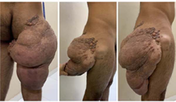
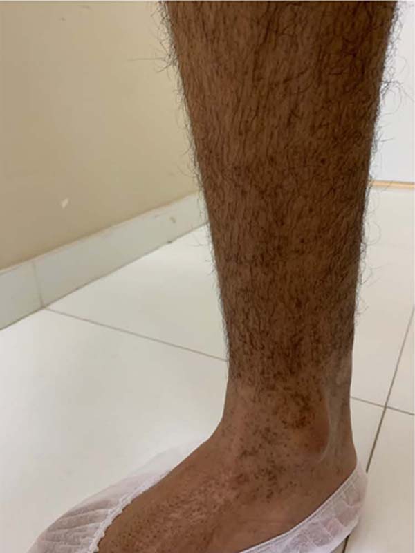
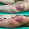
A male, brown, 23-year-old patient sought care at a basic health unit and later at a public hospital, complaining of a tumor in the buttocks and lateral surface of the right leg that had been growing for 11 years (Figure 1) in addition to café au lait spots on the distal third of legs 3 years ago (Figure 2).


The patient denied comorbidities and had no personal history of neoplasms or family history of similar NF1 conditions. Two years before the consultation, with the desire to remove the mass, he sought assistance from the hospital team, who carried out an anatomopathological examination, with a diagnosis of fibrolipoma, and a tomography, which concluded the presence of an infiltrative lesion, affecting the right gluteal region and proximal portion of the posterior face of the right thigh, measuring 22.2cm X 23.8cm (LxT), soft tissue density, fat content inside and no signs of muscle invasion in the area.
After the exams, the patient underwent a surgical procedure with only partially removed the tumor. The first surgery was performed by another team, with no data regarding the technique and the justification for the impossibility of removing the entire tumor. The patient had a great appeal for the total removal of the mass due to limited mobility (he could not perform his favorite activities, such as running and cycling) and the difficulty in maintaining interpersonal relationships, which isolated him from social interactions.
Therefore, with the more accelerated growth of the tumor and café-au-lait spots, he again sought the hospital for total mass removal. Based on clinical analysis and complementary exams, which did not indicate muscle invasion, we opted for the procedure for the total removal of the neurofibroma.
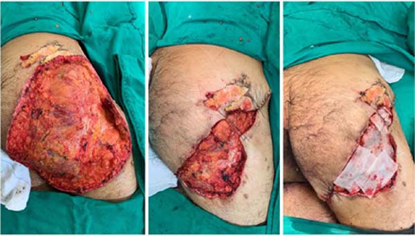
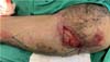
Surgery was performed under spinal and general anesthesia, with the patient in the left lateral decubitus position. The incision bordered the fibromatous lesion, which had a greasy appearance. Resection was performed in the plane above the fascia and hemostasis of the bleeding vessels, which resulted in non-significant intraoperative blood loss. After removing 6,920 kg of the surgical specimen, the edges of the lesion were brought together, and a bandage was applied with Rayon gauze and cotton gauze (Figure 3). There were no complications during the procedure and in the immediate postoperative period.

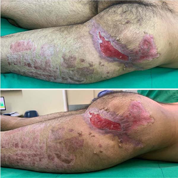
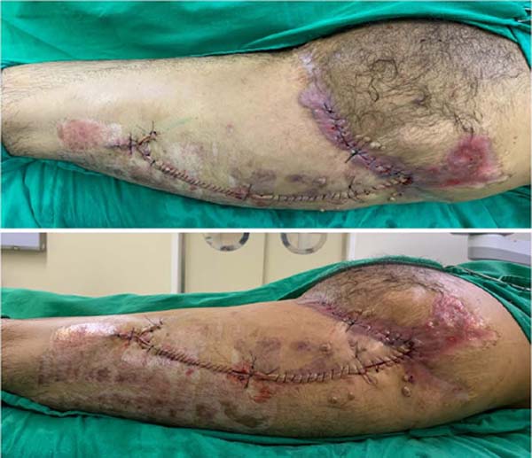
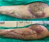
About two months after tumor removal, a skin graft was performed on the hip and thigh lesion on the right side from skin resection in the ipsilateral coxofemoral region, using a Brown dressing after grafting in the donor and recipient region (Figure 4). After another two months, a fasciocutaneous flap was performed in VY (Figure 5) in the affected hip and thigh region, with sutures in Donatti and continuous stitches (Figure 6).

DISCUSSION
The diagnosis of NF1 can be defined with clinical parameters only when two or more of the following conditions are present: 6 or more café-au-lait spots greater than 5mm in diameter in prepubertal individuals, and more than 15mm after puberty, 2 or more neurofibromas of any type or 1 plexiform, freckles in the axillary or inguinal region, an optic pathway tumor, two or more Lisch nodules, a distinct bone lesion or a first-degree relative with NF1 by the criteria described4.
The use of clinical criteria in the diagnosis evidences a possibility of earlier identification and management of the condition, preventing lesions, such as those of the patient in question, from being removed with exacerbated growth and causing limitations.
Regarding the characterization of neurofibroma, in a study by Rozza-de-Menezes et al.5, the presence of lipomatous dermal neurofibromas, as in the case described, is common (prevalence of 17.5%). This variant is more frequent in lesions of greater mass. It is believed that the NF1 mutation can lead to altered lipid metabolism and be related to the accumulation of lipid droplets in the cytoplasm.
Neurofibromatosis has no cure. Its only treatments are surgical excision or CO2 laser for cutaneous neurofibromas smaller than one centimeter6. In the patient in question, we opted for excision of the mass, with excellent results in the immediate postoperative period. Despite the great aesthetic benefit and the patient’s quality of life, the long-term gain of removing a large number of neurofibromas by surgery or laser, from the point of view of disease control, has not been proven. Likewise, laser removal of café au lait stains has not been fully evaluated7.
Regarding future prospects for NF1 control, some treatment alternatives, such as those related to p21Ras8 protein inhibitors (with the aim of inhibiting angiogenesis), cell differentiation inducers8, and hormone activity modulators9, are being studied. However, there is no effective treatment to prevent or even revert the characteristic lesions of NF1, which still represents a scientific delay in studies on the condition.
When performing the graft in a lesion on the back, there is an additional challenge, as this is the main decubitus area, providing shear force that compromises the skin graft integration10, as in the patient. However, despite the risks, grafting is widely recommended.
As for the flaps, in a study by Calil et al.11, 20 patients with lesions in the posterior region of the thigh were submitted to a fasciocutaneous flap in VY. There was no tissue necrosis among the patients, with few immediate complications in the procedures (three infections, one dehiscence, and one hematoma). The study confirms that this flap, the same used by the patient in question, can be used safely to treat isolated or multiple injuries in the gluteal region and posterior thigh.
NF1 is a disease with unpredictable evolution and can greatly impact the patient’s life. The deformity of the patient in question gave him extreme social isolation and a limitation of pleasurable daily activities, reducing his quality of life.
CONCLUSION
This study emphasizes the importance of early diagnosis and management of patients with neurofibromatosis type 1, intending to reduce the progression of lesions. In addition, neurofibromas can become extremely limiting and impair the quality of life of most individuals with NF1, and, despite not having a cure, research is needed into less invasive and preventive treatments for lesions.
1. Centro Universitário CESMAC, Maceió, AL, Brazil
2. Universidade Federal de Alagoas, Maceió, AL, Brazil
3. Hospital Universitário Professor Alberto Antunes, Maceió, AL, Brazil
Corresponding author Bárbara Miranda Martins Rua Cônego Machado, 984, Bairro Farol, Maceió, AL, Brazil. Zip code: 57051-160, E-mail: bmirandam94@gmail.com
 Read in Portuguese
 Read in English
 PDF PT
 Print
Print
 Send this article by email
 How to Cite
 Mendeley
 Pocket