


Case Report - Year 2022 - Volume 37 -
Arm reconstruction with myocutaneous flap of the latissimus dorsi muscle after resection of sarcomas: report of two cases and description of the surgical technique
Reconstrução do braço com retalho miocutâneo do músculo grande dorsal após ressecção de sarcomas: relato de dois casos e descrição da técnica cirúrgica
Leonardo Orletti1* ; Ana Luiza Miranda Cardona1; Ana Paula Mazzocco1; Marcio Diniz Barreto1; Luiz Augusto de Castro Fagundes1
ABSTRACT
Soft tissue sarcomas are rare malignant neoplasms arising from the mesenchyme, most commonly found in the limbs. Surgical resection with free margins greater than 1 cm is essential to obtain a cure for the patient. Radiation therapy can be combined with treatment in selected cases. Reconstruction of the upper limbs after extended resections is a challenge. The latissimus dorsi myocutaneous flap (LDMF) is an option in injuries to the upper limbs, especially the proximal and middle thirds of the arm, with preservation of limb function and primary closure of the resection area. We report two cases of arm resection and reconstruction using LDMF, focusing on the surgical technique description.
Keywords: Myocutaneous flap; Arm; Sarcoma; Soft tissue neoplasms; Reconstructive surgical procedures.
RESUMO
Os sarcomas de partes moles são neoplasias malignas raras originadas do mesênquima, mais comumente encontradas em membros. A ressecção cirúrgica com margens livres acima de 1cm é fundamental para obter a cura do paciente. A radioterapia pode ser combinada ao tratamento em casos selecionados. A reconstrução dos membros superiores após ressecções alargadas é um desafio. O retalho miocutâneo do músculo grande dorsal (RMGD) é uma opção em casos de lesões em membros superiores, sobretudo terço proximal e médio do braço, com preservação da função do membro e fechamento primário da área de ressecção. Relatamos dois casos de ressecção e reconstrução do braço utilizando RMGD com foco na descrição da técnica cirúrgica.
Palavras-chave: Retalho miocutâneo; Braço; Sarcoma; Neoplasias de tecidos moles; Procedimentos cirúrgicos reconstrutivos.
INTRODUCTION
Soft tissue sarcomas (STS) comprise a heterogeneous group of malignant neoplasms with different morphological patterns of the mesenchymal lineage, representing approximately 1% of malignant neoplasms in adults1. Most soft tissue sarcomas are located in the extremities, followed in order of frequency by the abdominal cavity, retroperitoneum, trunk wall and head and neck1,2. Despite having a peak incidence in childhood, STS is more common in adulthood, especially in people over 50 years of age1.
The staging defined by the TNM system of the International Union Against Cancer (UICC) mainly considers the size, depth, histological grade and the presence of lymph node metastases or distance for the composition of the stages1. The most common site of STS metastases is the lungs1,2.
The treatment of STS consists of performing a preoperative biopsy followed by resection of the primary lesion with a safety margin above 1 cm1-3. Radiotherapy can be used in a neoadjuvant or adjuvant manner in cases of lesions larger than 5 cm, when initially unresectable (neoadjuvant), after resections with compromised margins (adjuvant), recurrent lesions and in cases of high-grade lesions1,3-5.
Chemotherapy treatment is controversial and may be indicated for metastatic, recurrent, high-grade histological and locally advanced lesions5. Reconstruction after enlarged STS resections is a challenge, and this article aims to describe the surgical technique for arm reconstruction using the latissimus dorsi myocutaneous flap (LDMF) in our service.
CASE REPORT
Case 1
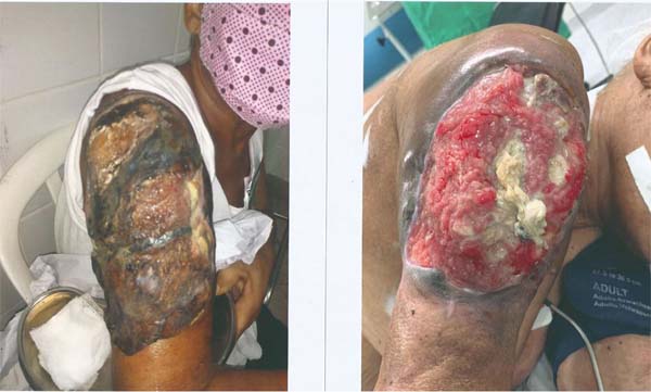
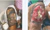
MFR, 61 years old, female, with a history of a progressively growing tumor on the posterior aspect of the right arm. The patient underwent an incisional lesion biopsy on 11/13/2020 with a histopathological result of spindle cell neoplasia with bone neoformation and osteoclastic activity associated with multinucleated giant cells. The positive immunohistochemical result for CD68 and SATB2 is compatible with osteosarcoma. The MRI of the arm performed on 12/31/2020 showed a solid lesion with an epicenter in soft tissues on the right arm’s posterior surface and a cleavage plane with the humerus, measuring 14x10x6cm. Due to the lesion being locally advanced, after multidisciplinary discussion, it was decided to perform neoadjuvant chemotherapy with doxorubicin followed by radiotherapy. The tumor lesion showed significant regression after neoadjuvant treatment (Figure 1).
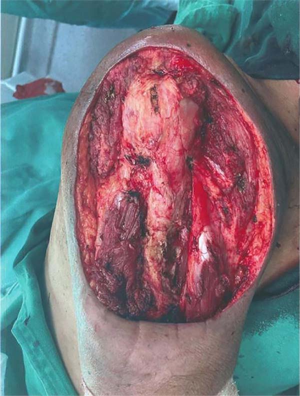
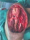
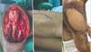
On 06/07/21, the patient underwent extensive resection of the lesion, with a safety margin above 1 cm, without requiring humeral bone resection (Figure 2).
The reconstruction was performed in the same surgical procedure using the LDMF (Figure 3). The patient progressed uneventfully, and the LDMF showed no signs of ischemia. Unfortunately, the patient evolved with lung metastases and is currently undergoing palliative treatment with chemotherapy until the present date (01/08/2021).

Case 2
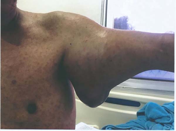
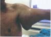
COS, 36 years old male with a history of presenting progressive growth nodulation in the anterior aspect of the left arm for about 1 year (Figure 4). He underwent an incisional biopsy on 02/11/2019, with a result of dermatofibrosarcoma. Immunohistochemistry was positive for CD10 and CD34 antibodies, confirming the diagnosis of dermatofibrosarcoma. The magnetic resonance imaging of the left arm performed on 08/15/2019 showed a voluminous solid formation on the anterior face of the arm measuring 16.2x15x11.6cm, involving the vascular-nervous bundle at 180°.
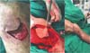
The patient underwent extensive resection of the lesion, with en bloc removal of the biceps and brachial muscles and exposure of the nerves and vessels of the anterior aspect of the arm (median nerve and brachial artery - Figure 5). The histopathological result was compatible with spindle cell mesenchymal neoplasia with chondrosarcomatous dedifferentiation and free surgical margins.

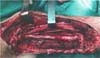
The reconstruction was performed in the same surgical procedure using the LDMF (Figure 6). The patient progressed uneventfully, and the LDMF did not show any signs of ischemia. Unfortunately, the patient evolved with lung metastases and underwent palliative chemotherapy with doxorubicin. He died on May 7, 2020.

Description of the surgical technique
The patient is submitted to general anesthesia and positioned in lateral decubitus with the forearm of the affected limb bandaged after rigorous antisepsis.
Resection of the primary lesion is performed following the basic oncological principles of surgical margins greater than 1 cm and, whenever possible, without violating the tumor pseudocapsule. Surgical margins smaller than 1 cm are allowed in cases of proximity to the periosteum, vessels and nerves that would lead to amputation of the limb3. Surgical margin freezing, whenever available, is performed intraoperatively to avoid compromised margins.
After resectioning the lesion in the arm with free margins, the demarcation of the cutaneous island of the LDMF is performed with methylene blue for subsequent synthesis of the resected skin segment on the arm. The size of this skin extension varies according to the defect generated in the limb after resection.
The isolation of the latissimus dorsi muscle is initiated by its inferior border with electrocautery, taking the entire available belly. The flap is released cranially, with limits inferior to the posterosuperior iliac spine, medially to the intersection of the thoracolumbar fascia with the trapezius muscle, laterally to the free edge of the latissimus dorsi muscle, and superiorly to the intersection with the humerus until the muscle insertion is identified. latissimus dorsi and the finding of the vascular pedicle (previously demarcated). The dissection is stopped at this point, and the flap is transported to the arm site without tension. The flap is transferred by creating a tunnel between the dorsal region and the anterior or posterior aspect of the arm.
Whenever possible, we leave a considerable amount of fat on the latissimus dorsi muscle to help fill the arm, as described in breast reconstruction techniques6.
When closing the donor area, whenever possible, separate stitches of vicryl 0 were applied to the deep planes to avoid the accumulation of seroma, and we performed closed drainage with a 6.4 mm suction drain.
The attachment of the latissimus dorsi muscle to the surgical bed in the arm (proximal and distal portions of the previously sectioned biceps and triceps muscles) helps to maintain limb function. We used separate vicryl 0 stitches to attach the latissimus dorsi muscle to the biceps and triceps brachii muscles.
The following surgical steps focus on rigorous hemostasis, washing and drainage of the pocket with a 6.4 or 4.8 mm suction drain, synthesis of the subcutaneous tissue, skin and, finally, dressing at the end of the procedure.
DISCUSSION
Surgery is the standard treatment for all patients with localized disease and should be performed by an experienced surgeon, with negative (R0) and wide (1cm) margins when possible1.
There are descriptions in the literature of the use of LDMF for upper limb reconstruction after trauma and injuries caused by electrical burns7. In this scenario, LDMF proved to be safe and with satisfactory results for reconstruction of the arm up to its distal third, either with a pedicled flap or a microsurgical technique. However, the reconstruction of upper limbs after resectioning sarcomas is rarely described in the literature8.
Behnamet al.8 published a series of 33 patients who underwent resection of sarcomas in the upper limbs and reconstruction of the LDMF, with only two cases of partial flap necrosis. Saba et al.9 reported a case of resection of a malignant fibrohistiocytoma in a shoulder that underwent immediate resection and reconstruction with LDMF, with satisfactory results.
The secondary objective of maintaining limb function using the LDMF was partially achieved in both cases. With the help of physical therapy, this result can be even better in the long term.
It is important to emphasize that in cases of STS resection with a diameter greater than 5 cm, adjuvant treatment with radiotherapy is indicated1,3. Thus, the closure of the primary defect with more robust tissues, as is the case of LDMF, is an additional safety since it is not uncommon to occur radionecrosis and wound dehiscence in cases of thin skin flaps over areas of bones and tendons10,11.
Therefore, this technique should be more widespread and applied in practice as it allows for adequate limb reconstruction and has a low rate of complications.
CONCLUSION
Larger studies are still needed to assess the effectiveness of arm reconstruction with LDMF since this technique is rarely described in the literature for reconstruction in cases of STS resection. In both cases presented, LDMF was an excellent option for reconstruction, as it allowed the primary synthesis of the lesion without complications such as dehiscence and infection. As a secondary objective, it allowed the partial maintenance of upper limb motor function in both cases.
REFERENCES
1. Manoel WJ, Sarmento BJQ, Silveira Júnior LP, Abreu DCB, Abreu Neto IP, Ferreira EC. Sarcomas de partes moles: resultados do tratamento dos tumores de baixo grau. Rev Bras Cancerol. 2008;54(1):17-24.
2. Bourcier K, Le Cesne A, Tselikas L, Adam J, Mir O, Honore C, de Baere T. Basic Knowledge in Soft Tissue Sarcoma. Cardiovasc Intervent Radiol. 2019;42(9):1255-61. DOI: 10.1007/s00270-19-02259-w
3. Hoefkens F, Dehandschutter C, Somville J, Meijnders P, Van Gestel D. Soft tissue sarcoma of the extremities: pending questions on surgery and radiotherapy. Radiat Oncol. 2016;11(1):136. DOI: 10.1186/s13014-016-0668-9
4. Ray-Coquard I, Serre D, Reichardt P, Martín-Broto J, Bauer S. Options for treating different soft tissue sarcoma subtypes. Future Oncol. 2018;14(10s):25-49. DOI: 10.2217/fon-2018-0076
5. Dujardin F, Debled M, Guillemet C, Simonet J, Hamidou H, Cambon-Michot C, et al. Diagnosis and treatment of soft-tissue tumors]. Rev Chir Orthop Reparatrice Appar Mot. 2006;92(7):637-50. DOI: 10.1016/s0035-1040(06)75924-4
6. Tavares-Filho JM, Franco D, Moreto L, Porchat C, Franco T. Utilização do retalho miocutâneo de grande dorsal, com extensão adiposa, nas reconstruções mamárias: uma opção para preenchimento do polo superior. Rev Bras Cir Plást. 2015;30(3):423-8. DOI: 10.5935/2177-1235.2015RBCP0174
7. Lazaro HA, Dupin AE, Leão CEG, Viel DO, Souza CB. Retalho de grande dorsal para reconstrução de perda de substância por queimadura elétrica em membro superior. Rev Bras Queimaduras. 2014;13(4):265-6.
8. Behnam AB, Chen CM, Pusic AL, Mehrara BJ, Disa JJ, Athanasian EA, et al. The pedicled latissimus dorsi flap for shoulder reconstruction after sarcoma resection. Ann Surg Oncol. 2007;14(5):1591-5. DOI: 10.1245/s10434-006-9292-5
9. Saba SC, Shaterian A, Tokin C, Dobke MK, Wallace AM. The pedicled myocutaneous flap as a choice reconstructive technique for immediate adjuvant brachytherapy in sarcoma treatment. Curr Oncol. 2012;19(6):e491-5. DOI: 10.3747/co.19.1141
10. Davis AM, O’Sullivan B, Turcotte R, Bell R, Catton C, Chabot P, Wunder J, et al; Canadian Sarcoma Group; NCI Canada Clinical Trial Group Randomized Trial. Late radiation morbidity following randomization to preoperative versus postoperative radiotherapy in extremity soft tissue sarcoma. Radiother Oncol. 2005;75(1):48-53.
11. O’Sullivan B, Davis AM, Turcotte R, Bell R, Catton C, Chabot P, et al. Preoperative versus postoperative radiotherapy in soft-tissue sarcoma of the limbs: a randomised trial. Lancet. 2002;359(9325):2235-41.
1. Associação Feminina de Educação e Combate ao Câncer, Vitória, ES, Brazil
Corresponding author: Leonardo Orletti Av. estudante José Júlio de Souza, 670, Apto 502, Praia de Itaparica, Vila Velha, ES, Brazil. Zip Code: 29102-010, E-mail: leomedufes@yahoo.com.br
Article received: August 18, 2021.
Article accepted: April 7, 2022.
Conflicts of interest: none.
 Read in Portuguese
 Read in English
 PDF PT
 Print
Print
 Send this article by email
 How to Cite
 Mendeley
 Pocket