


Original Article - Year 2022 - Volume 37 -
Evaluation of the data contained in the requests for the anatomopathological examination of the breast in the surgery of exchange or explantation of silicone breast implants: analysis of 251 cases
Avaliação dos dados contidos nas requisições do exame anatomopatológico da mama na cirurgia de troca ou explante de implantes mamários de silicone: análise de 251 casos
Jaime Anger1* ; Gerusa Biagione Tiburzio1; Giuliana Dilay Oba1; Daniela Antunez1; Fernando Augusto Soares1
ABSTRACT
Introduction: The description of the Anaplastic Large Cell Lymphoma and the explantation surgery resulted in an increase of histopathological exams in breast implant removing surgery.
Methods: 251 pathology requests were studied. The following data from the medical requests were analyzed: gender, age, type of surgery, number of specimens containers sent, laterality, anatomical and spatial location, clinical history, signs and symptoms, previous radiotherapy, previous chemotherapy, diagnostic hypothesis, previous surgeries, and reference to previous breast exams.
Results: The mean age was 43 years old. Laterality was not mentioned in 16 requests. The surgery performed was mentioned in 15.94% requests. The number of containers varies from 1 to 5, with a median of 2. The containers include capsules in 242 cases, 161 as isolated capsule, 27 mammary tissue, and capsule in the same specimen, 54 mammary tissues sent in a separate container, anatomical and spatial location was mentioned in 6.33% cases. The detailed clinical data was included in 19.12%, signs and symptoms 13.94%, contracture as the only item mention in 64 of them. In 27 requests, lymphoma evaluation was requested. 15 included seroma and from nine of those, liquid was sent with a request for immunohistochemical and cytology analysis. None of the requests had any data on implant type or brand.
Conclusion: The amount of information contained in the medical request forms is minimal. The authors recommend the need for a protocol to standardize the surgical removal of the capsule and the adjacent mammary tissue. Surgical specimens should be spatially oriented.
Keywords: Implant capsular contracture; Silicone elastomers; Pathology, surgical; Rupture, spontaneous; Breast neoplasms; Mammaplasty; Breast implants.
RESUMO
Introdução: A descrição do linfoma anaplásico de células T e o recente aumento das cirurgias de explante resultou na elevação do número de exames anatomopatológicos nas cirurgias de retirada de implantes mamários de silicone. O objetivo desta pesquisa é analisar a qualidade e quantidade de dados contidos na requisição do exame histopatológico.
Métodos: Foram estudados 251 casos. Os seguintes dados foram analisados: sexo, idade, localização anatômica e espacial, lateralidade, história clínica, sinais e sintomas, quimioterapia e radioterapia prévia, hipótese diagnóstica, cirurgias prévias, tipo e marca do implante, exames de imagem prévios e número e características dos espécimes enviados.
Resultados: A idade média foi de 43 anos. A lateralidade não foi mencionada em 16 (0,84%). A localização anatômica foi citada em 15 casos. O tipo de cirurgia foi mencionado por 40 (15,94%). O número de contêineres variou de 1 a 5, com mediana de 2. A cápsula foi enviada em 242 casos, em 161 de forma isolada, tecido mamário em conjunto com cápsula em 27, tecido mamário e cápsula em contêineres diferentes em 54 casos. A história clínica foi incluída em 19,12%, sinais e sintomas em 13,94%, em que a contratura foi o único termo inserido em 64. Em 27 requisições foi citado linfoma. Em 15 pacientes a presença de seroma foi referida e destes nove foram enviados. O tipo e marca do implante não foi citado.
Conclusão: Os dados são escassos. Recomenda-se a criação de protocolos na retirada da cápsula e tecido adjacente, incluindo a orientação anatômica e espacial.
Palavras-chave: Contratura capsular em implantes; Elastômeros de silicone; Patologia cirúrgica; Ruptura espontânea; Neoplasias da mama; Mamoplastia; Implantes de mama.
INTRODUCTION
In the 1960s, when silicone breast implants began to be used in breast augmentation surgery, the histopathological and immunohistochemical evaluation of the capsule formed around the implants was performed only for research purposes, to study the body’s reaction to different types of envelopes and, eventually, detect silicone leakage. Recently, with the increase in patients requesting implant removal due to different clinical complaints and the concern for the diagnosis of breast implant-associated anaplastic T-cell lymphoma (BI-ALCL), which is usually located in the peri-implant fibrous capsule region, the shipment of specimens for anatomopathological examination has increased1.
The diagnosis of the pathophysiological changes that may occur around the implants is essentially based on the change in the patient’s clinical status and imaging tests. However, it is the association of histology results and the immunohistochemical profile of the lesion that determine the diagnosis2,3.
The quality of the histopathological report depends on the precise execution of the multiple steps of this examination, from the surgeon’s sampling to the histopathological interpretation of the image. One of the steps in this process is the correlation with the patient’s clinical and surgical data. These data should be obtained by accessing the patient’s chart and requesting the surgeon in charge. However, access to patient clinical, surgical and laboratory data by the pathologist is not always easy; the specialized pathology service is not always part of the hospital complex where the patient was operated on, making access to medical records difficult. The surgery may have been performed in another hospital with a different chart or in another city.
The examination request by the surgeon represents a consultation request, and communication between the surgeon and the pathologist is an essential step in the diagnosis. The content of this request made by the surgeon must provide as much information as possible associated with the histopathological and histochemical findings, allowing the pathologist to prepare the report accurately. Errors and discrepancies in the pathological report may occur due to insufficient clinical information4,5.
OBJECTIVE
This study aims to evaluate the quantity and quality of data provided by the surgeon when requesting histopathological examination in patients undergoing definitive removal surgery or exchange of gelatinous silicone breast implants.
METHODS
From December 15, 2018, to April 30, 2020, 3,043 consecutive medical requests were studied for the histopathological study of samples obtained from breast surgeries performed in surgical centers of six hospitals and in the invasive radiology service that were sent to the service of clinical pathology, all members of Rede D’Or São Luiz.
The data contained in the institution’s standardized anatomopathological examination request form, which the requesting physician must complete, were recorded, including gender, age, laterality, location in the breast, type of surgery, specimen description, number of sample recipients sent, history clinic, signs and symptoms and diagnostic hypothesis.
It was also noted whether there were data on the previous radiotherapy, previous chemotherapy, type of breast implant removed, previous surgeries and previous imaging exams with the BI-RADS classification (Breast Imaging Data Reporting System). Finally, the specialty of the requesting physician was noted. The specialties of gynecology and mastology were grouped as gynecology.
The surgeries performed were classified into five categories of indications: breast cancer, benign breast pathologies, gynecomastia, breast reduction and need for removal or replacement of breast implants.
The research project was submitted to the Research Ethics Committee of Rede D’Or São Luiz under registration CAAE 05678918.1.0000.0087.
RESULTS
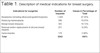
All 3043 studied cases were included. Surgeries involving the removal or replacement of implants totaled 251 (8.24%), 250 replacements and one explant (Table 1). The mean age of patients was 43 years; laterality was not mentioned in 16 requests (0.84%). The anatomical and spatial location was noted in 15 cases (6.33%). The type of surgery performed was mentioned in 40 (15.94%). Previous imaging examination was reported in one case (Table 2).
| Indications for surgeries | Number of requests | Values in Percentage of Total |
|---|---|---|
| Neoplasms (including ultrasound-guided biopsies) | 1,444 | 47.45% |
| Reducing mammoplasty | 586 | 19.25% |
| Benign breast diseases | 583 | 19.15% |
| Removal and/or replacement of silicone breast implants | 251 | 8.24% |
| Gynecomastia | 179 | 5.88% |
| Total | 3043 | 100% |
| Information contained in the request | Number | Percentage |
|---|---|---|
| Laterality | 235 | 99.16% |
| Anatomical and/or spatial location | 15 | 6.33% |
| BIRADS | 1 | 0.40% |
| Clinical history | 48 | 19.12% |
| Signals and symptoms | 35 | 13.94% |
| Prior imaging exam | 12 | 4.78% |
| Radiotherapy | 1 | 0.40% |
| Chemotherapy | 0 | 0.00% |
| Dimensions | 1 | 0.40% |
| Diagnostic hypothesis | 131 | 55.27% |
| Type of surgery performed | 40 | 15.94% |
| Type of Implant removed | 0 | 0.00% |
| Presence of liquid collection | 15 | 6.33% |
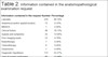
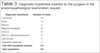
Clinical history data were missing in 48 (19.12%), and signs and symptoms in 35 (13.94%). In 15 requests, there was a reference to the current presence of fluid around the implant, referred to in all of them with the term “chronic seroma” (Table 2). The clinical hypothesis was present in 131 requests, and in 64, there was only the word contracture. And in 15, the term rupture. The requests mentioned the clinical hypothesis of lymphoma on 27 occasions; 20 wrote the term BI-ALCL, and the other seven lymphomas. Forty requests included the surgery that was performed (Table 3).
| Diagnostic hypothesis | Number of cases |
|---|---|
| Contracture | 64 |
| Break | 15 |
| Infection | 5 |
| Malignant neoplasm | 16 |
| BI-ALCL* | 21 |
| BI-ALCL* with seroma present | 6 |
| Late seroma | 3 |
| Organized hematoma | 1 |
| Total with hypothesis inserted | 131 |
| Total requisitions | 151 |
* BI_ALCL - Anaplastic T-cell lymphoma
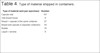
The number of containers sent ranged from 1 to 5, with a median of 2. Of the 251 cases, 242 sent the capsule, 161 sent only the capsule, 27 contained capsule and breast tissue in the same container without mentioning whether they were connected or separated, and, in 54 patients, capsules and breast tissue from the same breast were shipped in different containers. In 17 patients, an immunohistochemical study of CD30 was requested for the tissue sent. In nine cases, fluid was collected and sent for examination representing a specimen (Table 4).
| Type of material sent (per specimen) | Number |
|---|---|
| Capsule only | 161 |
| Only breast tissue | 9 |
| Breast + capsule in the same container | 27 |
| Breast and capsule in separate containers | 54 |
| Nodule | 16 |
| Liquid = seroma | 9 |
None of the requests contained data on the type of implant, brand, anatomical location concerning the pectoralis major muscle, and the length of stay of the implants.
The 251 patients underwent surgery by 57 requesting physicians, 29 of whom were certified plastic surgeons, five gynecologists, three anesthesiologists, and 20 physicians with no defined specialty (Table 5).
| Medical specialty | Total of 57 different |
|---|---|
| Plastic surgery | 29 |
| Gynecology | 5 |
| Anesthetist | 3 |
| No specialty | 20 |
DISCUSSION
The description, in 1997, of the BI-ALCL resulted in changes in the conduct of the removal of silicone breast implants6. The concepts of removing periprosthetic tissues have been discussed, whether en bloc together with the capsule or limited to total or partial capsulectomy. Histopathological examination, considered non-essential in most cases7-9, has become indispensable on many occasions. The importance of exchanging information between the surgeon in charge and the physicians involved in the diagnosis, especially radiologists and pathologists10, has also increased.
Another recent change factor is the surgery of definitive removal of the implants by demand of the patient, labeled as explant surgery, which brings with it the concept of the complete removal of the capsule that involves the implant. Studies have been carried out to evaluate the histological alterations in these cases to find evident correlations between the histological and histochemical findings and the diseases reported by the patients, making it essential to pay greater attention to the evaluation of the removed tissues11-13.
The lack of quantity and quality of the information in the medical request forms can compromise the quality of the surgical pathology report and, consequently, the definitive diagnosis. It should be noted that in this study, no request contained information about the type and brand of the implant, even though the relationship between a particular manufacturer and the high-texture envelopes with BI-ALCL14 was recognized.
Of the 251 requests, only two indicated the anatomical and spatial location of the submitted specimens. In cases with suspected tumors, the need to determine an exact location is well-defined15. However, it is also essential when the implant rupture, as the leaked material can infiltrate adjacent tissues and cause acute and long-term inflammatory reactions.
Surgical samples should be demarcated to allow for their anatomical and spatial location. With this simple practice, it would be possible to determine the region of the affected breast, facilitating surveillance and interpretation in future examinations. It would also make it possible to guide the exact location for any surgical revisions, particularly when other tissues are involved, such as muscles or ribs.
Grubstein et al.16 warned about the difficulty of differentiating, in imaging diagnosis, siliconoma from other conditions, especially breast cancer. Incorrect diagnosis leads to unnecessary examinations and interventions. The same happens when implants with an external envelope covered with polyurethane are used, which presents gradual degradation of this material and the presence of a chronic inflammatory reaction. When it is necessary to remove them, the capsule and the adjacent tissue must be resected, as the presence of residual fragments can result in the formation of nodules and lead to a new surgical revision17. Therefore, the tissue sent must be very well-identified regarding the spatial position and the relationship between the tissue fragments removed, even more so if sent in more than one container.
In the present study, in 242 cases, the capsule was sent for anatomopathological examination, in 161 in isolation and in 54, the breast tissue was also removed but sent in separate containers. This may have occurred due to the surgical technique and the need to correct the contour of the breast tissue associated with the replacement of implants, but the surgeon should inform this fact. However, only 15.40% (40) of the requests contained information about the surgical technique performed.
This finding indicates, as already warned by Lapid et al.8 in 2014, the need to create a protocol to standardize the surgical removal of the capsule and the adjacent breast tissue, preferably en bloc, even in purely aesthetic breast procedures.
In some cases, even when the implant appears to be visually intact, gel leakage may occur17. In these cases, during the surgical procedure to remove the implant, changes in the appearance of the implant may be detected, or a loss in the measured weight of the implant may also occur. This information could be present in medical requests.
Of the 27 cases in which a diagnostic evaluation for BI-ALCL was requested, in 15, the presence of fluid around an implant was reported. In nine of them, the liquid was sent simultaneously with the other specimens. Reports show that 100% of cases diagnosed with BI-ALCL present chronic fluid collection around the implant, and prior needle aspiration guided by ultrasound examination is recommended for material collection and cytology and immunohistochemical studies for research of CD 30 to ALK in order to determine clinical and surgical management18,19.
The data obtained in this study suggest that the surgeons failed to provide information in the request about the existence of some previous imaging exams followed by a puncture. It is also possible to infer that the clinical history researched may have been insufficient. It should be noted that not performing aspiration prior to surgery is not following current protocols3,19.
Information on clinical history was present in 34.52% of the 3043 requests for breast surgeries; however, in cases of implant removal, the rate was significantly lower, only 19.12%. The diagnostic hypothesis, an item contained in the exam requisition form, was only completed in 131 (52.19%), and in 64, only the mention of contracture, which is a physical sign or a finding on physical examination, which can be accompanied by other signs or symptoms such as pain.
This conceptual error seems to indicate, for the pathologist, that the understanding by plastic surgeons of the possible causes of contracture and the clinical evolution of this process is insufficient. The search for causal factors of contracture follows a line of reasoning that should be transmitted to the pathologist synthetically and objectively.
The lack of information in the medical request and, in particular, the lack of mention of laterality in 16 cases makes direct communication between the pathologist and the surgeon necessary, which results in a demand for time and cost for the laboratory. This can delay the definition of the diagnosis and the issuance of reports4,5,20.
When reviewing articles that discuss errors and discrepancies in surgical pathology, this lack of communication was not pointed out as a significant factor compared to all the failures detected in the steps of the histopathological examination. The interpretation of findings in the histopathological and histochemical examination has progressed a lot due to the increasing specialization of pathologists and the change in the work environment, composed of more specialized and larger teams, with the possibility of personal exchange of information or employing information technology.
However, the lack of completed data found in this research demonstrates that when a new clinical condition arises, as with BI-ALCL or explant surgery, a new process of adaptation and medical education is necessary for all specialists involved. . The information deficit in the medical request could be improved through the implementation of protocols and information technology to communicate the surgeon to the pathologist better.
CONCLUSIONS
Our results demonstrate that the quantity and quality of information contained in the medical request are scarce, which could compromise the pathological report. Most requesting physicians were certified plastic surgeons, and plastic surgery societies have gone to great lengths to report and clarify the recent adverse effects of silicone breast implants, especially regarding BI-ALCL or explant surgery. However, our findings suggest that these mechanisms still need improvement. Surgeons and pathologists should be encouraged to create means of communication through protocols and the use of information technology.
REFERENCES
1. Miranda RN, Feldman AL, Soares FA. Breast implant-associated anaplastic large cell lymphoma. In: Allison KH, ed. World Health Organization breast tumours. Lyon: IARC; 2019. p. 245-8.
2. Kim B, Predmore ZS, Mattke S, van Busum K, Gidengil CA. Breast Implant-associated Anaplastic Large Cell Lymphoma: Updated Results from a Structured Expert Consultation Process. Plast Reconstr Surg Glob Open. 2015;3(1):e296.
3. Clemens MW, Jacobsen ED, Horwitz SM. 2019 NCCN Consensus Guidelines on the Diagnosis and Treatment of Breast Implant-Associated Anaplastic Large Cell Lymphoma (BIA-ALCL). Aesthet Surg J. 2019;39(Suppl_1):S3-13.
4. Nakhleh RE, Gephardt G, Zarbo RJ. Necessity of clinical information in surgical pathology. Arch Pathol Lab Med. 1999;123(7):615-9.
5. Burton JL, Stephenson TJ. Are clinicians failing to supply adequate information when requesting a histopathological investigation? J Clin Pathol. 2001;54(10):806-8.
6. Keech JA Jr, Creech BJ. Anaplastic T-cell lymphoma in proximity to a saline- filled breast implant. Plast Reconstr Surg. 1997;100(2):554-5.
7. Roth FS, Felder JM, Friedman JD. Friedman JD. Breast capsulectomy specimens and their clinical implications. Plast Reconstr Surg. 2010;126(6):1848-52.
8. Lapid O, Noels EC, Meijer SL. Pathologic Findings in Primary Capsulectomy Specimens: Analysis of 2531 Patients. Aesthet Surg J. 2014;34(5):714-8.
9. Fisher M, Alba B, Bhuiya T, Kasabian AK, Thorne CH, Tanna N. Routine Pathologic Evaluation of Plastic Surgery Specimens: Are We Wasting Time and Money? Plast Reconstr Surg. 2018;141(3):812-6.
10. Anger J, Elias PE, Moraes PC, Hamerschlak N. A review of data in medical request and the patient questionnaire for magnetic resonance evaluation of silicone breast implants. Einstein (Sao Paulo). 2017;15(4):465-9.
11. Rohrich RJ, Kaplan J, Dayan E. Silicone Implant Illness: Science versus Myth? Plast Reconstr Surg. 2019;144(1):98-109.
12. de Boer M, Colaris M, van der Hulst RRWJ, Cohen Tervaert JW. Is explantation of silicone breast implants useful in patients with complaints? Immunol Res. 2017;65(1):25-36.
13. Kaplan J, Rohrich R. Breast implant illness: a topic in review. Gland Surg. 2021;10(1):430-43.
14. Loch-Wilkinson A, Beath KJ, Knight RJW, Wessels WLF, Magnusson M, Papadopoulos T, et al. Breast Implant-Associated Anaplastic Large Cell Lymphoma in Australia and New Zealand: High-Surface-Area Textured Implants Are Associated with Increased Risk. Plast Reconstr Surg. 2017;140(4):645-54.
15. Tokin CA, Wallace AM. Breast cancer presenting within or adjacent to the breast implant capsule: a case series and clinical recommendations. Clin Breast Cancer. 2012;12(4):296-9.
16. Grubstein A, Cohen M, Steinmetz A, Cohen D. Clin Imaging. 2011;35(3):228-31.
17. Hillard C, Fowler JD, Barta R, Cunningham B. Silicone breast implant rupture: a review. Gland Surg. 2017;6(2):163-8.
18. Di Napoli A. Achieving Reliable Diagnosis in Late Breast Implant Seromas: From Reactive to Anaplastic Large Cell Lymphoma. Plast Reconstr Surg. 2019;143(3S A Review of Breast Implant-Associated Anaplastic Large Cell Lymphoma):15S-22S.
19. Lyapichev KA, Piña-Oviedo S, Medeiros LJ, Evans MG, Liu H, Miranda AR, et al. A proposal for pathologic processing of breast implant capsules in patients with suspected breast implant anaplastic large cell lymphoma. Mod Pathol. 2020;33(3):367-79.
20. Ali SMH, Kathia UM, Gondal MUM, Zil-E-Ali A, Khan H, Riaz S. Impact of Clinical Information on the Turnaround Time in Surgical Histopathology: A Retrospective Study. Cureus. 2018;10(5):e2596.
1. Rede D’Or São Luiz S.A., Departamento de Patologia, São Paulo, SP, Brazil
JA Analysis and/or data interpretation, Conception and design study, Conceptualization, Final manuscript approval, Investigation, Methodology, Project Administration, Writing - Original Draft Preparation, Writing - Review & Editing.
GBT Analysis and/or data interpretation, Conception and design study, Data Curation, Final manuscript approval, Investigation, Realization of operations and/or trials.
GDO Analysis and/or data interpretation, Data Curation, Investigation, Methodology, Software.
DA Analysis and/or data interpretation, Conception and design study, Data Curation, Investigation.
FAS Conception and design study, Methodology, Project Administration, Supervision, Writing - Review & Editing.
Corresponding author: Jaime Anger Av. Brigadeiro Luiz Antônio, 3889, São Paulo, SP, Brazil. Zip code: 01401-001, E-mail: anger@uol.com.br
Article received: November 3, 2021.
Article accepted: April 7, 2022.
Conflicts of interest: none.
 Read in Portuguese
 Read in English
 PDF PT
 Print
Print
 Send this article by email
 How to Cite
 Mendeley
 Pocket