


Case Report - Year 2022 - Volume 37 -
Polypropylene and poliglecaprone mesh implant for lifting muscles in facial paralysis
Implante de tela de polipropileno e poliglecaprone para elevação da musculatura na paralisia facial
Pedro Celso de Castro Pita1,* ; Rafael Ximenes Bandeira de Morais2; Marcella Ferreira Barros3; Eduarda Augusta de Lucena Caldas3; Caroline Silva Costa de Almeida1; Kyldery Wendell Moura Cavalcante1; Vanderson Lamartine de Lima Silva1
ABSTRACT
Introduction: Facial paralysis is a condition that can cause some sequelae, and sometimes only aesthetics can be improved. Based on this, the objective was to report a case in which a polypropylene and poliglecaprone mesh was used in order to raise the structures of the right hemiface.
Case Report: The patient had paralysis in the right hemiface, with incompetent eyelid sealing, deviation of the labial commissure and no movement of the temporal musculature. An incision was made below the cutlet, pre and retroauricular, with detachment of the skin flap across the right hemiface. After lifting the superficial musculoaponeurotic system and fixing it with mononylon threads, the polypropylene and poliglecaprone mesh was placed in the middle third region and fixed with monocryl. Canthotomy and lateral canthopexy of the right eyelid were performed. In the immediate postoperative period, the patient evolved without edema, retraction or bulging, and after one year and seven months, she had complete integration of the mesh, with no retraction, fibrosis or recurrence.
Discussion: The choice of aesthetic treatment for facial paralysis depends on the cause and duration of the injury, but there are several ways to do it. Among the newer ideas are the use of stem cells and alloplastic materials, and following this second line, the polypropylene and poliglecaprone canvas can be thought of as a viable technique, as was reported in this case.
Keywords: Reconstructive surgical procedures; Facial paralysis; Esthetics; Face; Facial asymmetry.
RESUMO
Introdução: A paralisia facial é um quadro que pode gerar algumas sequelas, e às vezes apenas as estéticas podem ser melhoradas. Com base nisso, objetivou-se relatar um caso em que foi utilizada uma tela de polipropileno e poliglecaprone com finalidade de elevar as estruturas da hemiface direita.
Relato de Caso: A paciente apresentava paralisia em hemiface direita, com selamento palpebral incompetente, desvio de comissura labial e sem movimento da musculatura temporal. Foi realizada uma incisão abaixo da costeleta, pré e retroauricular, com descolamento do retalho cutâneo em toda a hemiface direita. Após levantar o sistema musculoaponeurótico superficial e fixá-lo com fios de mononylon, foi colocada a tela de polipropileno e poliglecaprone na região do terço médio e fixada com monocryl. Foi realizada cantotomia e cantopexia lateral da pálpebra direita. No pós-operatório imediato a paciente evoluiu sem edemas, retrações ou abaulamentos, e após um ano e sete meses apresenta total integração da tela, sem retração, fibrose ou recidiva.
Discussão: A escolha do tratamento estético de paralisia facial depende da causa e duração da lesão, mas existem diversas formas de fazê-lo. Entre as ideias mais novas, estão o uso de células tronco e materiais aloplásticos e, seguindo essa segunda linha, a tela de polipropileno e poliglecaprone pode ser pensada como uma técnica viável, como foi neste caso relatado.
Palavras-chave: Procedimentos cirúrgicos reconstrutivos; Paralisia facial; Estética; Face; Assimetria facial
INTRODUCTION
Facial paralysis has a wide range of etiologies and can result from injuries to the facial nerve and, depending on the level of this injury, changes occur in the muscles of facial mimicry. It is usually a reversible phenomenon spontaneously or through clinical or surgical treatment. Even so, about 20% of cases evolve with some sequel, ranging from a mild degree to complete paralysis of facial muscle movements1,2.
The treatment will depend on some variants, mainly the time, place and mechanism of injury. One can try to reanimate the mimic musculature for early injuries through neuroanastomoses or nerve repairs. For example, in late paralysis, one may try dynamic suspension with a muscle flap or static suspension when the mimic function is not recovered, only the aesthetics3.
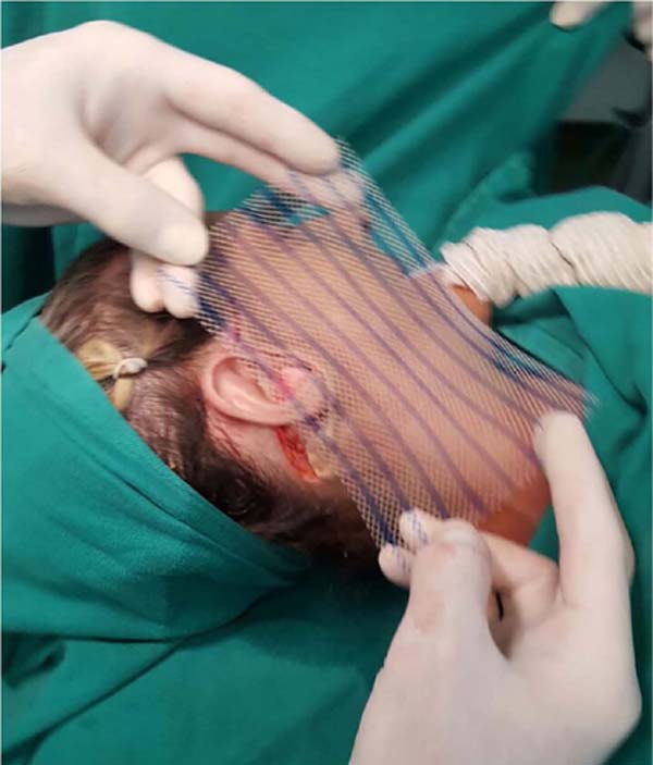
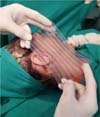
In surgery, the polypropylene and poliglecaprone mesh, commercially called ULTRAPROTM (Figure 1), is partially absorbable, has interesting space, and is mainly required for abdominal hernia repair. It generates tissue fibrosis, providing support but simultaneously stimulating a flexible scar and promoting multidirectional elasticity, providing the abdominal wall with normal dynamics and physiology4.
OBJECTIVE
Thus, this work aims to present a case report at Hospital das Clínicas de Recife-PE, in which the polypropylene and poliglecaprone mesh was used to treat the aesthetic sequel of a patient with facial paralysis, with the function of raising the right hemifacial structures.
CASE REPORT
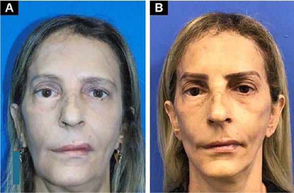
S.RS, female, 55 years old, presents with facial paralysis of unknown etiology for approximately 27 years, having already performed another surgery in an attempt to correct the paralysis in another center, but without satisfactory results. At the initial physical examination-June 2016 (Figure 2), she had an incompetent eyelid seal, scleral show, labial commissure deviation, and no right temporal musculature movements. After evaluation by the neurology service, it was decided to correct the asymmetry by employing a facelift with the elevation of the musculature and fixation of the entire hemiface structure with the ULTRAPRO® mesh due to its support characteristic associated with flexibility and elasticity.


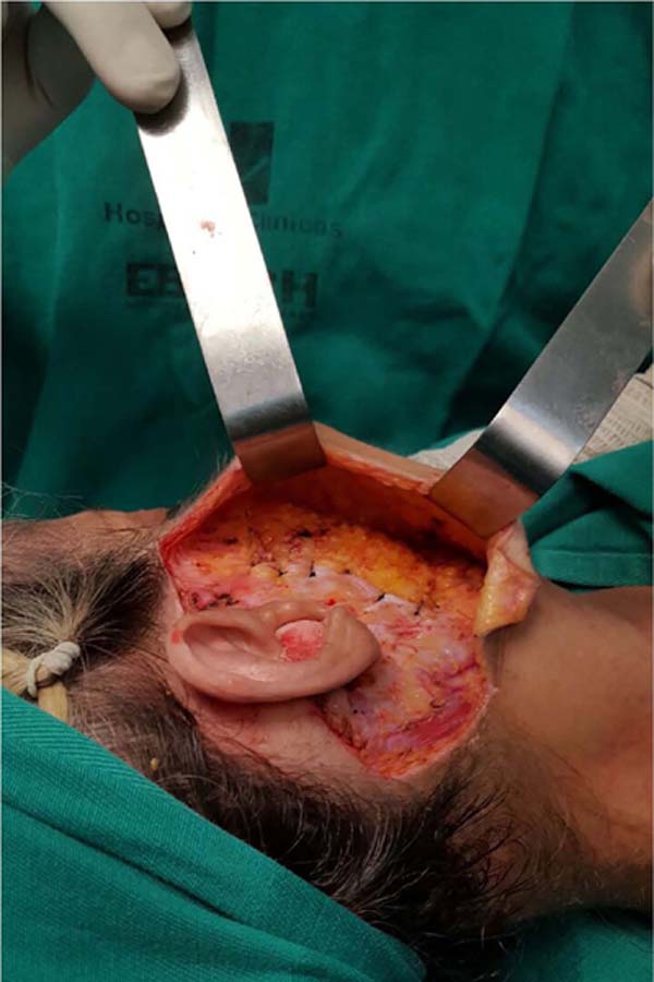
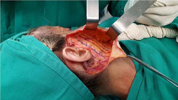
During surgery - in May 2017, with the patient in dorsal decubitus and under general anesthesia, marking was performed with bright green using a compass, and then started with an incision below the sideburn, pre and retroauricular with a detachment of the skin flap on the entire right hemiface. After lifting the superficial musculoaponeurotic system (SMAS) and fixing it with mononylon threads (Figure 3), the ULTRAPRO® mesh was placed in the middle third of the right hemiface (Figure 4). The mesh was fixed with monocryl over the SMAS.


The canthotomy and lateral canthopexy of the right eyelid were performed as a complement. On the left hemiface, the same incision was made below the pre- and retroauricular sideburns over the previous marking with skin flap detachment and treatment of the SMAS. Suspension of the tail of the left eyebrow was performed according to the Castanares technique. After a review of hemostasis, suturing was performed in layers. Tubular drains were introduced in both hemifaces.
In the immediate postoperative period, the patient evolved without edema, retractions or bulges. One year and eight months after the surgery, she presents complete integration of the mesh, remaining with the fixation of the musculature in its new location, showing no fibrosis or recurrence of sagging (Figure 5).

DISCUSSION
Facial paralysis is a disorder that involves the nerves of the face region; the etiology is quite wide, with more than 75 causes described, such as congenital, idiopathic, traumatic and tumoral. However, the etiological diagnosis is often difficult to give, thus increasing the idiopathic casuistry known as Bell’s palsy5.
For surgical treatment, the advantages and disadvantages of each proposal must be informed, stating that it is not possible to restore sufficient voluntary movements to restore facial mimicry6.
The choice of treatment will also depend on the cause and duration of the injury7. Numerous techniques have already been described to improve the function and appearance resulting from facial nerve injuries, such as immediate reconstruction by direct or indirect sutures or by the interposition of nerve grafts8. In these cases, an attempt is made to restore nerve function through neurorrhaphy, as reported by Viterbo et al.9 in several cases.
Another, more recent way of trying to recover nerves is using stem cells, as has been studied for some time. These cells must act in nerve regeneration10.
In long-term paralysis, the static suspension is the simplest surgical treatment. In 1934, Gillies11 described the use of temporal muscle transposition for facial resuscitation, which showed good results, but was impractical for the patient in question.
Some new static techniques for correcting long-term paralysis continue to be proposed, many with alloplastic materials, as presented by Alam12, in which “Gore-tex strip” was used, with a good aesthetic response, especially when analyzing the nasolabial fold.
Following the use of new materials, the use of ULTRAPROTM mesh can be considered a viable technique, as it was in this case. The main characteristic of generating tissue fibrosis, providing support and, at the same time, an elastic, flexible scar, motivated the choice of this product, which made that the result of the suspension, by the traction of the SMAS, was maintained even after absorption of the mesh4.
Therefore, it can be concluded that using polypropylene and poliglecaprone mesh can correct the muscle flaccidity resulting from facial paralysis and maintain its surgical result without presenting skin deformities, even with the mesh placed in the subcutaneous tissue, not causing retraction.
REFERENCES
1. Vlastou C. Facial paralysis. Microsurgery. 2006;26(4):278-87. PMID: 16628742 DOI: https://doi.org/10.1002/micr.20240
2. Lazarini PR, Fouquet ML. Paralisia facial: avaliação, tratamento e reabilitação. São Paulo: Lovise; 2006.
3. Brown S, Isaacson B, Kutz W, Barnett S, Rozen SM. Facial Nerve Trauma: Clinical Evaluation and Management Strategies. Plast Reconstr Surg. 2019;143(5):1498-512. DOI: 10.1097/ PRS.0000000000005572 PMID: 30807496 DOI: https://doi.org/10.1097/PRS.0000000000005572
4. Utrabo CAL, Czeczko NG, Busato CR, Montemór-Netto MR, Lipinski L, Malafaia O. Estudo tensiométrico de telas utilizadas na correção de defeito na parede abdominal ventral de ratos. ABCD Arq Bras Cir Dig. 2017;30(3):165-8. DOI: 10.1590/0102- 6720201700030001
5. Balogh B, Frühwald F, Millesi W, Millesi H, Firbas W. Sonoanatomy of the muscles of facial expression. Surg Radiol Anat. 1988;10(2):101-6. PMID: 3135614 DOI: https://doi.org/10.1007/BF02307817
6. Medina HS, Ramalho R, Biscotto R, Chveid M. Tela de Prolene - Utilização em Reconstruções de Cabeça e Pescoço. Rev Bras Cir Plást.1997;12(1):7-16.
7. Gur E, Kedar DJ, Zaretski A, Arad E, Meilik B, Yanko R, et al. Facial nerve paralysis - therapeutic approach, facial reanimation and adjunctive treatment. Harefuah. 2020;159(8):612-7. PMID: 32852164
8. Batista KT, Cauhi AF. Reabilitação Cirúrgica da Face Paralisada. Rev Bras Cir Plást. 2007;22(4):253-60.
9. Viterbo F, Romão A, Brock RS, Joethy J. Facial reanimation utilizing combined orthodromic temporalis muscle flap and end-to-side cross-face nerve grafts. Aesthetic Plast Surg. 2014;38(4):788-95. PMID: 24943646 DOI: https://doi.org/10.1007/s00266-014-0357-8
10. Wang TV, Delaney S, Pepper JP. Current state of stem cellmediated therapies for facial nerve injury. Curr Opin Otolaryngol Head Neck Surg. 2016;24(4):285-93. PMID: 27379549 DOI: https://doi.org/10.1097/MOO.0000000000000292
11. Baker DC. Facial paralysis. In: McCarthy JG, ed. Plastic Surgery. Philadelphia: W. B. Saunders; 1990. p. 2237-319.
12. Alam D. Rehabilitation of long-standing facial nerve paralysis with percutaneous suture-based slings. Arch Facial Plast Surg. 2007;9(3):205-9. PMID: 17515497 DOI: https://doi.org/10.1001/archfaci.9.3.205
1. Hospital das Clínicas, Universidade Federal de Pernambuco, Recife, PE, Brazil.
2. Universidade de Pernambuco, Faculdade de Ciências Médicas, Recife, PE, Brazil.
3. Universidade Federal de Pernambuco, Faculdade de Medicina, Recife, PE, Brazil.
PCCP Analysis and/or data interpretation, Conception and design study, Conceptualization, Data Curation, Final manuscript approval, Formal Analysis, Funding Acquisition, Investigation, Methodology, Project Administration, Realization of operations and/or trials, Resources, Software, Supervision, Validation, Visualization, Writing - Original Draft Preparation, Writing - Review & Editing.
RXBM Analysis and/or data interpretation, Conception and design study, Final manuscript approval, Investigation, Methodology, Project Administration, Validation, Visualization, Writing - Original Draft Preparation, Writing - Review & Editing.
MFB Analysis and/or data interpretation, Conception and design study, Conceptualization, Data Curation, Final manuscript approval, Formal Analysis, Funding Acquisition, Investigation, Methodology, Writing - Original Draft Preparation, Writing - Review & Editing.
EALC Analysis and/or data interpretation, Conceptualization, Data Curation, Final manuscript approval, Formal Analysis, Investigation, Project Administration, Realization of operations and/or trials, Resources, Software, Visualization, Writing - Original Draft Preparation, Writing - Review & Editing.
CSCA Analysis and/or data interpretation, Conceptualization, Data Curation, Final manuscript approval, Investigation, Methodology, Resources, Validation, Visualization, Writing - Original Draft Preparation, Writing - Review & Editing.
KWMC Analysis and/or data interpretation, Conceptualization, Final manuscript approval, Visualization, Writing - Review & Editing.
VLLS Analysis and/or data interpretation, Conceptualization, Final manuscript approval, Supervision, Visualization, Writing - Review & Editing.
Corresponding author: Pedro Celso de Castro Pita Praça Miguel de Cervantes, nº 60, sala 301, Ilha do Leite, Recife, PE, Brazil Zip Code: 50070-520 E-mail: pedro.pitta@hotmail.com
Article received: March 10, 2021.
Article accepted: July 14, 2021.
Conflicts of interest: none.
Institution: Universidade Federal de Pernambuco, Hospital das Clínicas, Recife, PE, Brazil.
 Read in Portuguese
 Read in English
 PDF PT
 Print
Print
 Send this article by email
 How to Cite
 Mendeley
 Pocket