


Original Article - Year 2022 - Volume 37 -
Microbiological profile in patients hospitalized for burns
Perfil microbiológico em pacientes hospitalizados por queimaduras
Carlos Manuel Collado Hernández1,* ; Arbenys Alexis Blanco Machado1; Vivian Pérez Núnez2
ABSTRACT
Introduction: Injuries caused by burns are a global health problem that affects all age groups, not only because of the frequency in which they occur but according to their severity. They can be disabling and have high mortality, and at the same time, generate an unfavorable economic impact on the country. The objective is to describe the microbiological profile of burned hospitalized patients.
Methods: A descriptive, longitudinal, and prospective study was carried out at the Provincial Clinical Surgical Celia Sánchez Manduley Hospital from July 2017 to June 2020. Microbiology culture, positivity, isolated microorganisms, and antimicrobial sensitivity were studied.
Results: The study showed that the culture microbiology of skin lesions due to burns (130 samples) were predominant, and of them, 58.46% were positive, Staphylococcus aureus with 51.73% was the most isolated germ; sensitivity to amikacin of 55.56% and vancomycin 51.11%; survival was high.
Conclusions: The positivity in the culture microbiology of burned skin, mainly Staphylococcus aureus, and high sensitivity to few antimicrobials predominated.
Keywords: Burns; Anti-infective agents; Burn units; Microbiology; Infections.
RESUMO
Introdução: As lesões por queimaduras são um problema de saúde global que atinge todas as faixas etárias, não só pela frequência com que ocorrem, mas também pela gravidade. Podem ser incapacitantes, ter alta mortalidade e, ao mesmo tempo, gerar um impacto econômico desfavorável para o país. O objetivo é descrever o perfil microbiológico de pacientes hospitalizados por queimaduras.
Métodos: Estudo descritivo, longitudinal e prospectivo no Hospital Provincial Clínico Cirúrgico Celia Sánchez Manduley, Cuba, de julho de 2017 a junho de 2020. Foram estudadas amostras de cultura, positividade, microrganismos isolados e sensibilidade antimicrobiana.
Resultados: O estudo mostrou que as amostras de lesões cutâneas por queimaduras (130) foram predominantes e delas 58,46% foram positivas; Staphylococcus aureus, com 51,73%, foi o germe mais isolado; sensibilidade à amicacina de 55,56% e vancomicina 51,11%; a sobrevida foi alta.
Conclusão: Predominou a positividade nas amostras de cultura de pele queimada, principalmente Staphylococcus aureus, e uma alta sensibilidade a poucos antimicrobianos.
Palavras-chave: Queimaduras; Anti-infecciosos; Unidades de queimados; Microbiologia; Infecções
INTRODUCTION
Injuries caused by burns are a global health problem that affects all age groups, not only because of the frequency with which they occur, but because of their severity, which can be disabling and have high mortality and, at the same time, generate an unfavorable economic scenario for the country1. It is a serious injury and is responsible for indelible consequences and death. Severe burns produce physical suffering, requiring treatments that last months or years2.
According to 2014 world health statistics, the rate of years of life lost due to burns is up to 29 times higher in the African region, and the lowest is found in Latin America and the Caribbean3. Worldwide, burns are responsible for approximately 265,000 deaths per year4,5, mainly in low- and middle-income countries6; and were among the main causes of disability7.
The risk of infection is particularly high in some parts of the world. Many projects in developed and developing countries have shown that applying available interventions and strategies can significantly reduce the disease burden of healthcare-associated infections8.
In general, infections produced in hospitalized patients have been of concern and are manifested by more than 1.4 million people worldwide. Between 5% and 10% of patients admitted to modern hospitals in the developed world will contract one or more infections. The risk of healthcare-associated infection in developing countries is 2 to 20 times higher than in developed countries8.
In the United States, one in 136 hospital patients becomes seriously ill from a hospital-acquired infection; this equates to 2 million cases and approximately 80,000 deaths per year. In England, more than 100,000 cases of healthcare-associated infection each year cause more than 5,000 deaths directly related to the infection. In Mexico, an estimated 450,000 cases of healthcare-associated infection, with 32 deaths per 100,000 inhabitants per year8.
In the case of burn patients, the infection can be aggravated by the characteristics of this type of injury. Regardless of etiology, clinical classification, therapeutic management and individual response, sepsis complicates the patient’s evolution and constitutes an important cause of mortality. Therefore, its control becomes one of the most important goals of treatment. In burn patients, skin and soft tissue infection are the starting point for bacteremia, which worsens the clinical course9,10.
The growing interest in adequate treatment, especially in infection control, and adequate and timely follow-up, motivated the study.
OBJECTIVE
To describe the microbiological profile of patients hospitalized for burns at the Hospital Provincial Clínico Cirúrgico “Celia Sánchez Manduley,” Manzanillo, Cuba.
METHODS
A descriptive, longitudinal and prospective study was carried out at the Plastic Surgery and Burns Service of the Hospital Provincial Clínico Cirúrgico “Celia Sánchez Manduley” from July 2017 to June 2020. The Research Ethics Committee evaluated and approved it under number 218.
The following variables were determined: types of culture samples performed according to their positivity, isolated microorganism, and antimicrobial sensitivity.
For data collection, a form was prepared after reviewing the medical records of all patients hospitalized for burns. The form was designed to be processed on a computer using descriptive statistics in the Microsoft Office Excel 2007 program, obtaining absolute numbers and percentages expressed in tables prepared for this purpose.
The study considered the provisions of the Declaration of Helsinki. Ethical standards of discretion, reliability of information and honesty were respected.
RESULTS
Table 1 shows the different types of samples for culture and their positivity, highlighting that the majority corresponded to skin lesions caused by burns, with 130 samples, and of these, 76 (58.46%) were positive, followed by the catheter tip, with 33 samples; only six (18.18%) were positive and, less frequently, blood cultures, with 25 samples, and positivity in only five (20%).
| Culture Samples | Positive | Negatives | ||
|---|---|---|---|---|
| No | % | No | % | |
| Burnt Skin (n=130) | 76 | 58.46 | 54 | 41.54 |
| Catheter tip (n=33) | 6 | 18.18 | 27 | 81.82 |
| Blood culture (n=25) | 5 | 20 | 20 | 80 |
| Total (n=188) | 87 | 46.28 | 101 | 53.72 |

In a general analysis, we found that from a total of 188 samples (skin lesions from burns, catheter tip and blood cultures), the negative result predominated in 101 (53.72%), that is, the non-growth of bacteria.
The most isolated microorganisms in the different samples (skin lesions from burns, catheter tip and blood culture) are identified in Table 2 and were Staphylococcus aureus in 45 samples (51.73%), and of these, they predominated in the burned skin samples, in 39 (44.83%).
| Microorganisms | Culture samples | |||||||
|---|---|---|---|---|---|---|---|---|
| Skin Burned | Blood culture | Catheter tip | Total | |||||
| No | % | No | % | No | % | No | % | |
| Staphylococcus aureus | 39 | 44.83 | 2 | 2.30 | 4 | 4.60 | 45 | 51.73 |
| Enterobacter aerogenes | 18 | 20.68 | 3 | 3.45 | 1 | 1.15 | 22 | 25.28 |
| Pseudomona aeruginosa | 11 | 12.64 | 0 | 0 | 1 | 1.15 | 12 | 13.79 |
| Proteus mirabilis or vulgaris | 5 | 5.75 | 0 | 0 | 0 | 0 | 5 | 5.75 |
| Escherichia coli | 2 | 2.30 | 0 | 0 | 0 | 0 | 2 | 2.30 |
| Providence | 1 | 1.15 | 0 | 0 | 0 | 0 | 1 | 1.15 |
| Total | 76 | 87.35 | 5 | 5.75 | 6 | 6.90 | 87 | 100 |
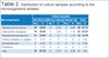
Enterobacter aerogenes continues in frequency in 22 samples (25.28%), of which 18 (20.68%) were found in samples of burned skin; Pseudomonas aeruginosa appears in third place, with 12 (13.79%), and of these, 11 (12.64%) corresponded to samples of burned skin.
Samples from burned skin had more pathogenic microorganisms isolated, with 76 (87.35%).
Table 3 shows the different pathogenic microorganisms isolated and their antimicrobial sensitivity, emphasizing sensitivity greater than 50%.
| Antimicrobial sensitivity | Microorganisms | |||||
|---|---|---|---|---|---|---|
| Staphylococcus aureus (n=45) | Enterobacter aerogenes (n=22) | Pseudomona aeruginosa (n=12) | Proteus mirabilis or vulgaris (n = 5) | Escherichia coli (n=2) | Providence (n=1) | |
| No. (%) | No (%) | No (%) | No (%) | No (%) | No (%) | |
| Gentamicin | 2 (4.44) | 0 (0) | 1 (8.33) | 0 (0) | 0 (0) | 0 (0) |
| Kanamycin | 0 (0) | 0 (0) | 0 (0) | 2 (40) | 0 (0) | 0 (0) |
| Amikacin | 25 (55.56) | 10 (45.45) | 9 (75) | 3 (60) | 0 (0) | 1 (100) |
| Sulfamethoxazole + Trimethoprim | 5 (11.11) | 2 (9.09) | 1 (8.33) | 3 (60) | 0 (0) | 0 (0) |
| Azithromycin | 7 (15.56) | 6 (27.27) | 0 (0) | 2 (40) | 0 (0) | 0 (0) |
| Penicillin | 4 (8.89) | 0 (0) | 0 (0) | 0 (0) | 0 (0) | 0 (0) |
| Oxacillin | 1 (2.22) | 0 (0) | 0 (0) | 0 (0) | 0 (0) | 0 (0) |
| Triphamox | 4 (8.89) | 0 (0) | 0 (0) | 0 (0) | 2 (100) | 0 (0) |
| Aumentin | 3 (6.67) | 0 (0) | 2 (16.67) | 0 (0) | 0 (0) | 0 (0) |
| Cephalexin | 1 (2.22) | 0 (0) | 0 (0) | 0 (0) | 0 (0) | 0 (0) |
| Cefazolin | 7 (15.56) | 0 (0) | 0 (0) | 2 (40) | 0 (0) | 0 (0) |
| Ceftriaxone | 16 (35.56) | 4 (18.18) | 0 (0) | 4 (80) | 0 (0) | 1 (100) |
| Cefotaxime | 10 (22.22) | 3 (13.64) | 0 (0) | 1 (20) | 0 (0) | 0 (0) |
| Ceftazidime | 0 (0) | 1 (4.55) | 4 (33.33) | 5 (100) | 0 (0) | 0 (0) |
| cefuroxime | 5 (11.11) | 1 (4.55) | 0 (0) | 1 (20) | 0 (0) | 0 (0) |
| Cefepime | 8 (17.78) | 6 (27.27) | 1 (8.33) | 2 (40) | 0 (0) | 0 (0) |
| Piperacillin | 9 (20) | 3 (13.64) | 6 (50) | 2 (40) | 2 (100) | 1 (100) |
| Piperazine | 1 (2.22) | 4 (18.18) | 0 (0) | 2 (40) | 0 (0) | 0 (0) |
| Tazobactam | 0 (0) | 0 (0) | 1 (8.33) | 0 (0) | 0 (0) | 0 (0) |
| Doxycycline | 2 (4.44) | 1 (4.55) | 0 (0) | 0 (0) | 2 (100) | 0 (0) |
| Ciprofloxacin | 10 (22.22) | 0 (0) | 1 (8.33) | 2 (40) | 0 (0) | 0 (0) |
| Phosphomycin | 19 (42.22) | 16 (72.73) | 10 (83.33) | 4 (80) | 0 (0) | 0 (0) |
| Vancomycin | 23 (51.11) | 0 (0) | 0 (0) | 0 (0) | 0 (0) | 0 (0) |
| Aztreonam | 1 (2.22) | 4 (18.18) | 4 (33.33) | 4 (80) | 0 (0) | 0 (0) |
| Meronem | 10 (22.22) | 3 (13.64) | 1 (8.33) | 2 (40) | 0 (0) | 0 (0) |
| Imipenem | 2 (4.44) | 0 (0) | 0 (0) | 0 (0) | 0 (0) | 0 (0) |
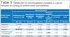
In the case of Staphylococcus aureus, only two antimicrobials showed sensitivity above 50%, amikacin in 25 samples (55.56%) and vancomycin with 23 (51.11%).
Enterobacter aerogenes only showed sensitivity above 50% with fosfomycin, represented in the 16 samples in which it was found (72.73%) of the 22 positive ones.
Pseudomonas aeruginosa was found in 12 samples and was sensitive to fosfomycin in 10 samples (83.33%), amikacin in nine samples (75%) and piperacillin in six samples (50%).
Proteus mirabilis or vulgaris were found in only five samples and had sensitivity with ceftazidime in five samples (100%), ceftriaxone, fosfomycin and aztreonam each, with four samples (80%), and amikacin and sulfamethoxazole + trimethoprim in three samples. (60%).
Escherichia coli was present in only two samples; its important antimicrobial sensitivity was represented by Trifamox, piperacillin and doxycycline, each one in the two samples (100%).
Providence, isolated from only one sample, was sensitive to amikacin, ceftriaxone and piperacillin; each showed effective sensitivity.
In general, amikacin, piperacillin and fosfomycin were the most effective antimicrobials against most pathogenic microorganisms isolated in the different samples collected for culture and antibiogram. Other antimicrobials had sensitivity below 50%.
DISCUSSION
Infections in patients hospitalized for burns are currently one of the main challenges related to a significant rate of complications that can lead to death in these patients. Multiple risk factors predispose burned patients to develop an infection during their stay in burn patient care units.
It is important to know the conditions of the arrival of the burned patient at the hospital, including some risk factors before the burn. However, once hospitalized, it is necessary to evaluate some clinical and epidemiological characteristics that can contribute to complications and an unfavorable evolution.
The different samples and cultures performed on burn patients were represented in more than 2/3 of all cases in the burnt skin samples, and the positivity in these was greater than in the negative ones. This was not the case for catheter tip samples and blood cultures, in which negative samples predominated. If a global analysis is carried out, we will observe that negative results predominated.
When comparing with studies on burn patients in other places, it was not possible to find them in the reviewed bibliography. However, some were performed in other skin conditions and/or in which deep venous catheterizations and blood cultures were used. For example, in a study by Rosanova et al. in Argentina in patients with other pathologies, low incidences of positivity were found in blood cultures and catheter tips, showing an incidence of 3.1 per 1000 tests performed11.
Another study, conducted by Zayas Martínez et al.12 in Camagüey, Cuba, showed significant rates of negative results in catheter tip cultures; therefore, positivity is low.
The findings of this research respond to the fact that the first and main affected area was at the skin level. Logically, it is necessary and according to the patients’ local and general clinical characteristics to prioritize the performance of microbiological studies to diagnose possible local infections.
In the case of samples from catheter tips and blood cultures, they were usually in a lower proportion for several reasons; among them, most patients hospitalized for burns did not need deep venous catheterization because they were classified as small burns and with favorable evolution from a clinical and local point of view.
Strict monitoring of clinical and local elements at the burn level and prompt treatment may be responsible for the low positivity in the case of culture samples performed at the tip of catheters and blood cultures.
The presence of Staphylococcus aureus as the most representative in the different samples taken from the patients of our investigation coincides with studies carried out in Colombia13 and other regions of Cuba10, in which they are found in the same way that it is the most isolated in the samples, mainly in burnt skin. In a second order are Enterobacter aerogenes and Pseudomonas aeruginosa.
Pseudomonas aeruginosa, in other investigations such as that by Morales et al.14 at the San Vicente de Paul University Hospital in Medellín, Colombia, was the most frequent, although the difference with Staphylococcus aureus was not relevant.
In a study by Dávalos et al.15, in Ecuador, Pseudomonas aeruginosa was also found as the main source of infection and Staphylococcus aureus second.
It is possible that the higher frequency of isolation of Staphylococcus aureus in this investigation in burn patients, especially in burnt skin, is explained by the frequent presence of this microorganism in healthy skin, which, once injured, favors the appearance of local infections. Another possible cause would be the presence it normally has in the nose, infecting the burned skin through the patient’s manipulation.
On the other hand, the presence of a smaller number of patients with gram-negative germs, which, despite being generally found in the hospital, are not predominant due to the compliance with hygienicsanitary measures by the personnel who care for patients with burn wounds, reducing the risk of infection by these germs.
Likewise, the decrease in the number of cases admitted to our Burn Unit helps avoid cross-infection between patients since it is not crowded at any time and, of course, separated into individual rooms, especially those with large burns.
The susceptibility to antimicrobials behaved considering the different microorganisms isolated in the burnt skin cultures, catheter tip and blood cultures samples.
Some studies, such as the one carried out by Rodríguez et al.10, in Villa Clara, Cuba, reported high sensitivity of Staphylococcus aureus to methicillin, ciprofloxacin, chloramphenicol, gentamicin and sulfamethoxazole + trimethoprim, not coinciding with this study, since most of them presented low susceptibility, with amikacin and vancomycin being the most susceptible. However, they coincide with the investigation carried out by Zayas Martínez et al.12, in Camagüey, Cuba.
In the case of Enterobacter aerogenes, this research shows high sensitivity only to Phosphomycin, not coinciding with studies such as those carried out by Troche et al.16 at a National Burn Center in Paraguay, which showed high sensitivity to ceftazidime, vancomycin, amikacin and ciprofloxacin.
Pseudomonas aeruginosa showed antimicrobial sensitivity above 50% only for fosfomycin, amikacin and piperacillin, which coincides with Morales et al.14, at Hospital Universitario San Vicente de Paul, in Medellín, Colombia, which showed amikacin and piperacillin with high antimicrobial sensitivity.
The rest of the isolated germs in the analyzed samples are not representative due to the little appearance in the different results.
The result of this investigation shows a high antimicrobial sensitivity to a few antimicrobial agents. It is the criterion that could be related to the frequent use at an institutional level of other antimicrobials, even if the use of some of them is more recent. For this reason, the clinical evaluation of the patient is important, and the institution and the Burn Unit’s microbiological map must be remembered to control the burn patient’s infection effectively.
CONCLUSIONS
The predominance of positivity was demonstrated in burnt skin culture samples more frequently in the isolation of Staphylococcus aureus, Enterobacter aerogenes and Pseudomona aeruginosa, respectively, and high antimicrobial sensitivity to few antimicrobials. The Burn Unit’s microbiological map and antimicrobial susceptibility should be periodically evaluated to continue reducing possible complications of infectious origin and maintain a timely, effective and updated policy for antimicrobials.
REFERENCES
1. Wiegering Cecchi GM, Rios Hidalgo E, Córdova Orrillo JV, Ludeña Muñoz JR, Medina CA. Características clínico-epidemiológicas y patrones de prescripción para quemaduras en tres hospitales de Lima, Perú. Rev Peru Med Exp Salud Publica. 2019;36(1):68-73. DOI: https://doi.org/10.17843/rpmesp.2019.361.3649
2. Arruda CN, Braide ASG, Nascimento MCA, Lima Júnior EM, Nations M. Tentativa de suicídio pós-queimadura: uma experiência humana inscrita na pele. Rev Bras Queimaduras. 2016;15(1):54-7.
3. Gandaria Marsillí A, Lozada Chinea M, Miquet LM, Gómez Zayas O. Quemaduras. En: Soler Vaillant R, Mederos Curbelo ON. Cirugía. Tomo VI. Lesiones graves por traumatismo. La Habana: Editorial Ciencias Médicas; 2018. p. 469-502.
4. Lara R, Del Rocío L, Andrade P, Andrade MTP. Causas de quemaduras en población adulta en el estado de Guanajuato 2011- 2016. Rev Divulg Cient. 2017;3(2):362-6.
5. Polo Andrade S, Mendoza Polo VA. Epidemiología, manejo inicial y análisis de morbimortalidad del Gran Quemado en un Hospital de tercer nivel de atención del municipio de La Paz. Arch Boliv Med. 2018;29(97):7-15.
6. Atwell K, Bartley C, Cairns B, Charles A. The epidemiologic characteristics and outcomes following intentional burn injury at a regional burn center. Burns. 2020;46(2):441-6. DOI: https://doi.org/10.1016/j.burns.2019.08.002
7. Sadeghian F, Saeedi Moghaddam S, Saadat S, Niloofar P, Rezaei N, Amirzade-Iranaq MH, et al. The trend of burn mortality in Iran - A study of fire, heat and hot substance-related fatal injuries from 1990 to 2015. Burns. 2019;45(1):228-40. PMID: 30274812 DOI: https://doi.org/10.1016/j.burns.2018.09.006
8. Arce Padilla EC. Infecciones nosocomiales en la unidad de quemados del Hospital Baca Ortiz [Tesis]. Ambato: Universidad Regional Autónoma de los Andes; 2017. Disponível em: https://rraae.cedia.edu.ec/Record/UNIANDES_366122d3298246aad30aa768128cd3fe
9. Greenhalgh DG. Sepsis in the burn patient: a different problem than sepsis in the general population. Burns Trauma. 2017;5:23. PMID: 28795054 DOI: https://doi.org/10.1186/s41038-017-0089-5
10. Rodríguez JA, García Urquijo A, Manzanas RL, García González ME. Susceptibilidad y patrones fenotípicos antimicrobianos de Staphylococcus aureus en la piel de quemados hospitalizados. Acta Méd Centro. 2018;12(4):422-8.
11. Rosanova MT, Mussini MS, Arias AP, Sormani MI, Mastroianni A, García ME, et al. Análisis epidemiológico y de factores de riesgo de mortalidad en bacteriemia por Pseudomonas aeruginosa en niños. Arch Argent Pediatr. 2019;117(2):128-31.
12. Zayas Martínez IG, Romero González A, Bouza López D. Evaluación de los resultados de cultivos de catéteres en pacientes graves. AMC. 2003;7(1):39-49.
13. Ferrada R, Aragón N, Becerra C. Cultivo biopsia en quemaduras. Rev Colomb Cir. 1992;17(3):151-3.
14. Morales CH, Gómez AF, Herrera JO, Gallego MC, Usuga YA, Hoyos MA, et al. Infección en pacientes quemados del Hospital Universitario San Vicente de Paúl, Medellín, Colombia. Rev Colomb Cir. 2010;25:267-75.
15. Dávalos Dávalos P, Lorena Dávila J, Alexandra Meléndez S. Manejo de morbimortalidad del paciente pediátrico quemado en el hospital “Baca Ortiz” de Quito, Ecuador. Cir Plást Iberolatinoam. 2007;33(3):163-70.
16. Troche Zaracho M, Maidana de Larrosa G, Lugo Rodríguez G, Vera Galván Z, Samaniego Silva L. Utilización de antibióticos en el Centro Nacional del Quemado, Paraguay. Mem Inst Investig Cienc Salud. 2017;15(2):97-103.
1. Hospital Provincial Clínico Cirúrgico “Celia Sánchez Manduley”, Cirugía Plástica
y Caumatología Manzanillo, Granma, Cuba.
2. Hospital Provincial Psiquiátrico “Manuel Fajardo Rivero”, Psiquiatría, Manzanillo,
Granma, Cuba.
CMCH Analysis and/or data interpretation, Conception and design study, Conceptualization, Data Curation, Final manuscript approval, Formal Analysis, Investigation, Methodology, Project Administration, Resources, Supervision, Validation, Visualization, Writing - Original Draft Preparation, Writing - Review & Editing.
AABM Conception and design study, Conceptualization, Final manuscript approval, Formal Analysis, Investigation, Validation, Visualization, Writing - Original Draft Preparation, Writing - Review & Editing.
VPN Analysis and/or data interpretation, Conceptualization, Data Curation, Final manuscript approval, Formal Analysis, Supervision, Validation, Visualization, Writing - Original Draft Preparation, Writing - Review & Editing.
Corresponding author: Carlos Manuel Collado Hernández Hospital Provincial Clinico Cirúrgico Celia Sánchez Manduley, Circunvalación, Manzanillo, Cuba Zip Code: 87510 E-mail: vivicollado2013@gmail.com
Article received: March 13, 2021.
Article accepted: April 07, 2022.
Conflicts of interest: none.
Institution: Hospital Provincial Clinico Cirúrgico Celia Sánchez Manduley, Manzanillo, Cuba.
 Read in Portuguese
 Read in English
 PDF PT
 Print
Print
 Send this article by email
 How to Cite
 Mendeley
 Pocket