

Case Report - Year 2022 - Volume 37 -
Total reconstruction of the skin coverage of the penis with myocutaneous flap: case report
Reconstrução total da cobertura cutânea do pênis com retalho miocutâneo: relato de caso
ABSTRACT
Introduction: The penis is an important structure of the male body, and its reconstruction is a challenge. Several diseases and deformities affect this organ, being necessary, in certain cases, for the total reconstruction of the cutaneous coverage of the penis, having already been described in the literature several techniques, such as the use of total grafts, scrotal flap, myocutaneous flaps of the fasciae latae and others.
Case Report: In this report, a reconstruction of the total coverage of the penis is presented using a myocutaneous flap of the cremaster muscle with skin from the scrotum, achieving good vascularization and maintaining urethral permeability.
Conclusion: This technique was not found in any of the databases researched in this study, only similar ones, and it proved to be a good option for the total reconstruction of penile skin coverage.
Keywords: Penis; Scrotum; Male urological surgical procedures; Reconstructive surgical procedures; Surgical flaps.
RESUMO
Introdução: O pênis é uma importante estrutura do corpo masculino, sendo sua reconstrução um desafio. Existem diversas doenças e deformidades que acometem este órgão, sendo necessário, em certos casos, a reconstrução total da cobertura cutânea do pênis, tendo já sido descritas na literatura diversas técnicas, tais como o uso de enxertos totais, retalho escrotal, retalhos miocutâneos da fáscia lata e outros.
Relato de Caso: Neste relato é apresentada uma reconstrução da cobertura total do pênis por meio do uso de retalho miocutâneo do músculo cremaster com pele da bolsa escrotal, conseguindo prover uma boa vascularização e mantendo a permeabilidade uretral.
Conclusão: Tal técnica não foi encontrada em nenhuma das bases de dados pesquisadas no trabalho, apenas semelhantes, e mostrouse como uma boa opção para a reconstrução total da cobertura cutânea peniana.
Palavras-chave: Pênis; Escroto; Procedimentos cirúrgicos urológicos masculinos; Procedimentos cirúrgicos reconstrutivos; Retalhos cirúrgicos
INTRODUCTION
The penis is an important structure of the male body, and damage to its anatomy is always dramatic, functionally, and psychologically for the patient. Among the lesions that can affect the penis and its coverage, there are congenital deformities, inflammatory diseases, infections, lymphedema, traumatic and iatrogenic lesions due to benign and malignant tumors and, as in this case, radiodermatitis. The cases in which it is necessary to perform the wide excision of the penile coverage are a great challenge regarding reconstructing the affected region, often requiring skin grafts and flaps. Therefore, specific anatomical, physiological, and surgical knowledge is required for such procedures for adequate treatment1,2.
CASE REPORT
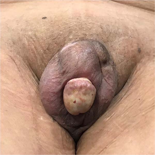
A 78-year-old male patient was diagnosed with penile cancer 40 years ago and was treated with radiotherapy. He reports that, about 1 year ago, ulcerated lesions began surrounding the entire penile skin. As diagnostic hypotheses, previous tumor recurrence, SCC and radiodermatitis were raised, whose biopsy was inconclusive. Due to the extensive lesion and radiotherapy sequelae, the cremaster muscle myocutaneous flap was chosen for reconstruction.
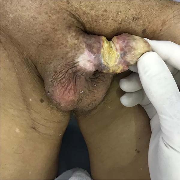
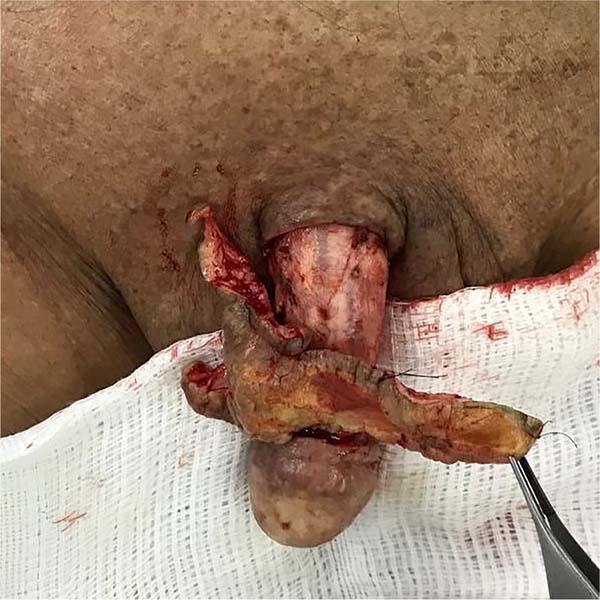
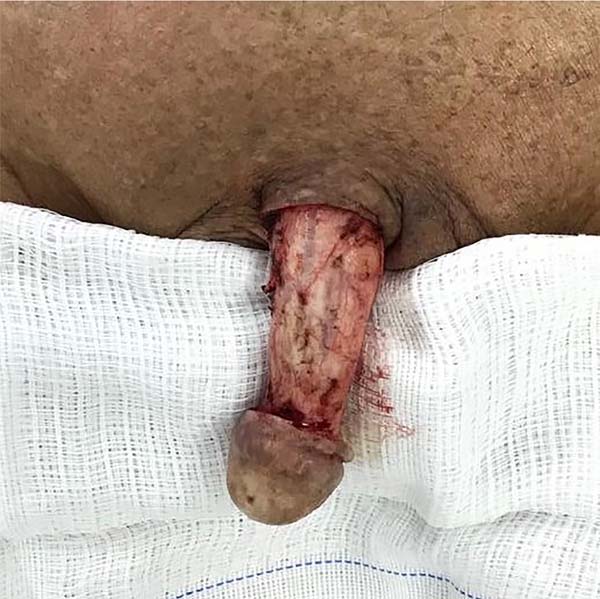
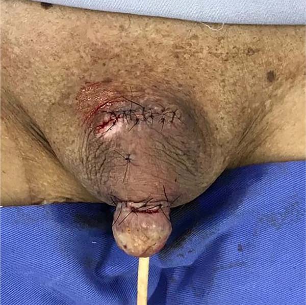
At the intraoperative moment (November 31, 2019), the entire penile coverage (Figure 1) and the lesion (Figure 2) were resected, which was referred for frozen section biopsy. Then, to reconstruct the penile coverage (Figure 3), a 4 cm incision was made in the ventral root of the penis, followed by creating a tunnel in the scrotum. A region of approximately 5 cm was detached from below the cremaster muscle, whose irrigation was made by the right and left lateral pedicles. The penis was inserted through the tunnel, with the glans exiting through a 3cm incision in the scrotum, 5cm after entry (Figure 4), thus resulting in a new structuring of the penile body, with the flap modeled on the cylindrical shape.
The postoperative period was uneventful, progressing with good permeability of the urethral canal, well-irrigated flaps, and no fibrosis or scar retraction. The biopsy did not show malignancy at any site of the lesion (Figure 5).
DISCUSSION
Several techniques have been described to reconstruct the cutaneous coverage of the penis, the most common being the use of total skin grafts for partial reconstruction3,4, with good aesthetic and functional results. For large lesions, techniques are used with fasciocutaneous flaps from the medial region of the thighs, described by Hirschowitz in 19824, scrotal flap (dartos flap) described by Goodwin and Thelen in 1950 5, used for partial cutaneous loss of the genitalia, preputial flap 6, and myocutaneous flaps of the rectus abdominis muscle, fasciae latae and gracilis7.
A bibliographic review on the subject was carried out, with searches in the SciELO, PubMed and Revista Brasileira de Cirurgia Plástica (Brazilian Journal of Plastic Surgery), using the following descriptors: Penis; Scrotum; Urogenital surgical procedures; Reconstructive Surgical Procedures; Surgical flaps. No report was found in the literature on the technique performed. However, a technique has been described with implanting the degloved penis in a subcutaneous tunnel between the dermis and the dartos fascia, using scrotal skin flaps8. In a second step, the pedicles of the flap used are released; coverage of the penis is performed only with the skin of the scrotum9,10. The technique used in this study used a bipedicled myocutaneous flap of the cremaster muscle with the skin of the scrotum to create the tunnel, as it is more irrigated and causes less scar retraction to cover an area with radiotherapy sequelae.
Because the patient was 78 years old and had other comorbidities, it was decided not to perform the second surgical procedure, which would be the release of the lateral pedicles with the release of the ventral region of the penis.
CONCLUSION
Despite the various techniques already described in the literature, the reconstruction of the penis and its coverage remains a functional, anatomical, and aesthetic challenge because it is an area with unique characteristics of the body, such as elasticity, sensitivity and texture.
The myocutaneous skin and cremaster flap proved to be a good option for total reconstruction of the penile skin coverage, achieving good vascularization and maintaining urethral permeability.
REFERENCES
1. Garaffa G, Sansalone S, Ralph DJ. Penile reconstruction. Asian J Androl. 2013;15(1):16-9. PMID: 22426595 DOI: https://doi.org/10.1038/aja.2012.9
2. Garaffa G, Raheem AA, Ralph DJ. An update on penile reconstruction. Asian J Androl. 2011;13(3):391-4. PMID: 21540867 DOI: https://doi.org/10.1038/aja.2011.29
3. Thakar HJ, Dugi DD 3rd. Skin grafting of the penis. Urol Clin North Am. 2013;40(3):439-48. PMID: 23905942 DOI: https://doi.org/10.1016/j.ucl.2013.04.004
4. Hirshowitz B, Peretz BA. Bilateral superomedial thigh flaps for primary reconstruction of scrotum and vulva. Ann Plast Surg. 1982;8(5):390-6. PMID: 6287901
5. Goodwin WE, Thelen HM. Plastic reconstruction of penile skin; implantation of the penis into the scrotum. J Am Med Assoc. 1950;144(5):384. PMID: 14774109
6. Tolba AM, Azab AAH, Nasr MA, Salah E. Dartos fascio-myo-cutaneous flap for penile skin loss: A simple flap with an immense potential. Surg Sci. 2014;5(1):6-9. DOI: https://doi.org/10.4236/ss.2014.51002
7. Bergamo JMO, Silva LD, Correa JHS, Dinali AC, Gallardo CMH, Gomes JRP. Reconstrução de pênis após desenluvamento traumático com retalho prepucial em espiral: uma nova abordagem. Rev Bras Cir Plást. 2018;33(Suppl.2):58-60. DOI: https://doi.org/10.5935/2177-1235.2018RBCP0124
8. Barreira MA, Lima LO, Alves Júnior JJ, Silva LFG, Lima MVA. Experiência do Hospital Haroldo Juaçaba com Reconstrução Utilizando Retalhos Miocutâneos em Cirurgia para Tratamento do Câncer de Pênis locorregionalmente Avançado. Rev Bras Cancerol. 2014;60(1):43-50. DOI: https://doi.org/10.32635/21769745.RBC.2014v60n1.494
9. Guo L, Zhang M, Zeng J, Liang P, Zhang P, Huang X. Utilities of scrotal flap for reconstruction of penile skin defects after severe burn injury. Int Urol Nephrol. 2017;49(9):1593-603. PMID: 28589215 DOI: https://doi.org/10.1007/s11255-017-1635-6
10. Ziylan O, Acar Ö, Özden BC, Tefik T, Dönmez Mİ, Oktar T. A practical approach for the correction of iatrogenic penile skin loss in children: Scrotal embedding technique. Turk J Urol. 2015;41(4):235-8. PMID: 26623155 DOI: https://doi.org/10.5152/tud.2015.17048
1. Hospital Esperança, Recife, PE, Brazil
2. Universidade Federal de Pernambuco, Recife, PE, Brazil
3. Universidade de Pernambuco, Recife, PE, Brazil
PCCP Final approval of the manuscript, Carrying out operations and/or experiments, Supervision.
ACAA Writing - Preparation of the original, Writing - Proofreading and Editing.
RFR Writing - Preparation of the original, Writing - Proofreading and Editing.
VOPD Writing - Preparation of the original, Writing - Proofreading and Editing.
LRC Methodology, Writing - Preparation of the original.
RXBM Writing - Preparation of the original, Writing - Proofreading and Editing.
Corresponding author: Pedro Celso de Castro Pita Praça Miguel de Cervantes, n°60, Sala 301, Ilha do leite Recife, PE, Brazil, Zip Code: 50070-520, E-mail: pedro.pitta@hotmail.com
Article received: February 12, 2021.
Article accepted: April 19, 2021.
Conflicts of interest: none.







 Read in Portuguese
 Read in English
 PDF PT
 Print
Print
 Send this article by email
 How to Cite
 Mendeley
 Pocket
 Twitter