


Case Report - Year 2022 - Volume 37 -
Technique with double muscle and fasciocutaneous flap in the correction of sciatic pressure ulcers
Técnica com retalho muscular duplo e fasciocutâneo na correção de úlceras isquiáticas por pressão

ABSTRACT
Pressure ulcers are lesions caused on the skin and underlying tissues due to local pressure force, usually at points of bony prominences. Here, we mention the case of a bedridden patient due to a spinal cord injury caused by myelomeningocele who developed an ulcer in the right ischial region, treated with the technique of muscular fasciocutaneous flaps on the posterior aspect of the thigh. As it is a lesion routinely found in these patients with functional limitations, it is essential to carry out adequate treatments aimed at the patient's clinical improvement and minimizing the rate of relapses. In addition, the implementation of new surgical techniques is extremely important, given the enormous variety of pressure injuries.
Keywords: Pressure injury; Meningomyelocele; Spina bifida cystica; Myocutaneous flap; immobilization.
RESUMO
Úlceras por pressão são lesões ocasionadas na pele e tecidos subjacentes devido à força de pressão local, geralmente em pontos de proeminências ósseas. Cita-se aqui o caso de uma paciente acamada devido à lesão medular por mielomeningocele que evoluiu com úlcera em região isquiática à direita, tratada com técnica de retalhos muscular e fasciocutâneo de face posterior da coxa. Por ser lesão rotineiramente encontrada nesses pacientes portadores de limitações funcionais, é fundamental a realização de tratamentos adequados que visem a melhora clínica do paciente e minimizar índice de recidivas. Além disso, é de suma importância a implementação de novas técnicas cirúrgicas, haja vista a enorme variedade de lesões por pressão.
Palavras-chave: Úlcera por pressão; Meningomielocele; Espinha bífida cística; Retalho miocutâneo; Imobilização
INTRODUCTION
Myelomeningocele, or spina bifida cystica, is characterized by a defect in closing the neural tube during the 20th week of gestation. Amniotic fluid exerts deleterious effects on unprotected neural tissue. It is the most common neural tube defect and the second cause of chronic deficiency of the locomotor system in the pediatric age group. Its cause is multifactorial, with risk factors such as folic acid deficiency, maternal hyperthermia during the early stages of pregnancy, or antiepileptic drugs.1.2.
In Brazil, the incidence of myelomeningocele is 2.28 per 1000 births. It is highly disabling, resulting in tetraparesis or paraparesis, neurogenic bladder, and cognitive impairment. It is estimated that among those with spinal cord injury, 85% will develop pressure ulcers. These are formed due to the constant pressure exerted by bony prominences on the skin, leading to local ischemia and ulcerated lesion formation.
The treatment of this type of injury aims to maintain tissue functionality, eliminate devitalized tissues and infectious processes, avoid secondary complications and allow aesthetic improvement of the affected region when possible.3.
The technique presented uses two types of locoregional flaps to correct ulcers in the ischial region. The first is a muscle flap made with the semimembranosus and biceps femoris muscles, which generate a protective cushion and avoid local dead space. To cover the muscle flap’s fixation area, we chose to use a fasciocutaneous flap from the posterior aspect of the thigh.
Due to the high incidence of pressure injuries in bedridden patients and for generating health problems and worsening the quality of life of these patients, it is necessary to implement adequate prophylaxis and treatments with a lower recurrence rate.
OBJECTIVE
Therefore, this work covers the initial experience with the double flap technique, muscular and fasciocutaneous, at the Plastic Surgery Service of Hospital São Paulo de Muriaé, MG, Brazil.
CASE REPORT
Female, 23 years old, with multiple sequelae due to myelomeningocele, with cystostomy and a deep ulcer in the right ischial region. According to the mother’s reports, the lesion has about seven years of evolution, with several unsuccessful attempts at treatments. Approximately one and a half years ago, the patient developed daily evening fever and right gluteal hyperemia. Since then, under the guidance of an infectious disease specialist, treatment with ciprofloxacin 500 mg orally twice a day and topical calcium alginate has been implemented.
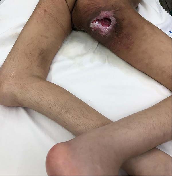
The patient had a deep and extensive lesion in the right ischial region, measuring approximately eight centimeters in its greatest diameter and with underlying muscle exposure, without signs of devitalized tissues or secretions, but with a chronic evolution and no possibility of healing without appropriate surgical intervention. On laboratory tests, she did not have anemia or leukocytosis (Figure 1).

The procedure was performed on April 8, 2020.
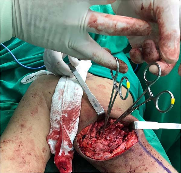
Patient in prone position under general anesthesia, asepsis and antisepsis were performed, surgical marking with methylene blue, infiltration of an anesthetic solution containing epinephrine and debridement with complete resection of the underlying bursa, since its permanence is associated with a high rate of lesion recurrence (Figure 2).

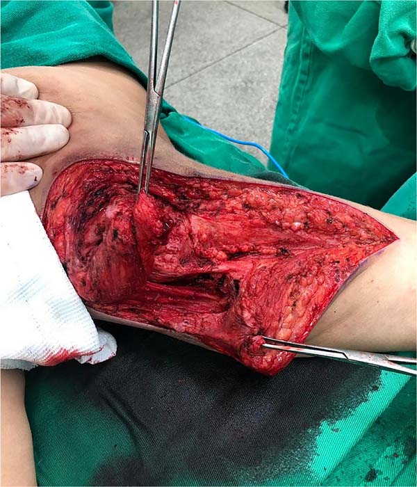
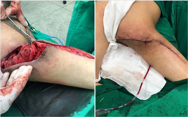
An incision extended from the lower margin of the right gluteus to the lateral face of the ipsilateral thigh, and a muscle flap was later made using the semimembranosus and biceps femoris muscles. The flap was rotated to accommodate redundant tissue in the trochanteric prominence to avoid trochanteric ulcers. Throughout the procedure, careful hemostasis was maintained. There were no bony prominences in need of removal. Afterward, the muscle flap was fixed over the ischial region (Figure 3).
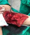
To cover the exposed area, a fasciocutaneous flap was used by advancing the posterior region of the thigh. We chose to insert a 4.8 mm Hemovac drain with its fixation by counter-opening on the medial face of the right thigh. The synthesis was performed in planes and ended with a local dressing (Figure 4).
Prophylactic antibiotic therapy was maintained during hospitalization with intravenous cefazolin, and vascular flow optimization was performed with oral administration of pentoxifylline 400 mg twice a day.
On the first postoperative day, the patient had an operative wound in good appearance, with no signs of hematoma or secretions, a drain volume of 250 ml, laboratory tests without a significant drop in hematocrit. On the second postoperative day, she was discharged, and the family was instructed to keep the drain at home and remove it after eight days in a hospital environment. The drainage through the drain showed a progressive decrease, and this was removed with a flow of 25 ml of serous secretion in 24 hours on the eighth postoperative day. The externalization hole of the drain healed by the second intention. The patient presented adequate surgical wound healing without removing sutures since the synthesis was performed with absorbable threads.
DISCUSSION
Patients with myelomeningocele sequelae are, in most cases, bedridden with severe neurological and orthopedic impairments. Thus, they are more prone to the development of pressure ulcers. Among the affected sites, the ischial region is the most frequent.4.
Several procedures are used for the surgical management of this type of injury. However, the reduced mobility of bedridden patients and techniques that are sometimes inadequate contribute to the high rate of recurrence.5.6.
The technique demonstrated has been used in two patients, with the case described being the most recent. In the first experience, the patient had similar clinical conditions, such as not being able to walk, and the results, in a year and a half of follow-up, were satisfactory.
A key point for long-term success is adequate debridement, with tissue resection up to the viable bone. Attention should be paid to the total resection of the bursa underlying pressure ulcers since its permanence is a favorable factor for local recurrence. Also, performing careful hemostasis is essential for the good evolution of the flaps and for not causing an anemic condition in already debilitated patients. In addition, this technique relies on the association of two types of flaps, muscular and fasciocutaneous. As this is a bedridden patient, who does not walk and, therefore, maintains constant pressure on the skin surfaces, a surgical technique was chosen that provides muscle protection, preserving its arterial irrigation, associated with the fasciocutaneous flap to form a bigger cushion,7.
CONCLUSION
The surgical technique above, using a double muscle flap and fasciocutaneous flap, concomitantly with the total resection of the bursa under the ischial ulcer, is effective and reproducible, with satisfactory results in the short and long term.
More patients are being submitted to the same technique to improve the statistics and confirm the results presented here.
REFERENCES
1. Batista KT, Pereira ICC, Romano ACL. Tratamento cirúrgico de úlcera por pressão na unidade de Pediatria de hospital de reabilitação. Rev Bras Cir Plást. 2017;32(4):570-8.
2. Borghardt AT, Prado TN, Bicudo SDS, Castro DS, Bringuente MEO. Úlcera por pressão em pacientes críticos: incidência e fatores associados. Rev Bras Enferm. 2016;69(3):460-7.
3. Ferreira FR, Bexiga FP, Martins VVM, Favero FM, Sartor CD, Artilheiro MC, et al. Independência funcional de crianças de um a quatro anos com mielomeningocele. Fisioter Pesqui. 2018;25(2):196-201.
4. Flávio Júnior WF, Bernardes BR, Silva TAS, Capanema HXM, Nishimoto F, Costa PR. Reconstrução complexa de regiões glútea e perineal após ressecção de extenso carcinoma escamocelular de borda anal: relato de caso. Rev Bras Cir Plást. 2016;31(4):586-90.
5. McKinley WO, Jackson AB, Cardenas DD, DeVivo MJ. Long-term medical complications after traumatic spinal cord injury: a regional model systems analysis. Arch Phys Med Rehabil. 1999;80(11):1402-10.
6. Milchelski DA, Mendes RRS, Freitas FR, Zaninetti G, Moneiro Júnior AA, Gemperli R. Protocolo de internação breve para tratamento cirúrgico de lesões por pressão: preparo ambulatorial e cobertura em tempo único. Rev Col Bras Cir. 2017;44(6):574-81.
7. Salomão RM, Cervante TP, Salomão JF, Leon SV. The mortality rate after hospital discharge in patients with myelomeningocele decreased after implementation of mandatory flour fortification with folic acid. Arq Neuropsiquiatr. 2017;75(1):20-4.
1. Hospital São Paulo, Departamento de Cirurgia Plástica, Muriaé, MG, Brazil
| COLLABORATIONS | |
|---|---|
| FMS | Analysis and/or data interpretation, Conception and design study, Conceptualization, Data Curation, Formal Analysis, Investigation, Methodology, Project Administration, Realization of operations and/or trials, Resources, Supervision, Visualization, Writing - Original Draft Preparation, Writing - Review & Editing |
| RBC | Conceptualization, Final manuscript approval, Investigation, Resources, Supervision, Validation, Visualization |

Corresponding author: Flávia Mesquita Soares, Rua João Marcelo Santoni, 296, Parque Renato Maia, Guarulhos, SP, Brazil, Zip Code 07114-120, E-mail: flavia.soares.m93@gmail.com
Article received: September 11, 2020.
Article accepted: October 15, 2021.
Conflicts of interest: não há.
 Read in Portuguese
 Read in English
 PDF PT
 Print
Print
 Send this article by email
 How to Cite
 Mendeley
 Pocket