


Case Report - Year 2022 - Volume 37 -
Correction of bifid nose with tongue-in-groove technique in patient with Tessier fissure 0-14
Correção de nariz bífido com técnica tongue-ingroove em paciente com fissura 0-14 de Tessier
Balduino Ferreira de Menezes1,* ; Murilo Sgarbi Secanho1; Laisa Brandão Carvalho1; Weber Ribolli Moragas1; Paulo Victor Cunha Costa1; Aristides Augusto Palhares1
ABSTRACT
Introduction: Craniofacial anomalies are usually identified by their appearance. Over time, several scales and classifications were proposed based on clinical and anatomical aspects. In 1976, Tessier made an association between soft tissue and underlying bone. With this concept, he created a numeral system starting from the clockwise orbit of 0 - which he called the zero line, a vertical line of the face - to 14. Rare and with multiple presentations, its conduction is a challenge even for more experienced professionals.
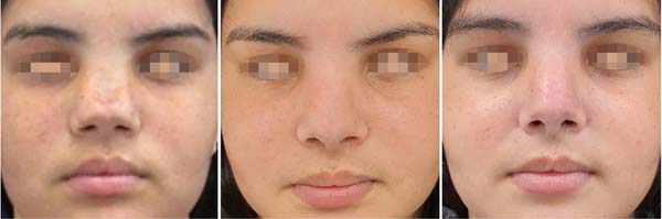
Case Report: Female patient who underwent open structured rhinoplasty at the age of 15 to correct a bifid nose, using costal cartilage and the tongue-in-groove technique.
Discussion: Bifid nose is one of the main presentations of cleft 0. Structured open rhinoplasty has already been successfully applied in other studies, and the tongue-in-groove technique is especially useful for the projection and rotation of the nasal tip. Conclusion: Craniofacial anomalies vary in their presentations, and it is up to the plastic surgeon to identify the problems and propose therapeutic solutions that alleviate these changes. Their treatment will require a thorough preoperative assessment, careful surgical planning, and meticulous surgical technique.
Keywords: Nose; Rhinoplasty; Costal cartilage; Congenital abnormalities; Craniofacial abnormalities.
RESUMO
Introdução: As anomalias craniofaciais são identificadas usualmente pela sua aparência. Ao longo do tempo, diversas escalas e classificações foram propostas a partir de aspectos clínicos e anatômicos. Em 1976, Tessier fez uma associação entre os tecidos moles e o ósseo subjacente. Com esse conceito, criou um sistema numeral a partir da órbita em sentido horário de 0 - que chamou de linha zero, uma linha vertical da face - a 14. Rara e com múltiplas apresentações, sua condução segue um desafio mesmo para profissionais mais experientes.
Relato de Caso: Paciente feminina, submetida a rinoplastia aberta estruturada aos 15 anos de idade para correção de nariz bífido, utilizando cartilagem costal e técnica de tongue-in-groove.
Discussão: O nariz bífido é uma das principais apresentações da fissura 0. A rinoplastia aberta estruturada já foi aplicada em outros trabalhos com grande sucesso, sendo a técnica tongue-in-groove especialmente útil para projeção e rotação da ponta nasal.
Conclusão: As anomalias craniofaciais variam em suas apresentações e cabe ao cirurgião plástico identificar os problemas e propor soluções terapêuticas que amenizem essas alterações. Seu tratamento irá necessitar de uma avaliação pré-operatória completa, planejamento cirúrgico cuidadoso e técnica cirúrgica meticulosa.
Palavras-chave: Nariz; Rinoplastia; Cartilagem costal; Anormalidades congênitas; Anormalidades craniofaciais
INTRODUCTION
Craniomaxillofacial clefts are rare malformations but with disfiguring potential. The incidence is estimated at 1.4-4.9% for every 100,000 live births. Tessier developed a classification system based on facial lines, starting from 0 to 141.
Cleft number 0 has unique characteristics due to the possibility of having normal tissue, in excess or even deficient, in addition to the possibility of association with other clefts, such as cranial number 14, generating a wide spectrum of phenotypes. The variability of presentations is expressed from a simple lip notch or a slight bifid nose to a complete division of the structures of the midline of the face2.
When there is the involvement of the nasal tip, it is important that there is an improvement in support, gaining projection and, when necessary, also greater rotation. For this purpose, the tongue-in-groove technique is an arsenal to create a connection between the septum and medial crura, which can be very useful in certain presentations of median clefts3.
OBJECTIVE
This article aims to present a report of a bifid nose approach in a patient with 0-14 Tessier cleft.
CASE REPORT
AJPO, 15 years old, female, being followed up since she was 8 years old at the Plastic Surgery outpatient clinic, complaining of an enlarged nose associated with episodes of bullying at school. She had no other complaints, such as hyperteleorbitism. She had no other comorbidities.
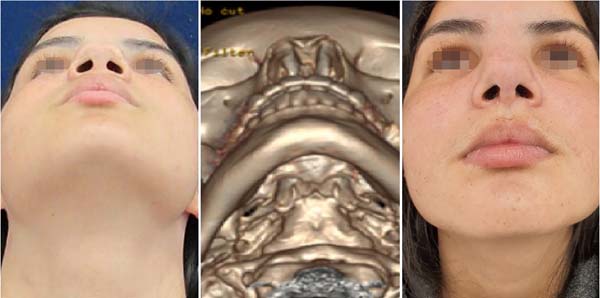
Surgical planning involved radiological evaluation with computed tomography in thin sections and 3D reconstruction, and the ideal time was planned for after the patient had completed the phases of puberty and bone development.
She underwent a one-time procedure on December 9, 2019, lasting 4 hours, under general anesthesia at the Hospital das Clínicas of the Faculty of Medicine of Botucatu - UNESP (HC FMB UNESP).
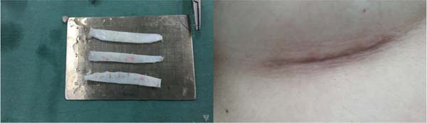
Patient in the horizontal supine position, under orotracheal intubation. A 4 cm subcostal incision was made in the right inframammary fold with bright green infiltration with a solution containing 2% lidocaine, 0.9% saline solution and 1:200,000 adrenaline. This site was incised, followed by dissection until the identification of the sixth costal arch and opening of the perichondrium to collect cartilage 5 cm in length. The closure was done by planes and with an occlusive dressing.
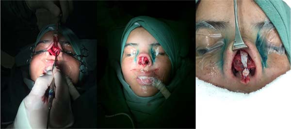
Nasal access was made with a transcolumellar “W” and infracartilaginous incision after previous local infiltration, with 1:100,000 epinephrine and 0.75% ropivacaine added to 0.9% saline. The covering flap was lifted after careful dissection with exposure of the osteocartilaginous framework, where it was noted: nasal bone with double contour, in the shape of a gull’s wing; absence of conventional cartilaginous septum; distant triangular cartilages; and distant alar cartilages with thick and unusually curved ligaments.
The medial crus were separated, and the bone cartilages of the nasal dorsum were separated, allowing three osteotomies on each side, the first being paramedian close to the center; the second, oblique from lateral to medial, which removed a triangular slice that contained bilateral bone defect; and the third, low-high completed by digital pressure. After hemostasis, spreader grafts measuring 40 mm x 4 mm on each side were implanted using 5-0 polypropylene thread.
Closure made in 5 layers: septum (osteofibrous remnant), two grafts and two triangular with three stitches. Afterward, the medial crura of the wings were sutured with a columellar cartilage strut measuring 25 mm x 3 mm and fixed with 5-0 polypropylene thread and the strut with a tongue-in-groove spreader. The cephalic margins of the alar cartilages were then transectioned, maintaining 6 mm in length, followed by transdomal and interdomal sutures with 5-0 polypropylene thread, maintaining a diamond shape in the nasal neodome.
Subsequently, a shield-type cartilage graft was performed for projection and camouflage of the nasal tip and a graft of crushed cartilage on the back, covered by perichondrium. Finally, the transcolumellar and intracartilaginous incisions were synthesized with 6-0 mononylon and 4-0 catgut, respectively, with a bilateral nasal splint implant fixed by a 3-0 mononylon thread. The dressing was made with micropore, aquaplast and nasal packing.
She was discharged 24 hours after the procedure, removing the nasal pack, prescription of non-steroidal anti-inflammatory for 5 days, amoxicillin with clavulanate for 7 days and nasal solution with a saline solution daily. On the 8th postoperative day, the nasal splint and aquaplast and the external non-absorbable sutures were removed.
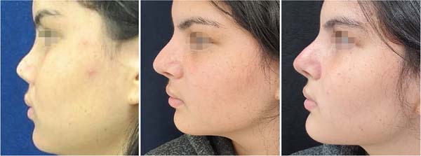
The outpatient return took place on the 4th, 8th, 30th, 6th and 15th postoperative month. She was asymptomatic and satisfied with the aesthetic and functional results, with nasal projection gain and absence of the bifid appearance (Figure 1). The radiological and pre, intra and postoperative images are shown below (Figures 2 to 5).


DISCUSSION
The mechanism of Tessier’s cleft 0 is not fully understood, but its onset in the third week of gestation is recognized4. It results in failure of fusion of the two medial nasal processes, and when this occurs, the presence of a bifid nose is frequent. Eventually, this error in embryogenesis can cause agenesis or even duplication of these structures in the midline. Meanwhile, cleft 14 is characterized by cranial alteration, with marked hyper or hypoteleorbitism5.
In our report, the patient presents, in addition to a bifid nose, hypertelorbitism. However, the patient did not present this complaint during the follow-up, and we opted for an exclusive approach to the nose6.
The treatment of bifid nose has been sought for a long time7, and in previous articles, there is a report of an open approach, resecting the tissues that were in excess and with associated cartilage grafts4. Due to the rare incidence, studies are scarce, even though the craniofacial cleft is the most common. The great phenotypic variation of the face in this syndrome is also present in the nose: short and wide columella, duplicated or absent nasal septum, flat dorsum, also variable nasal tip. The case series shows that this diversity also alters the standardization of treatment, which must be individualized8.
The technique chosen for rhinoplasty was the open structure, with nasal tip reconstruction being the most challenging step. Based on previous good results, the tongue-in-groove technique was chosen for predictable and permanent projection and tip rotation, based on previous good results9.
The treatment still proves successful when performed as a single procedure in most series, obviously varying depending on the number of associated procedures to correct common defects, especially labral defects10.
CONCLUSION
Open structure rhinoplasty using the tongue-in-groove technique can provide a satisfactory esthetic result in a patient with a bifid nose secondary to a 0-14 Tessier cleft.
REFERENCES
1. Tessier P. Anatomical classification facial, cranio-facial and latero-facial clefts. J Maxillofac Surg. 1976;4(2):69-92.
2. da Silva Freitas R, Alonso N, Shin JH, Busato L, Ono MC, Cruz GA. Surgical correction of Tessier number 0 cleft. J Craniofac Surg. 2008;19(5):1348-52.
3. Antunes MB, Quatela VC. Effects of the Tongue-in-Groove Maneuver on Nasal Tip Rotation. Aesthet Surg J. 2018;38(10): 1065-73.
4. Mazzola RF, Mazzola IC. Facial clefts and facial dysplasia: revisiting the classification. J Craniofac Surg. 2014;25(1):26-34.
5. Nam SM, Kim YB. The Tessier number 14 facial cleft: a 20 years follow-up. J Craniomaxillofac Surg. 2014;42(7):1397-401.
6. Pidgeon TE, Flapper WJ, David DJ, Anderson PJ. From birth to maturity: midline tessier 0-14 craniofacial cleft patients who have completed protocol management at a single craniofacial unit. Cleft Palate Craniofac J. 2014;51(4):e70-9.
7. Miller PJ, Grinberg D, Wang TD. Midline cleft. Treatment of the bifid nose. Arch Facial Plast Surg. 1999;1(3):200-3.
8. Spataro EA, Most SP. Tongue-in-Groove Technique for Rhinoplasty: Technical Refinements and Considerations. Facial Plast Surg. 2018;34(5):529-38.
9. Tawfik A, El-Sisi HE, Abd El-Fattah AM. Surgical correction of bifid nose. Int J Pediatr Otorhinolaryngol. 2016;86:72-6.
10. Kolker AR, Sailon AM, Meara JG, Holmes AD. Midline Cleft Lip and Bifid Nose Deformity: Description, Classification, and Treatment. J Craniofac Surg. 2015;26(8):2304-8.
1. Hospital das Clínicas, Faculty of Medicine of Botucatu, Plastic Surgery, Botucatu,
SP, Brazil.
| COLLABORATIONS | |
|---|---|
| BFMN | Analysis and/or data interpretation, Conception and design study, Data Curation, Final manuscript approval, Methodology, Project Administration, Supervision, Writing - Original Draft Preparation, Writing - Review & Editing |
| MSS | Analysis and/or data interpretation, Data Curation, Writing - Review & Editing |
| LBC | Data Curation |
| WRM | Data Curation |
| PVCC | Realization of operations and/or trials |
| AAP | Supervision |
Corresponding author: Balduino Ferreira de Menezes Neto, Avenida Professor Mário Rubens Guimarães Montenegro, S/N - Jardim Sao Jose, Botucatu - SP, Brazil, Zip Code 18618-970, E-mail: balduinofmneto@gmail.com
Article received: December 02, 2020.
Article accepted: May 18, 2021.
Conflicts of interest: none.
 Read in Portuguese
 Read in English
 PDF PT
 Print
Print
 Send this article by email
 How to Cite
 Mendeley
 Pocket