


Ideas and Innovation - Year 2022 - Volume 37 -
Surgical rejuvenating for the aged earlobe: a technical innovation
Rejuvenescimento cirúrgico do lóbulo senil: uma inovação técnica
Miguel Marques de Oliveira1, ; Gustavo de Sousa Marques de Oliveira1
ABSTRACT
The authors present a new surgical approach to the aging earlobe. The technique is based on the classic marginal reduction technique of the earlobe contour that has been improved. In essence, it seeks to preserve the subdermal fat layer of the distal flap created for the purpose of raising the earlobe while being shortened, thickened and smooth, in a hidden scar. It is a tripartite resolution to this problem never seen in the relevant literature. Four 68-yearold women were treated: the first with the classic manner in 2009 and the others with this modification to improve the earlobe withered appearance in 2017-2019. The study shows a rejuvenating eutrophic reduction technique.
Keywords: Surgery, plastic; Ear; Ear, external; Reconstructive surgical procedures; Rejuvenation.
RESUMO
Os autores apresentam técnica corretiva conservadora para o lóbulo senil. A literatura pertinente é rica em técnicas redutoras de lóbulos longilíneos ou rasgados. Os autores creem que a cirurgia para os lóbulos deve poupar tecidos in situ, a despeito de usuais técnicas de preenchimento com material orgânico e biocompatíveis. A presente técnica se baseia na clássica redução lobular periférica da borda livre, que agrega ao componente do face lifting para reduzir o lóbulo e ao mesmo tempo poupar tecidos e encorpá-lo na cicatriz oculta retrolobular. Resolução tripartite até hoje não apresentada na literatura. Com oito casos operados: seis tipo pec-man no passado; um na forma clássica marginal e três recentes com esta modificação técnica.
Palavras-chave: Cirurgia plástica; Orelha; Orelha externa; Procedimentos cirúrgicos reconstrutivos; Rejuvenescimento
INTRODUCTION
The earlobe is considered an important attribute of beauty in many western societies. Its lateral aspect is a key detail in facial aesthetic appreciation. Today, women and men are adept at wearing earrings in their ears. Normal lobes of good shape and appearance have a decorative sociocultural role through pendant jewelry and an erogenous function in the facial region.
The proposition of corrective lobe techniques attests to the psychosocial importance of this segment, normally small and showy. The basic defect is in lobular hypertrophy for many authors, meaning longitudinal enlargement. The study sees its basic pathology in physiological atrophy that generates stretching and wrinkling. There are young women whose earlobes are elongated from wearing heavy, fashionable earrings.
This work presents a simple technique that combines the free edge’s ideal peripheral incision with the vascular pedicle’s basic reconstructive principle by reusing excess tissue. A free margin resection is chosen since it combines a hidden scar with the pleasant and tricky facial insertion of the free (pedicled) lobe, a universal female cosmetic preference. Here it is a question of reducing the length employing an elevator flap that simultaneously increases the lobular thickness.
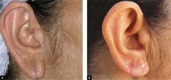
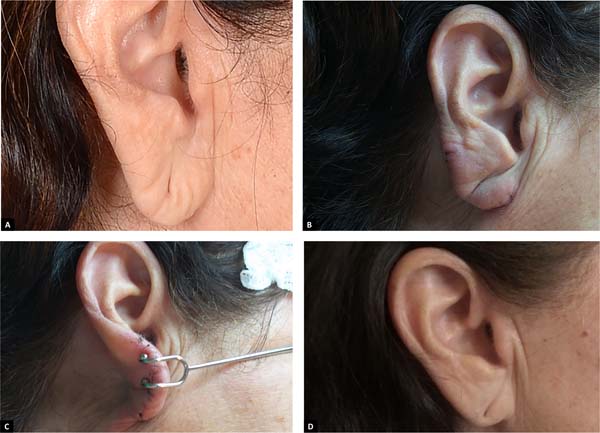
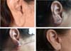
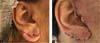
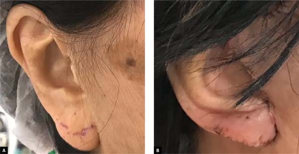
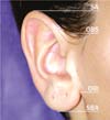
Using the modified Tipton’s marginal method, its performance aims to hide the scar (Figures 1A and 1B). Given our current small series of 1 case (Figures 2A-D), we requested the partnership for technical proof in 2019. The colleague reports a safe corrective process in concomitant facial rejuvenation (Figures 3A and 3B and 4A and 4B). Esthetic earlobe correction surgery involves obtaining:


METHODS
Our experience of lobular correction concomitant with facial surgery is 6 cases using the Guerrero-Santos technique (pec-man). In the last 12 years, the Tipton-mode graded resection free edge technique has been chosen, with 1 case. In the current technical improvement, there were 3 cases, two of which were kindly provided by the distinguished surgeon from São Paulo, Dr. A Bersou.
Four white women aged 58 to 74 years, three Brazilians and one Palestinian, underwent surgery at the Interclínicas-Interplástica in Campo Grande, MS, Brazil, from 2007 to 2019, with follow-up from 1 month to 12 years. The technique performs a lifting by shortening and thickening the lobe simultaneously in a retrolobular skin resection. Preserves subcutaneous fat to reintroduce it to the lobular body medially.
Anatomy
As for the lobular morphology, it can be said with absolute conviction that its aesthetically correct position depends on the cartilaginous skeleton of the normal external ear. The study of auricular anatomy is well described in the literature1, attesting to the remarkable vascular supply of the segment. As the pinna has almost all the lateral skin intrinsically attached to the perichondrium, its normal or non-normal skeletal anatomy becomes visible, except for the low antitragus in the lobe domain.
Therefore, this is the only part of the ear suffering from premature facial skin aging. Once there is no greater skeletal adhesion at this point, the lobe is at the mercy of its helix tail helm2. An important facet is the attached (sessile) or loose (pedunculated) lobe, the latter being a universal female aesthetic preference. The following are anatomical repairs3 for aesthetic appreciation in lobular reduction: otobasion inferius (oi) to the subaural (sa) and otobasion superius (os) to the superaurale (spa) that must obey certain reciprocity (Figure 2A).
Technical Layout (Figure 5)
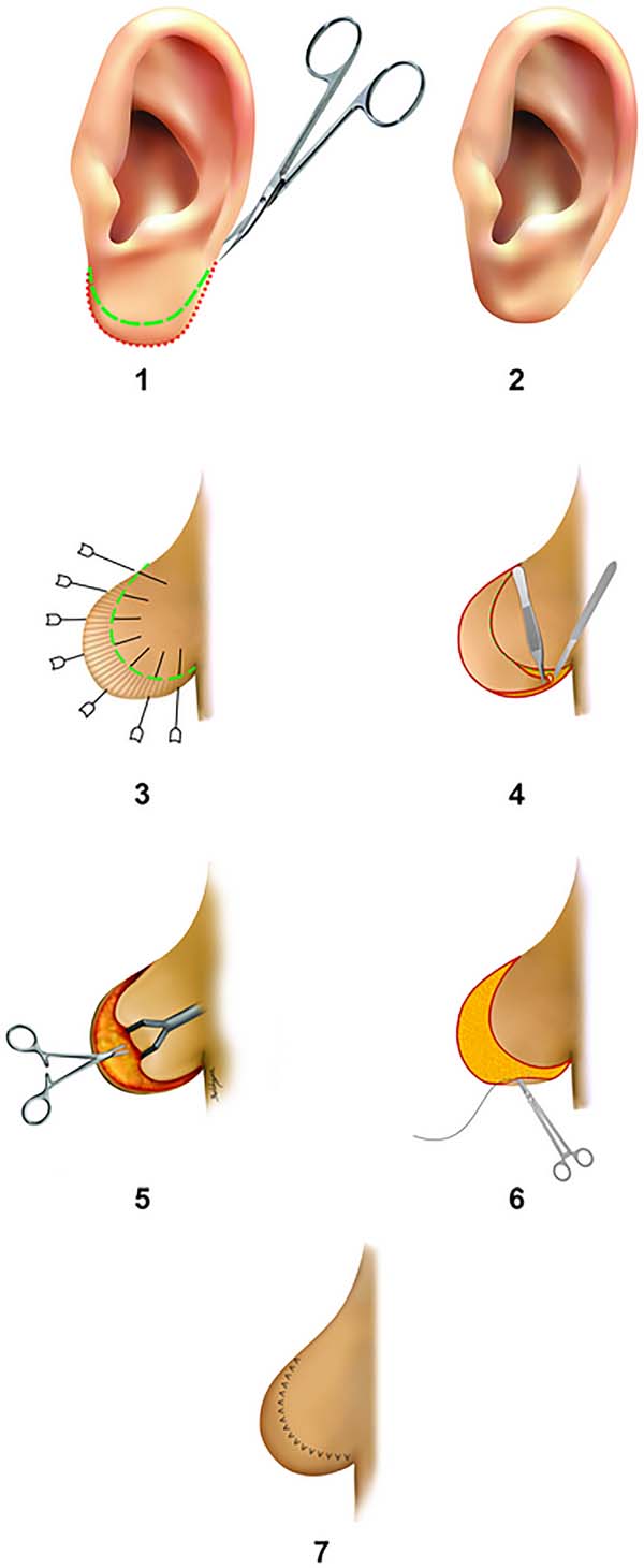

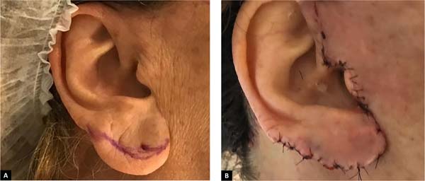
Preoperative front view of the original lobe and the demarcation in front of it of the ideal size in green and red the tracing of access to the lobular flap with scissors along the free edge. Anesthetic post-infiltration and firm handling make a small prick the scalpel for the introduction of fine curved Stevens scissors. It is the marginal incision only in the skin.
The final postoperative front view of the short and full-bodied lobe thanks to the reintroducing adipose to the lobe, reviving the continent through increased content.
Transoperative back view with demarcations: the drawing of the ideal size traced in green by transfer with insulin needles to the retrolobule and in marginal red after cutting with scissors.
Intraoperative rear view with the line already incised to the size of the lobular reduction. Cutaneous resection of thin skin decorticated by the edge of the upper incision is started with forceps and a scalpel. Adipose tissue is preserved.
Transoperative back view of the decorticated area in yellow and the fine detachment of the mosquito forceps and hook to create space for the fat of the elevator flap.
Transoperative back view and application of sutures in the elevation of the lobular ridge made with silk or Nylon 6-0 sutures.
Postoperative back view of the short, fat lobe, and hidden scar in the simple stitches on the retrolobular inner part. Once the anterior ostium is spared, it must be recanalized using the retrolobular flap.
DISCUSSION
The lobular deformities of aging are: stretching, wrinkling and thinning. The pertinent literature is rich in ear lobe reduction techniques from a distant past to the present day4-10. The preference is for free contour surgery, which simultaneously reduces length and width, leaving for future fat grafts the gain in thickness in an inconspicuous scar5,7. The wrinkles mark its surface due to senile atrophy in the connective-adipose tissue. Social appearance is a decisive factor in joint facial rejuvenation, especially in women.
Modern surgeries for the senile lobe began in Brazil, with Loeb and Pitanguy, and in Mexico, with Guerrero-Santos4-6. Techniques that treat the lobe through facial implantation have the inconvenience of altering its expression and natural insertion, which are often difficult to regularize and have a visible scar4,8,10. Many authors have proposed techniques that reduce continent and content simultaneously, to the detriment of thickness, which is vitally important for females8,10. Many reduce the lobular extension globally, leaving it smaller, thinner and with an apparent scar. Today, successful lobular rejuvenation techniques using fatty tissue are a reality3.
Our Tipton7 modification represents an evolution in facial rejuvenation: smaller lobe, revitalized and natural. The present technique aims to reduce length and width while increasing lobular thickness. The hidden scar behind the lobe has a pleasant contour line and a natural insertion on the face.
It is a lobular anti-aging technique, as it reduces the flaccid and aged skin continent, preserving the fatty content to give it body. The aesthetic surgical technique of the earlobe has always been relegated to a deforming secondary plane as if the cosmetic objective contraindicated aesthetic reconstructive principles. It is a surgery performed against the law of the strongest, most famous and experienced surgeon, to the detriment of this peripheral ear’s weakest and smallest lobe.
CONCLUSION
The technique simultaneously shortens and thickens the lobe in a retrolobular skin resection and inconspicuous scar. Preserves subcutaneous fat to reintroduce it to the lobular body medially.
Acknowledgment
The authors thank Prof. JM Psillakis (In memoriam) and Ms. Eliana from GrafiQx for invaluable assistance. And to Dr. A. Bersou, a brilliant surgeon from the capital of São Paulo, for his helpful collaboration in two cases.
REFERENCES
1. Park C, Roh TS. Anatomy and embryology of the external ear and their clinical correlation. Clin Plast Surg. 2002;29(2):155-74.
2. Oliveira MM, Oliveira DS, Oliveira GS. The Existence of a Natural Plica at the Anatomical Base of the Antihelix and its Surgical Importance to Address Protruding Ears: An Anatomicosurgical Study. Aesthetic Plast Surg. 2017;41(2):321-6.
3. Pi H, Kurlander DE, Guyuron B. Earlobe Rejuvenation: A Fat Grafting Technique. Aesthet Surg J. 2016;36(8):872-6.
4. Loeb R. Earlobe tailoring during facial rhytidoplasties. Plast Reconstr Surg. 1972;49(5):485-9.
5. Pitanguy I, Müller P, Kauk LK, Freitas LFP. Incisões remodelantes no lóbulo da orelha. Rev Bras Cir. 1988;78(2):155-62.
6. Guerrero-Santos J. Correction of hypertrophied earlobes in leprosy. Plast Reconstr Surg. 1970;46(4):381-3.
7. Tipton JB. A simple technique for reduction of the earlobe. Plast Reconstr Surg. 1980;66(4):630-2.
8. Constant E. Reduction of the hypertrophic earlobe. Plast Reconstr Surg. 1979;64(2):264-7.
9. Connell BF. Correcting deformities of the aged earlobe. Aesthet Surg J. 2005;25(2):194-6.
10. Van Putte L, Colpaert SD. Earlobe Reduction with Minimally Visible Scars: The Sub-Antitragal Groove Technique. Aesthetic Plast Surg. 2017;41(2):335-8.
1. General Hospital of Santa Casa de Campo Grande, Division of Plastic Surgery, Campo
Grande, MS, Brazil.
| COLLABORATIONS | |
|---|---|
| MMO | Analysis and/or data interpretation, Conception and design study, Data Curation, Final manuscript approval, Investigation, Methodology, Project Administration, Realization of operations and/or trials, Supervision, Validation, Visualization, Writing - Original Draft Preparation, Writing - Review & Editing |
| GSMO | Conceptualization, Data Curation, Final manuscript approval, Realization of operations and/or trials, Writing - Review & Editing |

Autor correspondente: Miguel Marques Oliveira, Av. Afonso Pena, 3504, Conjunto 126, Campo Grande, MS, Brazil, Zip Code 79002-075, E-mail: miguelmar@hotmail.com.br
Article received: October 04, 2021.
Article accepted: December 13, 2021.
Conflicts of interest: none.
 Read in Portuguese
 Read in English
 PDF PT
 Print
Print
 Send this article by email
 How to Cite
 Mendeley
 Pocket