


Case Report - Year 2021 - Volume 36 - Issue 2
Bilateral eyelid necrosis with dermal matrix reconstruction and skin grafting
Necrose palpebral bilateral com reconstrução por matriz dérmica e enxertia cutânea
Carlos Miguel Pereira1,* ; Irene Daher Barra2; Kerly Abraão Badaró2
ABSTRACT
Skin and soft tissue infections are a group of pathologies of high prevalence. Necrotizing fasciitis is a rapid and destructive infection of the subcutaneous tissue and superficial fascia with high morbidity and mortality. It is frequent in the perineal region and of rare occurrence in the periorbital region. This report illustrates a case of bilateral eyelid necrosis after mild head trauma with abrasions. Intensive clinical treatment and surgical debridement of the affected area were performed. In the first phase of eyelid reconstruction, a dermal regeneration matrix was used. This cutaneous substitute initially described for burns is currently of great importance in plastic surgery, aiming to heal wounds better. Subsequently, cutaneous self-grafting was performed, and a good aesthetic and functional result was obtained.
Keywords: Necrosis; Skin abnormalities; Reconstructive surgical procedures; Acellular dermis; Orbital cellulitis.
RESUMO
As infeções de pele e tecidos moles constituem um grupo de patologias de elevada prevalência. A fasceíte necrotizante é a infeção rápida e destrutiva do tecido subcutâneo e fáscia superficial com elevada morbimortalidade. Mais frequente na região perineal é de ocorrência rara na região periorbitária. O relato deste caso ilustra um caso de necrose palpebral bilateral após traumatismo cranioencefálico leve com escoriações. Foi realizado tratamento clínico intensivo e desbridamento cirúrgico da área afetada. Na primeira fase da reconstrução palpebral foi usada matriz de regeneração dérmica. Este substituto cutâneo inicialmente descrito para queimaduras se reveste atualmente de grande importância em cirurgia plástica visando uma melhor e mais rápida cicatrização das feridas. Posteriormente, realizou-se a autoenxertia cutânea tendo-se obtido um bom resultado estético e funcional.
Palavras-chave: Necrose; Anormalidades da pele; Procedimentos cirúrgicos reconstrutivos; Derme acelular; Celulite orbitária.
INTRODUCTION
Skin and soft tissue infections are a group of high prevalence pathologies, with many clinical presentations and degrees of severity. These can go from folliculitis through cellulite to necrotizing fasciitis, depending on how deep the microbial invasion is.
The plastic surgeon’s challenge is to differentiate cases that require more aggressive treatment, such as necrotizing fasciitis. Necrotizing fasciitis is the rapid and destructive infection of the subcutaneous tissue and superficial fascia with a high rate of morbidity and mortality1.
Initially described by Hippocrates in the 5th century .C. became known as hospital gangrene during the American Civil War. In 1883, a French surgeon named it after his own name, gangrene Fournier. In 1924, Meleney designated it necrotizing fasciitis.
More frequent in the perineal region and lower extremities, it is rare in the periorbital region due to its rich vascular supply1.
The repair of the resulting defects follows the reconstruction ladder using the dermal regeneration matrix as an innovative aid.
A product of tissue engineering, dermal matrices are skin substitutes that imitate the dermis and/or epidermis, promoting better healing. They may have biological and/or synthetic origin, and their use may be temporary or permanent.
An example is the matrix used in the reported case: PELNAC®. This consists of a layer of collagen derived from porcine tendon and a thin layer of silicone.
Subsequently, reconstruction can be completed with cutaneous self-grafting on the neodermis.
CASE REPORT
Patient 66-year-old, obese, hypertensive and diabetic with a history of fall from a ladder with mild head trauma, which only resulted in abrasions on the face and occipital region.
After five days, she started to have pain, hyperemia, edema and heat and was diagnosed with facial cellulitis. He started intravenous antibiotic treatment with oxacillin.
After 48 hours, she was transferred to the Hospital Municipal Souza Aguiar (HMSA) in poor general condition, dehydrated, although hemodynamically stable. He presented edema and hyperemia of the upper half of the face. With intense leukocytosis with neutrophilia (leukocytes [leuc.] 38,000 with 8% sticks). Tomography of the face showed intense infiltration of the subcutaneous tissue with extension to the cervical region without the involvement of the eyeballs. He started treatment with vancomycin and meropenem.
She evolved unfavorably with worsening of the general state, increased leukocytosis (leuc. 60,400 with 22% sticks) and acute renal failure (creatinine 5.9), initiating hemodialysis under constant surveillance in the Intensive Care Unit (ICU), and antibiotic treatment with linezolid was prescribed.
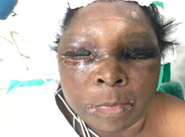
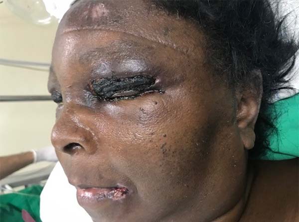
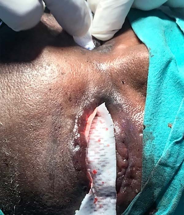
She evolved with the appearance of bilateral eyelid necrosis with fluctuation and adjacent violet area at the moment of hospitalization. After evaluation by plastic surgery and clinical and laboratory improvement of the patient, it was decided to perform surgical debridement of necrosis areas (Figures 1 and 2).


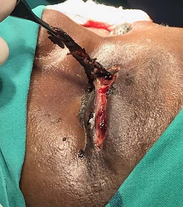
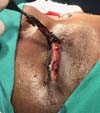
Necrosis involved the superficial layers of the bilateral upper eyelids and lowered to the right, compromising the muscle layer (Figure 3).
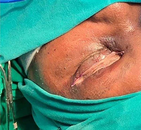
The upper eyelid defects were covered with a PELNAC® dermal regeneration matrix (Figure 4).

After 15 days of the initial surgery, cutaneous self-grafting of the upper eyelids was performed, with the left arm’s inner face as the donor area (Figure 5).
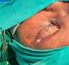
With a good clinical evolution and improvement of infectious parameters, the patient was discharged from the hospital thirty days after the first intervention by plastic surgery (Figure 6).

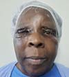
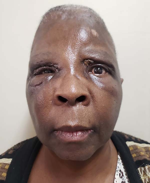
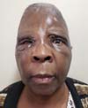
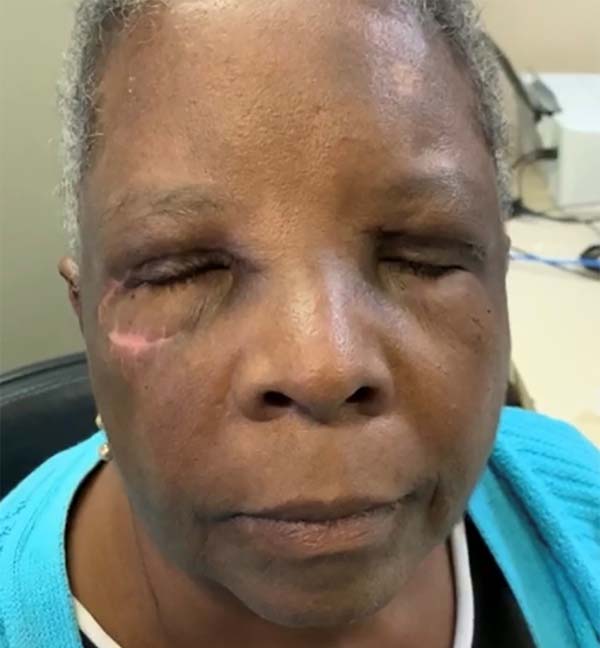
It is currently followed in the plastic surgery outpatient clinic of the HMSA with a good aesthetic and functional recovery (Figures 7 and 8).

DISCUSSION
Soft tissue infection results from two fundamental processes: microbial invasion and the interaction between the body’s defenses and the invader.
The breakage of the skin barrier may be caused by bites, lacerations, wounds or burns that allow bacterial invasion. After this, the inflammatory response of the organism occurs1,2.
Common to all skin infections are edema, heat, flushing and pain, making it difficult to differentiate between periorbital cellulitis and necrotizing fasciitis itself 2,3.
In the initial phase of hospitalization in the HMSA, the presumed diagnosis was periorbital cellulitis. In the present case, this was the patient’s initial presentation; although calculated a posteriori, we could verify that the LRINEC (laboratory risk indicator for necrotizing fasciitis) proposed by Wong was suggestive of necrotizing fasciitis. This point index includes leukocytosis, increased CRP, anemia, hyponatremia, hyperglycemia and creatinine increase4.
Necrotizing fasciitis also occurs more frequently in patients with some degree of immunosuppression due to alcoholism, diabetes, rheumatologic or malignant disease, or the use of corticosteroid therapy2-4. The patient had diabetes.
In a second moment, there was a worsening of his clinical condition with fever, hypotension, tachycardia and acute renal failure with the development of septic shock, which could occur as a response to deeper infections 2-5.
The infection progressed, producing a violet appearance of the skin, the appearance of fluctuation, and subsequent skin necrosis, which is compatible with the usual course of this condition; also, having a bilateral presentation corroborates this diagnosis4,5.
This pathology can be divided according to the causing microorganisms. Type I is due to a polymicrobial infection with a mixture of anaerobes, Gram-negative bacilli and enterococci, and type II is mainly represented by infection by beta hemolytic Streptococci2-4. Due to a certain diagnostic uncertainty, the excised material’s culture was not performed, which could have definitively confirmed this disease.
The resulting defect involved the entire upper eyelid bilaterally to the muscle layer, opting for placement of a matrix of dermal regeneration of porcine origin to facilitate healing, to obtain a better bed for subsequent skin grafting6,7.
In the ‘70s, of the 20th century, the INTEGRA® dermal matrix appeared to treat major burns through Yannas and Burke’s8 studies. Perfected over the 1980s, the FDA approved it in 19968 .
Suzuki et al.9, already in the early 1990s, published a study on improvements that led to INTEGRA® culminating in the PELNAC ®. It operated under the same principles but with advantages such as the lowest antigenicity and the highest porosity of the membrane10.
The PELNAC® dermal regeneration matrix consists of two layers: the first layer is formed by a silicone layer, which acts temporarily as the epidermis, preventing fluid loss and microbial invasion; the second layer consists of a porous structure, composed of cross-links of porcine collagen. This structure is infiltrated by fibroblasts that synthesize the new dermis, very similar to the human dermis6.
In a second step, skin autografting was performed on a viable dermis, which facilitated the grafts’ adhesion. Although with residual edema in the postoperative period, the patient did not present sequelae in the subsequent follow-up to achieve a good aesthetic and functional result.
CONCLUSION
Soft-tissue infections are common in plastic surgeon practice. A high degree of suspicion is required for the most severe cases with probable systemic involvement, such as necrotizing fasciitis.
When diagnosed, intravenous antibiotic therapy, hemodynamic stabilization and surgical debridement are essential.
In the defects resulting from this disease, we can resort to the dermal regeneration matrix’s help, obtaining better healing with lower morbidity for the patient and faster recovery of function.
The dermal regeneration matrix has been an important assistant in the plastic surgeon’s practice, enabling a better functional and aesthetic recovery in the most diverse areas, being imperative the universalization of its access.
REFERENCES
1. Lazzeri D, Lazzeri S, Figus M, Tascini C, Bocci G, Colizzi L, et al. Periorbital necrotising fasciitis. Br J Ophthalmol. 2010 Dez;94(12):1577-85.
2. Amrith S, Pai VH, Ling WW. Periorbital necrotizing fasciitis - a review. Acta Ophthalmol. 2013 Nov;91(7):596-603.
3. Costa I, Cabral ALSV, Pontes SS, Amorim JF. Fasceíte necrosante: revisão com enfoque nos aspectos dermatológicos. An Bras Dermatol. 2004;79(2):211-24.
4. Hakkarainen T, Kopari NM, Pham TN, Evans HL. Necrotizing soft tissue infections: review and current concepts in treatment, systems of care, and outcomes. Curr Probl Surg. 2014 Ago;51(8):344-62.
5. Overholt E, Flint PW, Overholt EL, Murakami CS. Necrotizing fasciitis of the eyelids. Otolaryngol Head Neck Surg. 1992 Abr;106(4):339-44.
6. Cruz LGB. Uso de matriz dérmica acelular heteróloga em cirurgia plástica reparadora. Rev Bras Cir Plást. 2016;31(1):88-94.
7. Ferreira M, Paggiaro AO, Isaac C, Teixeira Neto N, Santos GB. Substitutos cutâneos: conceitos atuais e proposta de classificação. Rev Bras Cir Plást. 2011;26(4):696-702.
8. Moiemen N, Vlachou E, Staiano J, Thawy Y, Frame JD. Reconstructive surgery with Integra dermal regeneration template: histologic study, clinical evaluation, and current practice. Plast Reconstr Surg. 2006 Jun;117(7 Supl 1):160-74.
9. Suzuki S, Matsuda K, Isshiki N, Tamada Y, Ikada Y. Experimental study of a newly developed bilayer artificial skin. Biomaterials. 1990 Jul;11(5):356-60.
10. Scuderi N, Fioramonti P, Fanello B, Fino P, Spalvieri C. The use of dermal regeneration template (Pelnac(r)) in a complex upper limb trauma: the first Italian case report. Eur Rev Med Pharmacol Sci. 2019 Jul;23(13):5531-4.
1 . Hospital Federal Ipanema, Plastic Surgery Service, Rio de Janeiro, RJ, Brazil.
2 . Hospital Souza Aguiar, Plastic Surgery Service, Rio de Janeiro, RJ, Brazil.
Corresponding author: Carlos Miguel Pereira, Avenida Veríssimo de Amaral, 580, Jardim Europa, Porto Alegre, RS, Brazil. Zip Code: 91360-470. E-mail: carlosmppereira@hotmail.com
Article received: December 09, 2019.
Article accepted: February 29, 2020.
Conflicts of interest: none
COLLABORATIONS
CMP Analysis and/or data interpretation, Conception and design study, Data Curation, Writing - Original Draft Preparation
IDB Supervision, Writing - Review & Editing
KAB Analysis and/or data interpretation, Data Curation, Supervision
 Read in Portuguese
 Read in English
 PDF PT
 Print
Print
 Send this article by email
 How to Cite
 Mendeley
 Pocket