


Case Report - Year 2020 - Volume 35 -
Fibroproliferative disorders: report and discussion on keloid management
Distúrbios fibroproliferativos: relato e discussão da conduta em queloide
MARCELUS VINICIUS DE ARAÚJO SANTOS NIGRO1 ; SARA MERLIN MASCHIETTO1; RENATA DAMIN1,*; GIOVANA LANDAL DE ALMEIDA LOBO1
ABSTRACT
Fibroproliferative disorders are expressed in hypertrophic scars and keloids, the latter being more aggressive and derived from an abnormal healing process. They are multifactorial and relate to physical, chemical, biological, and endogenous agents. They have a genetic predisposition, with a higher incidence in Asian and black people. The therapeutic modalities comprise most of the times: compression of the keloid, cryosurgery, application of silicone plates, surgical excision isolated or followed by radiotherapy, laser application, and intralesional injection of corticosteroids. The study aims to report a considerable keloidtype fibroproliferative disorder with a high therapeutic response and discuss its etiologies and various therapeutic modalities.
Keywords: Keloid; Scar; Plastic surgery; General surgery; Skin
RESUMO
Os distúrbios fibroproliferativos expressam-se pelas cicatrizes hipertróficas e pelos queloides, sendo estes últimos mais agressivos e derivados de um processo anormal da cicatrização. São multifatoriais relacionando-se com agentes físicos, químicos, biológicos e endógenos. Apresentam predisposição genética, com incidência maior em orientais e negros. As modalidades terapêuticas compreendem, na maioria das vezes: compressão do queloide, criocirurgia, aplicação de placas de silicone, exérese operatória isolada ou seguida de radioterapia, aplicação de laser e injeção intralesional de corticoesteroides. O objetivo do estudo é relatar um caso de distúrbio fibroproliferativo do tipo queloide de grandes dimensões com alta resposta terapêutica e discutir suas etiologias e diversas modalidades terapêuticas.
Palavras-chave: Queloide; Cicatriz; Cirurgia plástica; Cirurgia geral; Pele
INTRODUCTION
Fibroproliferative disorders are expressed in hypertrophic scars and keloids, the latter being more aggressive and derived from an abnormal healing process. In these cases, a longer inflammatory period is observed with more significant infiltration of fibroblasts with increased expression of the p632 gene and beta transforming growth factor (TGF-â1), leading to excessive deposition of the extracellular matrix. They differ from hypertrophic scars because they do not respect the scar’s limits and do not regress spontaneously or continue to progress after six months of evolution1-3.
Keloids are multifactorial, relating to physical, chemical, biological, and endogenous agents. There seems to be a genetic predisposition, with an exacerbated immune response related to emotional factors. Clinically, they may present with pain, pruritus of uncertain etiology, and significant aesthetic discomfort. The incidence of keloids is higher in Asian people and people with black skin, in the latter case ranging from 4.5% to 16%, approximately 15 times more than in whites. Its incidence is higher between 10 and 30 years of age, with no prevalence between genders(3-5.)
There is much discussion about the ideal treatment for keloids and, although there is evidence that combined therapy is more efficient than monotherapy, there is still no consensus on the characteristics of the lesion with the best therapeutic response. The therapeutic modalities comprise most of the time: compression of the keloid with bandages or elastic meshes, associated or not with silicone plates; cryosurgery; surgical excision followed or not by radiotherapy; isolated radiotherapy; laser application; and intralesional injection of corticosteroids4,6.
CASE REPORT
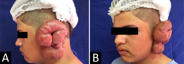
Male patient, 14 years old, black, was referred to the Plastic Surgery and Burns Service of the Hospital Universitário Evangélico de Curitiba (Curitiba/PR, Brazil) due to increased volume and itching in the left ear. The condition started mildly two years before, with a mass of 2 cm in diameter in the left lobe, just after placing earrings. He started treatment in another service with intralesional triamcinolone applications, in unknown number and dosage, with the case’s partial resolution. From then on, he showed an insidious evolution, with an increase in the speed of growth, reaching an expressive volume after about six months due to trauma with complete detachment of the mass, extending the anomalous healing process to the ear. He has a positive family history of scarring disorders and the presence of small inactive keloids elsewhere. At the time of the surgical intervention, the lesion was 12x8x3 cm in size, with a bright pinkish-light surface interspersed with areas of hypo and hyperchromia, nodular appearance, and stiff consistency (Figures 1A and 1B).
Ample surgical removal, skin suture with MonocrylTM 4-0 was performed, followed by ten radiotherapy sessions. The parameters used to analyze the therapeutic response were the patient’s opinion regarding the pruritus, reduced tissue hypertrophy upon inspection, and softening of the scar upon palpation. Signs of good therapeutic response occurred seven days after the operation (Figures 2A and 2B) with an acceptable aesthetic result in the sixth postoperative month (Figures 3A and 3B). As adverse events, irregularities and slight chromic heterogeneity were observed in the lower lobe.


DISCUSSION
In the present case, the evolution was compatible with the classic descriptions, expressing the most prevalent age and ethnic groups, combined with the previous traumatic episode and the positive family history.
Surgery as an isolated treatment modality is practically abandoned due to the high recurrence rates, varying between 45-100% in the first postoperative year. Surgical excision - combined with injection of corticosteroids, radiation therapy, or compression in the postoperative period - shows more encouraging results. Among intralesional corticosteroids, the drug of choice is triamcinolone (TCN). This association is based on these drugs’ mechanisms to promote a decrease in cytokine synthesis, the number, and activity of local fibroblasts1,6,7.
Due to the failure of intralesional corticosteroid applications, the association between surgical excision and radiotherapy was chosen. In the early 1970s, the therapeutic use of beta-therapy for complex keloids was recognized. It is based on the emission of radiation with little tissue penetration; it presents a mechanism of action to destroy the replicating cells’ nuclear genetic material, which explains the action proportional to cellular immaturity. This factor justifies its immediate use after excision, up to 24 hours, with a recurrence rate of around 10%1,7,8.
As a complementary modality, we can include the silicone plate, which acts on the lesions by increasing collagenase activity by raising the local temperature associated with the hydration of the stratum corneum by occlusion, and the negative electrical charge would guide the collagen fibers. Its effectiveness is proven in keloids only for the plates made 100% of silicone and used in association with excision, corticosteroids, and/or beta-therapy6,8.
The patient’s follow-up time after surgery is still short, but he remains under follow-up. The literature does not specify whether there is any necessary follow-up time or whether it should be uninterrupted, with 12 months being routinely adopted in the authors’ service. The treatment of keloids can be frustrating for both the plastic surgeon and the patient since its recurrence rate is high since the new wound will be susceptible to the same genetic, immunological, mechanical, and biochemical mechanisms as the initial wound9.
CONCLUSION
Thus, it is concluded that the result of the treatment was satisfactory, with significant dimensional reduction and almost complete restoration of the anatomy of the ear, obtaining patient satisfaction, and leading to better social reinsertion. The synergism between extensive surgical removal and radiotherapy sessions led to improved keloid scar’s clinical and aesthetic quality for lobe and auditory pavilion. However, due to the high rate of recurrence, the keloid-type fibroproliferative disorders are still a challenge for the medical community in general, given the need to deal with etiological mechanisms intrinsic to patients and inert to external conducts.
REFERENCES
1. Figueiredo JCA, Oliveira Junior FC, Zampar AG, Mélega JM. Quelóide: fatores de influência prognóstica. Rev Bras Cir Plást. 2008;23(4):274-80.
2. Gauglitz GG. Management of keloids and hypertrophic scars: current and emerging options. Clin Cosmet Investig Dermatol. 2013 Abr;6:103-14.
3. Mélega JM, Viterbo F, Mendes FH. Cirurgia plástica: os princípios e a atualidade. Rio de Janeiro: Guanabara Koogan; 2011.
4. Carvalhaes SM, Petroianu A, Ferreira MAT, Barros VM, Lopes RV. Assesment of the treatment of earlobe keloids with triamcinolone injections, surgical resection, and local pressure. Rev Col Bras Cir [Internet]. 2015 Feb; [citado 2015 Fev 4]; 42(1):9-13. Disponível em: https://www.scielo.br/scielo.php?script=sci_arttext&pid=S0100-69912015000200009
5. Hochman B, Farkas CB, Isoldi FC, Ferrara SF, Furtado F, Ferreira LM. Distribuição de queloide e cicatriz hipertrófica segundo fototipos de pele de Fitzpatrick. Rev Bras Cir Plást. 2012;27(2):185-9.
6. Wolwacz A, César EO, Ciufo MR, Wolwacz Júnior I, Kuyven CR, Deos MF. Opções terapêuticas nas cicatrizes queloidianas. Rev Bras Cir Plást. 2000;15(1):21-4.
7. Oliveira Junior B, Schellini SA, Lastória JC, Carvalho LR, Stolf HO, Oliveira P. Tratamento de queloides usando radioterapia pós-operatória com elétrons: estudo comparativo e randomizado com dois esquemas. Surg Cosmet Dermatol. 2013;5(1):16-26.
8. Fernandes WS, Ferreira RCA. Queloide: uma revisão dos tratamentos atualmente disponíveis. Rev Bras Ciênc Saúde [Internet]. 2014; [citado 2014 Dev 8]; 18(2):181-6. Disponível em: http://periodicos.ufpb.br/ojs/index.php/rbcs/article/view/18141/12925
9. Metsavaht LO, Garcia CAR. Infiltrações intralesionais de 5-FU no tratamento de queloides, cicatrizes hipertróficas e contraturas. Surg Cosmet Dermatol. 2015 Jan/Mar;7(1):17-24.
1. Evangelical Mackenzie University Hospital, Curitiba, PR, Brazil.
MVASN Analysis and/or data interpretation, Conception and design study, Conceptualization, Final manuscript approval, Project Administration, Supervision, Writing - Review & Editing
SMM Analysis and/or data interpretation, Conception and design study, Final manuscript approval, Methodology, Project Administration, Supervision, Validation, Visualization, Writing - Review & Editing
RD Analysis and/or data interpretation, Data Curation, Final manuscript approval, Formal Analysis, Methodology, Project Administration, Realization of operations and/or trials, Resources, Software, Supervision, Validation, Writing - Original Draft Preparation, Writing - Review & Editing
GLAL Analysis and/or data interpretation, Data Curation, Formal Analysis, Investigation, Methodology, Resources, Writing - Original Draft Preparation, Writing - Review & Editing
Corresponding author: Renata Damin, Rua Padre Anchieta, 1846, Conj. 103, Bigorrilho, Curitiba, PR, Brazil. Zip Code: 80730-000. E-mail: renatadamin@hotmail.com
Article received: June 14, 2019.
Article accepted: October 21, 2019.
Conflicts of interest: none
 Read in Portuguese
 Read in English
 PDF PT
 Print
Print
 Send this article by email
 How to Cite
 Mendeley
 Pocket