

Original Article - Year 2020 - Volume 35 -
Use of VaserTM plus liposuction in body contouring surgery
Uso de Vaser® mais lipoaspiração na cirurgia do contorno corporal
ABSTRACT
Introduction: Body contouring surgery is among the most requested surgical procedures in cosmetic surgery. Mentz was the first to perform superficial liposuction to define the abdominal muscles in male patients. However, Scuderi first popularized the use of continuous ultrasound to produce fat fragmentation in lipoplasty. Ultrasound, when applied internally to adipose tissue using a probe or metal cannula, breaks cells through three mechanisms: cavitation, thermal effect, and direct mechanical effect.
Methods: Since November 2018, 50 patients with an indication for body liposuction performed the procedure with the help of the third-generation ultrasound equipment (VASER).
Results: During the period between November 2018 and March 2019, 50 patients with a surgical indication underwent body contour liposuction using third-generation VASER technology. Of this universe of patients, 96% were women (47), with patients with an average age of 35 years (21-54).
Conclusion: The association of VASER with liposuction is a safe and reproducible technique that has the advantage of improving the result of liposculpture. Good aesthetic results were achieved, with an athletic and more natural contour.
Keywords: Lipectomy; Fat body; Subcutaneous fat; Surgery, Plastic; Ultrasonography; Adipose tissue.
RESUMO
Introdução: A cirurgia de contorno corporal está entre os procedimentos cirúrgicos mais solicitados em cirurgia estética. Mentz foi o primeiro a realizar lipoaspiração superficial para definição da musculatura abdominal de pacientes masculinos. Todavia, o uso de ultrassom contínuo para produzir fragmentação de gordura em lipoplastia foi popularizado, pela primeira vez, por Scuderi. O ultrassom, quando aplicado internamente ao tecido adiposo por uma sonda ou cânula metálica, realiza a quebra das células por meio de três mecanismos: cavitação, efeito térmico e efeito mecânico direto.
Métodos: A partir de novembro de 2018, 50 pacientes com indicação de lipoaspiração corporal realizaram o procedimento com a assistência do equipamento de ultrassom de terceira geração (VASER).
Resultados: Durante o período de novembro de 2018 a março de 2019, 50 pacientes com indicação cirúrgica foram submetidos à lipoaspiração de contorno corporal com uso da tecnologia VASER de 3a geração. Desse universo de pacientes, 96% eram mulheres (47), apresentando os pacientes idade média de 35 anos (21-54).
Conclusão: A associação do VASER na lipoaspiração é uma técnica segura e reprodutível, com a vantagem de melhorar o resultado das lipoesculturas. Bons resultados estéticos foram atingidos, com um contorno atlético e mais natural.
Palavras-chave: Lipectomia; Gordura abdominal; Gordura subcutânea; Cirurgia plástica; Ultrassom; Tecido adiposo
INTRODUCTION
The body contour surgery is among the most requested surgical procedures in aesthetic plastic surgery. Since the beginning of body contouring surgery, several authors around the world described significant advances in technique, with their cultural changes and scientific developments1.
In 2017, according to data from the International Society of Aesthetic Plastic Surgery (ISAPS), 1,573,630 liposuction procedures were carried out worldwide, with more than 210,000 procedures performed in Brazil2.
The introduction of lipoplasty into the surgical arsenal by Illouz, in 19853 and 19894, produced many changes in body contouring procedures with the use of white cannulas with holes for grease suction.
However, in 19935, Mentz was the first to perform a superficial liposuction to define the abdominal muscles of male patients, which he called abdominal etching. Regarding the treatment of the superficial layer in specific anatomical regions, this author says in his conclusions that it would be a technique for specific patients who want to have a muscular abdomen, with a need for results above the norm.
In 20076, Hoyos and Millard published the association of VASER (Vibration Amplification of Sound Energy at Resonance Lipo System) with high definition liposculpture. With his later work, Hoyos, in 20127 and 20188, consolidated the high definition liposuction with concrete results, creating standards and parameters for its correct performance.
However, it was Scuderi for the first time, in 19879, who popularized the use of continuous ultrasound to produce fat fragmentation in lipoplasty. The ultrasound, when it is applied internally to the adipose tissue using a probe or a metal cannula, breaks the cells by three mechanisms: cavitation, thermal effect, and direct mechanical effect10,11.
Sound Surgical Technologies LLC (Lafayette, CO) developed the VASERTM device, a surgical ultrasound system for the fat emulsion. This system uses solid probes of small diameter (i.e. 2.9mm and 3.7mm) with grooves around the point to increase fragmentation and efficiency. The rigid design of the probe redistributes the ultrasound energy, transferring part of the energy vibration from the point to a region near the point of the device.
OBJECTIVE
Therefore, the objective of the present study is to present our casuistry and body contouring experience with the aid of the VASER equipment.
METHODS
The study took place in 2 surgical centers in Florianópolis/SC, from November 2018 to March 2019.
As of November 2018, 50 patients with an indication for body liposuction underwent the procedure with the help of a third-generation ultrasound equipment (VASERTM).
The surgeries were performed by the same medical team, in different locations, using a standardized procedure sequence. During the postoperative visits, the photos of the evolution were taken, as well as the anamnesis and the physical examination for complications.
All patients signed the surgical consent form. This study was conducted following the Helsinki declaration.
Surgical technique
The preoperative marks of the liposuction areas were made with the patients in the orthostatic position. The muscular region is better defined with the active contraction performed by the patient. The authors use topographic marking in an attempt to present the places of highest projection and the presence of subcutaneous tissue. The topographic lines reinforce the places of greatest need for liposuction in the recumbent patient.
For infiltration, a super-wet anesthetic solution was prepared with lidocaine (2%) and adrenaline (1: 1,000,000). Due to the need for a humid environment to use the VASERTM, 4 liters of solution are prepared. The cutaneous opening points for the technique do not differ from the opening sites for classic liposuction; there is no need to enlarge the incision. The surgical irrigator (FagaTM) is used to infiltrate the super-wet anesthetic solution, in an attempt to compensate for the additional time generated by the use of ultrasound equipment.
The surgeon determined the selection of the VASERTM probe, the amplitude, and the pulse mode versus the continuous mode, according to the characteristics of the patient’s localized fatty deposits, which were individualized by physical examination. In the places where the equipment was introduced, a skin protector was used in an attempt to avoid skin damage by friction or thermal action of the device.
The protocol of the equipment employs the following parameters of the ultrasound of the third generation: for abdomen, 70% and time of use of 12 minutes; for the back, 70% and time of use of 12 minutes; y, for arms, 50% y time of use of 4 minutes. The additional time generated by using the device is approximately 30 minutes. The movement performed with the VASERTM tip is smooth and continuous, a movement very similar to that performed with the liposuction cannula.
Initially, the VASERTM is applied in a superficial fat plane, in an attempt to produce anatomical drawings of body structures, in women: mid/white line, semi-lunar lines and inguinal line. In the semi-lunar lines, we must mark the meeting point with the rib cage, where we will liposuction more superficially in an attempt to create local fatty depression. In men, in addition to the markings mentioned above, we can produce the design of the metamers of the rectus abdominis muscle. After covering the fatty superficial plane with the ultrasonic equipment, it is carried out in the entire area of deep liposuction. Only after completing the VASER, we start the liposuction of the fat solution.
The complete treatment (VASERTM + Liposuction) is performed in the initial position of decubitus, for the subsequent change of decubitus and the continuity of the procedure. Only dorsal and ventral decubitus is used, with the choice of initial decubitus based on surgical planning, especially when there is a need to obtain fat for gluteal fat grafting.
After using the VASERTM, it is performed liposuction of the fat solution, with the help of a pneumatic vibration equipment (VibrolipoTM) associated with the use of continuous aspiration equipment (LipoCoelhoTM). Superficial liposuction (anatomical drawing) is performed first, to complement the patient’s deep liposuction then.
All patients underwent a PortovacTM drainage placement with orientation for extraction on medical return in 1 week.
A local bandage was applied, and the patient remained with a compression mesh associated with 360º abdominal foam for one month after the operation. Lymphatic drainage began in the first postoperative week.
RESULTS
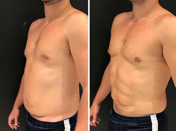
During the period between November 2018 and March 2019, 50 patients with a surgical indication underwent body contour liposuction using third-generation VASERTM technology.
Of this universe of patients, 96% were women (47), with patients with an average age of 35 years (21-54).
Among the associated procedures we had: 29 of gluteal fat grafting (58%), 24 of abdominoplasty (48%), 14 of mastopexy with prostheses (28%), 10 of augmentation prostheses (20%) and 6 of mastopexy without prosthesis (12%). For gluteal fat grafting, we performed it at the subcutaneous level, with the use of liposuction fat from where VASERTM was used.
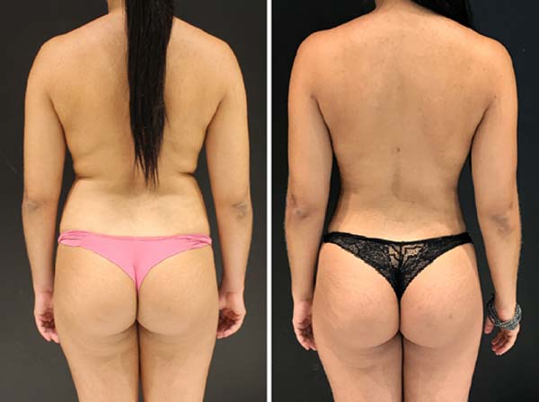
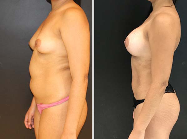
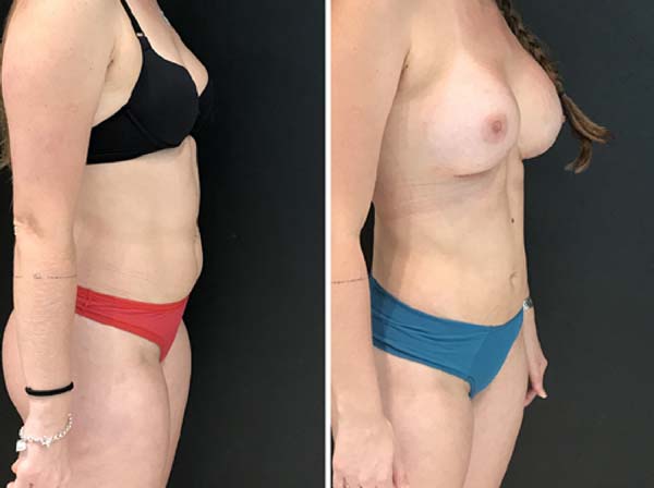
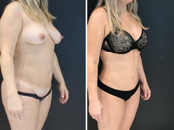
The average postoperative drainage explained by PortovacTM was 300 ml/day, with drainage elimination in 1 week (2 liters average in one week). We present patients photos for the comparison of pre and postoperative (Figures 1 to 5).
There were no postoperative complications, such as seroma, induration, altered sensitivity, portal burns, infections, skin necrosis, and dyschromia.
DISCUSSION
The refinement of surgical techniques in search for better results is a trend in plastic surgery. Numerous published studies contribute to a complete compilation of the vascular anatomy of the entire abdominal unit and back, providing critical directions for more advanced techniques12,13.
In liposuction, a relatively recent technique, the search for increasingly safer and aesthetically pleasing procedures is no different. The most recent publications and scientific events in plastic surgery have highlighted high definition liposuction or Lipo HD3,4.
Previous authors6,13 have already discussed and presented their results for VASERTM assisted high definition liposculpture: satisfaction in 84% of patients. There was a seroma in 6.5% of the cases that were solved with punctures. The use of drains was standardized for 48 to 72 hours. Of the 306 cases, 3.92% had a loss of definition.
One of the great fears about the use of ultrasound technology for the fragmentation of adipose tissue would be related to burns and necrosis caused by the energetic heat. We did not find this complication in our studio. In 20076, Hoyos and Millard, presented in their case series a burn of the liposuction portal during their learning curve, a complication that no longer appeared after the use of the portal infiltration associated with the skin protector14. The technology associated with the protocol brought security to the use. In the study by Danilla et al., in 201915 with 417 operated patients, the most frequent complication was hyperpigmentation (66%), followed by seroma (30%) and nodular fibrosis (20%), with the complications being transient in almost all of their entirety.
The work in superficial layers for the abdominal definition has always provoked discussions in the medical field. The aggression to the dermis can cause serious problems, such as dyschromia, fibrosis, adhesions, irregularities, retractions, and the dreaded epidermolysis and necrosis. The use of ultrasound technology in superficial layers is another advantage of the technique. In these layers, it detaches the most proximal fat from the dermis and makes the removal safer and less aggressive, with better conservation of the skin texture. There is a need for less movement of the liposuction cannula, after the use of VASERTM, to remove the same amount of fat as classic liposuction, causing less mechanical trauma to the patient’s dermis.
The use of third-generation ultrasound combined with the design of low trauma in cannulas allows us to achieve better results on the abdominal lateral surface and deep liposuction, creating a defined waist and lateral skin retraction6.
The use of VASERTM causes a better suction of fat by detaching it from attached tissues, facilitating its removal with less physical effort by the operator. The subsequent suction causes less bleeding, which can be checked by the color pattern of the liposuction in the collector (less blood). By facilitating the extraction of fat, it is possible to remove a higher amount of fat volume, and, by bleeding less, we can remove larger volumes, without decreasing the patient’s hematocrit/hemoglobin.
The increase in surgical time for the application of VASERTM is compensated by the decrease in the time required for liposuction associated with the use of surgical irrigator. Even so, the surgeon must plan to increase his surgical time with the use of technology, especially when implementing the technique, when the team’s processes are not well standardized. A large number of associated procedures in our study indicate that it is possible to optimize the use of technology to the point that combined surgeries do not exceed the programmed and recommended anesthetic time.
In the study by Nagy and Vanek, in 201216, the VASERTM-assisted lipoplasty method demonstrated a 53% improvement (17x11) in the retraction of the skin per cubic centimeter of aspirate removed compared to traditional suction-assisted lipoplasty and a reduction an average of 26% in blood loss compared to suction-assisted lipoplasty. Swanson in 201217, questioned the calculation methods of the study by Nagy and Vanek16, which had an N of 20 patients. In his own calculations, Swanson17 indicated that a difference of 6% would need a sample of 199 patients, concluding that it is a weak study in methodology and with conflicts of interest. Matarasso, in 201218, also questioned the results of the study by Nagy and Vanek16, noting that the likely great advantage of VASERTM would be to facilitate the removal of fat for the surgeon, especially in those with some degree of fibrosis due to previous liposuction.
Even with the discussions of quantitative evaluation of the VASERTM cutaneous retraction in liposuction, it is sure that it reduces skin flaccidity in the postoperative period and, in borderline cases, in which we have to choose a skin resection (abdominoplasty) or only liposuction, the use of ultrasound technology helps in deciding for a procedure with less scarring and a pleasant aesthetic result, with a uniform adherence to deep tissues, especially in young patients. When the procedure of choice is an abdominoplasty, the use of VASERTM produces less traumatic fat removal from the abdominal flap tissue, decreasing the chances of complications associated with lipoabdominoplasty19.
In order to assess the quality of fatty liposuction for fat grafting, Duscher et al., In 201620,21 and 201722, proved that the use of VASERTM does not impair the viability of the adipocyte derived from the stromal cell, vital information to increase graft retention. Therefore, all fat grafting performed in the technique is done with liposuction from the area where the VASERTM was used. The only process we use in the fat solution before grafting is a simple decanting.
We did not see any difference in graft integration or different fat reabsorption rate with the use of the technique. The fat grafted has a smaller diameter compared to the fat coming from a classic liposuction, which theoretically would facilitate its integration, as pointed out by the study by Eto et al., in 201223, who identified the size of the fatty tissue survival zone after its grafting as smaller than 300microns, so fat grafts greater than 600microns already have a regeneration zone and possibly a central necrosis zone.
We always recommend the subcutaneous plane for grafting, with large diameter cannulas, syringe, and supragluteal portals, thus reducing the chances of complications24,25.
In the postoperative period, we noticed a less traumatic evolution for the patient. Due to less aggression and less bleeding, we have a recovery with fewer symptoms and faster return of the patient to work activity.
As the postoperative period progressed, we did not have a patient with post-procedure weight recovery. However, given the use of a technology that facilitates fat removal, the possible results with the gain of new adipose tissue will be similar to those found with classical liposuction. The use of the protocol to create extremely athletic results, with an aspect of muscular hypertrophy, has its specific indication, being essential to inform the patient about possible unwanted aesthetic results in case of a significant weight gain.
CONCLUSION
The association of VASERTM in liposuction is a safe and reproducible technique, with the advantage of improving the result of liposculpture.
1. Clínica Vivva, Florianópolis, Florianópolis, SC, Brasil.
2. Hospital Universitário, Cirurgia Plástica e Queimados, Florianópolis, SC, Brasil.
Corresponding author: Caio Pundek Garcia, Av. Osvaldo Reis, 3385, Sala 501 Praia Brava, Itajaí, SC, Brazil. Zip Code: 88306-773 E-mail: caio_pgarcia@hotmail.com







 Read in Portuguese
 Read in English
 PDF PT
 Print
Print
 Send this article by email
 How to Cite
 Mendeley
 Pocket
 Twitter