


Review Article - Year 2019 - Volume 34 -
Burn lesions with progression to neoplasia: Marjolin's ulcer
Lesões por queimaduras com evolução para neoplasia: úlceras de Marjolin
Thiago Maciel Valente1,* ; Mateus Pinheiro Fernandes Feitosa Arrais1; Bárbara Matos De Carvalho Borges1; Samy Lima Carneiro1; Maressa Cavalcante Fernandes de Albuquerque1; Nelson Gurgel Simas de Oliveira1
ABSTRACT
Introduction: Marjolin's ulcer is defined as a malignancy within scars that is usually chronic and results from several lesion types, with burn injuries being the most common. Methods: A bibliographic survey was conducted of the Virtual Health Library, PubMed, Scientific Electronic Library Online, and Cochrane databases using the inclusion criteria of studies published in the last 5 years, human studies, and published in English or Portuguese. Results: A total of 31 studies were analyzed, of which only 6 were included in the final sample. Discussion: Marjolin's ulcer is found in old burn scars and can occur anywhere, but it is more common in the upper and lower limbs. The diagnosis begins with the clinical suspicion based on lesion characteristics: chronic unhealed ulcerative lesions with high and hardened edges, an unpleasant odor, and purulent discharge. However, the diagnosis can only be made histopathologically. The latency period between injury and malignancy is 30-35 years. Although treatment should be individualized since it depends on several factors, surgical excision is considered the gold standard. Conclusion: Knowledge about this condition is essential to better patient prognosis and prevent underestimation of possible cases of malignancy, allowing for appropriate therapy to minimize recurrence and enabling prophylactic measures to prevent burn injury and reduce risk factors for malignancy.
Keywords: Burns; Skin ulcer; Carcinoma; Healing; Plastic surgery
RESUMO
Introduction: Marjolin's ulcer is defined as a malignancy within
scars that is usually chronic and results from several lesion
types, with burn injuries being the most common. Methods: A
bibliographic survey was conducted of the Virtual Health Library,
PubMed, Scientific Electronic Library Online, and Cochrane
databases using the inclusion criteria of studies published in
the last 5 years, human studies, and published in English or
Portuguese.
Results: A total of 31 studies were analyzed, of which
only 6 were included in the final sample.
Discussion: Marjolin's
ulcer is found in old burn scars and can occur anywhere, but it
is more common in the upper and lower limbs. The diagnosis
begins with the clinical suspicion based on lesion characteristics:
chronic unhealed ulcerative lesions with high and hardened
edges, an unpleasant odor, and purulent discharge. However,
the diagnosis can only be made histopathologically. The latency
period between injury and malignancy is 30-35 years. Although
treatment should be individualized since it depends on several
factors, surgical excision is considered the gold standard.
Conclusion: Knowledge about this condition is essential to
better patient prognosis and prevent underestimation of
possible cases of malignancy, allowing for appropriate therapy
to minimize recurrence and enabling prophylactic measures
to prevent burn injury and reduce risk factors for malignancy.
Palavras-chave: Queimaduras; Úlcera cutânea; Carcinoma; Cicatrização; Cirurgia plástica
INTRODUCTION
Worldwide, about 6 million people require medical care because of burns; in Brazil, this number is about 1 million patients per year1,2. Such lesions can be caused by thermal, chemical, electrical, biological, or radioactive agents and are divided into three degrees according to their complexity.
In addition to the physical damage, many burn victims suffer from psychological and economic problems due to a prolonged recovery time3,4. Given that this pathology requires long and painful treatment, it may, in some cases, be neglected and evolve into a process of lesion malignancy.
Malignant skin lesions are the most frequent form of cancer in Brazil and worldwide, being subdivided into melanoma and non-melanoma, which is the most common, representing 95% of tumors. Exposure to solar radiation is a predisposing factor for squamous cell carcinogenesis and basal cell carcinomas5,6.
In 1828, the French surgeon Jean Nicolas Marjolin associated the inadequate treatment of Marjolin’s ulcer with burns that were not treated due to their malignancy. However, the term Marjolin’s ulcer was first recognized in 1903 in a description by Da Costa of a malignant process resulting from a burn8. The term is currently used more generally for any chronic malignancy in scars, although burns remain the main precursor lesion, with the incidence of burn scar evolving to carcinoma being 0.77-2%8.
Although it is possible to identify other malignant neoplasms, such as sarcoma, melanoma, and basal cell carcinoma, the main histopathological sample of the tumor is spinocellular9. Thus, 76% of patients with a history of burn scar have squamous cell carcinoma10.
Furthermore, this pathology is classified as acute or chronic, lesions that became malignant within or beyond a 12-month period after the injury, respectively7.
This study provides knowledge about the above-mentioned problems and the tools used for the effective diagnosis, treatment, and prognosis of Marjolin’s ulcer. Thus, it is necessary to use therapeutic tools and medical knowledge to better manage patients, prevent the development of this disease in burn patients, and awaken the interest of clinicians about its early identification.
OBJECTIVE
The objective of this study is to review the literature on Marjolin’s ulcer resulting from burn injuries.
METHODS
This is an integrative review of burn scar carcinoma, classically known as Marjolin’s ulcer. The Virtual Health Library (VHL), PubMed (Publisher Medicine), Scientific Electronic Library Online (SciELO), and Cochrane databases were used. The following controlled keywords were used: “Queimadura/Burn” and “Úlceras de Marjolin/Marjolin’s Ulcers.” The inclusion criteria established were studies published in the last 5 years of humans and full availability on the web in English or Portuguese.
Moreover, integrative reviews, literature reviews, systematic reviews, and letters to the editor were defined as the exclusion criteria.
RESULTS
By using the inclusion criteria, we found 31 studies in the databases, of which only six met the inclusion criteria after the analytical reading being included in the final sample of this study (Tables 1 and 2). Of the 11 studies found in the VHL, two letters to the editor, two studies that were not available on the web, and two articles that did not address burns were excluded. Thus, five studies were included in the sample. Eighteen studies were found in PubMed, eight of which were already found in the VHL, three were not available on the web, and six studies were excluded because they did not address burns, one of which addressed trauma-related injuries in general. Thus, only one study of this database was included in the sample. Two studies using the previously determined keywords were found in SciELO, but none of them addressed burns. No study was found in the search of the Cochrane database.
| Study Title | Study Type | Year | Language | Origin |
|---|---|---|---|---|
| Úlcera de Marjolin: relato de caso (Marjolin's ulcer: case report) | Case Report | 2015 | Portuguese | Brazil |
| Multiple Marjolin's ulcers arising from irradiated post-burn hypertrophic scars: a case report | Case Report | 2014 | English | Canada |
| Acute Marjolin's ulcers: a nebulous diagnosis | Case Report Series | 2014 | English | United States |
| Incidences of malignancy in chronic burn scar ulcers: experience from Bangladesh | Prospective and Observational Study | 2015 | English | Bangladesh |
| Epidemiology and predictors of recurrence of Marjolin's ulcer: experience from Mansoura University | Retrospective Study | 2017 | English | Egypt |
| Úlcera de Marjolin: revisão de literatura e relato de caso – (Marjolin's ulcer: literature review and case report) | Case Report | 2016 | Portuguese | Brazil |

| Study | Number of cases studied | Age at burn | Age at diagnosis | Sex | Treatment |
|---|---|---|---|---|---|
| Úlcera de Marjolin: relato de caso (Marjolin's ulcer: case report) | 2 | Not reported | Case 1: 59 years | Cases 1 and 2: Male | Cases 1 and 2: Exeresis with wide margin of safety |
| Case 2: 82 years | |||||
| Multiple Marjolin's ulcers arising from irradiated post-burn hypertrophic scars: a case report | 1 | 4 years | 61 years | Male | Exeresis with wide margin of safety |
| Graft for radial artery reconstruction | |||||
| Acute Marjolin's ulcers: a nebulous diagnosis | 3 | Not reported | Case 1: 42 years | Case 1: Female | Case 1: Exeresis with wide margin (3.5 × 1.8 cm) |
| Case 2: 50 years | Case 2: Male | Case 2: Surgical debridement | |||
| Case 3: 60 years | Case 3: Male | Case 3: Exeresis with wide margin | |||
| Úlcera de Marjolin: revisão de literatura e relato de caso (Marjolin's ulcer: literature review and case report) | 1 | 27 years | 52 years | Male | Wide margin exeresis and local graft |
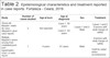
It should be noted that although the last selected study has the term “Literature Review” in its title, a type of study that was considered an exclusion criterion, the “Úlcera de Marjolin: Revisão de literatura e relato de caso (Marjolin’s Ulcer: Literature Review and Case Report)” article was actually a case report.
DISCUSSION
After analyzing the results, we decided to divide the study into the following topics to facilitate understanding.
Risk Factors and Topography
Although chronic pressure lesions are reported in 2.6% of cases of Marjolin’s ulcer, they are more common in old burn scars, representing 76% of cases, and venous stasis ulcers, representing 6.3% of cases11.
A study conducted of 140 patients showed that, on average, 80% of the malignant transformations of burns scars were caused by flames, mainly due to the ability of this etiological agent to deepen the lesion, causing greater stiffness and consequent cracking of the lesion scar12.
Moreover, although Marjolin’s ulcers may occur in any location, the upper and lower limbs were the most affected topographies, which is explained by the high incidence of venous stasis burns and ulcers, and the higher propensity to repeated damage in these sites, especially in the joints12.
In addition to burn scars with a long healing time, other important risk factors are reported in the literature, such as second intention scars and scars that are easily traumatized, confirming the relevance of topography for assessing lesion malignancy13. Skin grafts in deeper burns is mentioned as a protective factor against malignancy14.
A study conducted in China demonstrated that easily traumatized scars are a risk factor for the development of Marjolin’s ulcer. Of the 17 patients with Marjolin’s ulcer (35%) they assessed, six reported recurrent ulcers within the lesion15.
Etiology
Although Marjolin’s ulcer is not carcinogenic, its etiology appears to include several factors that are influenced by the trauma that occurs within the region. This trauma makes the lesion more malignant since it intensifies some carcinogenic factors, such as UV rays, due to the greater sensitivity of traumatized skin13. It also causes prolonged cell proliferation due to inefficient healing, which can cause mutations in the DNA of these cells16.
Another factor that may contribute to carcinogenesis of these lesions is the reduction in vascularization in the region where the healing process occurs, making the immune response to these cells with mutated DNA less efficient16. It also includes lymphatic obliteration due to healing, impairing the presentation of the antigen and the activation of the defense cells17.
At the molecular level, some studies have attributed the malignancy process of Marjolin’s ulcer to mutations in the p53 and FAS genes17.
Diagnosis and Latency
The diagnostic approach begins with clinical suspicion of Marjolin’s ulcer based on the lesion’s characteristics, i.e., chronic ulcerative lesions with unhealed high and hardened edges, an unpleasant odor, a budding appearance, and occasional purulent discharge18.
Furthermore, it is important to pay attention to the patient’s history since many reports show that the transformation to carcinoma was associated with poorly healing burns during childhood19.
Although the diagnosis can be directed by lesion characteristics and anamnesis, it is only made histopathologically18. Ulcers persisting for more than 3 months should be biopsied20.
A common problem found in health services is the patient’s delay in seeking care, which makes it difficult to make the early diagnosis that is essential for a favorable prognosis. Another problem eventually reported is underestimation of the patient’s condition, postponement of the therapeutic approach, and possible confusion with an infected ulcer18,14.
The latency period between injury and malignancy is 11-75 years, with a mean of 30-35 years. Moreover, the patient’s age when the first lesion occurred, such as the burn injury, is inversely proportional to the latency period, i.e., individuals who developed this lesion at an early age have a longer latency period21.
Treatment and Recurrence
Although treatment should be individualized, since it depends on several factors, such as age, comorbidity, and tumor characteristics, surgical excision is considered the gold standard when the tumor has a margin at least 2 cm18,22. However, limb amputation and radiotherapy or chemotherapy may also be indicated.
Amputation is convenient when Marjolin’s ulcer infiltrates more deeply, affecting bones and great vessels, with imaging examinations eventually being useful for detecting the degree of bone involvement14,20, infectious processes, and significant hemorrhage, and when excision of the lesion may generate greater functional disability17.
Radiotherapy may be indicated in cases of metastasis that cannot be surgically corrected, tumors larger than 10 cm with positive lymph nodes after dissection, and head and neck lesions with positive lymph nodes after dissection23.
Although chemotherapy is not always indicated, it may be convenient in situations in which the patient does not consent to surgical treatment, has distant metastasis, or experiences disease recurrence23.
A study analyzing 412 cases observed Marjolin’s ulcer after surgical excision in 16% of reports. This result may be associated with some factors, such as male sex, cooking oil burn, and treatment neglect during the first injury and recurrence13,24.
A study in Egypt analyzing 26 cases of Marjolin’s ulcer identified other factors as predictors of recurrence, such as young age at the time of diagnosis, malignancy in the nodal groups of lymphatic drainage, and the use of a flap or graft after wide excision20.
CONCLUSION
This study analyzed the literature on Marjolin’s ulcer resulting from malignancy of burn scars and discussed risk factors and topography, etiology, diagnosis and latency, and treatment and recurrence.
From the discussion of this review, it was possible to conclude that Marjolin’s ulcer presents as malignant skin neoplasms that appear most frequently after thermal burns that require specific and individualized treatment since several factors influence the therapeutic plan.
Despite the need for histopathological confirmation, clinical suspicion together with anamnesis becomes necessary for an early diagnosis, which is essential due to the highly aggressive nature, metastatic potential, and high recurrence rate of Marjolin’s ulcer, which in most cases is treated surgically. In more extreme cases of bone involvement, amputation becomes necessary to avoid worse conditions.
Thus, the knowledge of health professionals about Marjolin’s ulcers is essential for a better prognosis so that possible cases of malignancy are not underestimated, appropriate therapy is implemented, recurrence is avoided, and prophylactic measures are implemented to effectively avoid burn and mitigate risk factors for malignancy.
Limitations of the present study include the low number of articles describing an epidemiological analysis with a more considerable sample in the analyzed period and the consideration of only Portuguese and English articles, which may have limited the search. Therefore, clinical studies and trials are needed to gain more comprehensive knowledge of Marjolin’s ulcer.
COLLABORATIONS
|
TMV |
Data Curation, Methodology, Writing - Original Draft Preparation, Writing - Review & Editing |
|
MPFFA |
Writing - Original Draft Preparation |
|
BMC |
Writing - Original Draft Preparation |
|
SLC |
Writing - Original Draft Preparation |
|
MCFA |
Writing - Original Draft Preparation |
|
NGSO |
Review & Editing |
REFERENCES
1. Daga H, Morais IH, Prestes MA. Perfil dos acidentes por queimaduras em crianças atendidas no Hospital Universitário Evangélico de Curitiba. Rev Bras Queimaduras. 2015;14(4):268-72.
2. Ehrl D, Heidekrueger P, Ninkovic M, Broer PN. Effect of primary admission to burn centers on the outcomes of severely burned patients. Burns. 2018 May;44(3):524-530. PMID: 29463463 DOI: https://doi.org/10.1016/j.burns.2018.01.002
3. Soares LR, Barbosa FS, Santos LA, Mattos VCR, De-Paula CA, Leal PML, et al. Estudo epidemiológico de vítimas de queimaduras internadas em um hospital de urgência da Bahia. Rev Bras Queimaduras. 2016;15(3):148-52.
4. Moraes LP, Echevarría-Guanilo ME, Martins CL, Longaray TM, et al. Apoio social e qualidade de vida na perspectiva de pessoas que sofreram queimaduras. Rev Bras Queimaduras. 2016;15(3):142-7.
5. Souza RAL, Matos RRL, Ventura LM, Pinho L, Santos AAA, Marques MS. Câncer de pele: estratégias de fotoproteção e fotoexposição solar em agentes comunitários de saúde. Unimontes Científica. 2018;70-81.
6. Pires CAA, Fayal AP, Cavalcante RH, Fayal SP, Lopes NS, Fayal FP, et al. Câncer de pele: caracterização do perfil e avaliação da proteção solar dos pacientes atendidos em serviço universitário. J Health Biol Sci. 2018;6(1):54-59. DOI: https://doi.org/10.12662/2317-3076jhbs.v6i1.1433.p54-59.2018
7. Bazalinski D, Przybek-Mita J, Baranska B, Wiench P. Marjolin’s ulcer in chronic wounds - review of available literature. Contemp Oncol (Pozn). 2017;21(3):197-202.
8. Copcu E. Marjolin’s ulcer: a preventable complication of burns?. Plast Reconstr Surg. 2009 Jul;124(1):156e-64e. PMID: 19568055 DOI: https://doi.org/10.1097/PRS.0b013e3181a8082e
9. Iqbal FM, Sinha Y, Jaffe W. Marjolin’s ulcer: a rare entity with a call for early diagnosis. BMJ Case Rep. 2015 Jul;bcr2014208176. PMID: 26177995 DOI: https://doi.org/10.1136/bcr-2014-208176
10. Tacani PM, Tacani RE, Machado AFP, Montezello D, Góes JCGS, Marx AG, et al. High-frequency generator in wound healing of Marjolin’s Ulcer after carcinoma resection. Adv Wound Care (New Rochelle). 2018 May;7(5):165-170. DOI: https://doi.org/10.1089/wound.2017.0757
11. Kerr-Valentic MA, Samimi K, Rohlen BH, Agarwal JP, Rockwell WB. Marjolin’s Ulcer: modern analysis of an ancient problem. Plast Reconstr Surg. 2009 Jan;123(1):184-191. DOI: https://doi.org/10.1097/PRS.0b013e3181904d86
12. Das KK, Chakaraborty A, Rahman A, Khandkar S. Incidences of malignancy in chronic burn scar ulcers: experience from Bangladesh. Burns. 2015 Sep;41(6):1315-21. PMID: 25716761 DOI: https://doi.org/10.1016/j.burns.2015.02.008
13. Zuo KJ, Tredget EE. Multiple Marjolin’s ulcers arising from irradiated post-burn hypertrophic scars: a case report. Burns. 2014 Jun;40(4):e21-e25. DOI: https://doi.org/10.1016/j.burns.2013.10.008
14. Ochenduszkiewicz U, Matkowski R, Szynglarewicz B, Kornafel J. Marjolin’s ulcer: malignant neoplasm arising in scars. Rep Pract Oncol Radiother, 2006 Jan;11(3):135-138. DOI: https://doi.org/10.1016/S1507-1367(06)71058-6
15. Yu N, Long X, Lujan-Hernandez JR, Hassan KZ, Bai M, Wang Y, et al. Marjolin’s ulcer: a preventable malignancy arising from scars. World J Surg Oncol. 2013;11(1):313. DOI: https://doi.org/10.1186/1477-7819-11-313
16. Chang JB, Kung TA, Cederna PS. Acute Marjolin’s Ulcers a nebulous diagnosis. Ann Plast Surg. 2014 May;72(5):515-20. PMID: 24691319 DOI: https://doi.org/10.1097/SAP.0000000000000134
17. Chalya P, Mabula J, Rambau P, Mchembe M, Kahima K, Chandika A, et al. Marjolin’s ulcers at a university teaching hospital in Northwestern Tanzania: a retrospective review of 56 cases. World J Surg Oncol. 2012;10(1):38. DOI: https://doi.org/10.1186/1477-7819-10-38
18. Dinato SLM, Sigueta ML, Almeida JRP, Romiti N. Úlcera de Marjolin: relato de caso. Diagn Tratamento. 2015 Mar;20(1):4-7.
19. Vieira RRBT, et al. Úlcera de Marjolin: Revisão de literatura e relato de caso. Rev Bras Queimaduras. 2016;15(3):179-84.
20. Metwally IH, Roshdy A, Saleh SS, Ezzat M. Epidemiology and predictors of recurrence of Marjolin’s ulcer: experience from Mansoura Universityxs. Ann R Coll Surg Engl. 2017 Mar;99(3):245-249. DOI: https://doi.org/10.1308/rcsann.2016.0309
21. Pekarek B, Buck S, Osher L. A comprehensive review on Marjolin’s Ulcers: diagnosis and treatment. J Am Col Certif Wound Spec. 2011 Sep;3(3):60-64. DOI: https://doi.org/10.1016/j.jcws.2012.04.001
22. Elkins-Williams ST, Marston WA, Hultman CS. Management of the chronic burn wound. Clin Plast Surg. 2017 Jul;44(3):679-87. DOI: https://doi.org/10.1016/j.cps.2017.02.024
23. Saaiq M, Ashraf B. Marjolin’s ulcers in the post-burned lesions and scars. World J Clin Cases. 2014 Oct;2(10):507-514. DOI: https://doi.org/10.12998/wjcc.v2.i10.507
24. Kowal-Vern A, Criswell BK. Burn scar neoplasms: a literature review and statistical analysis. Burns. 2005 Jun;31(4):403-413. DOI: https://doi.org/10.1016/j.burns.2005.02.015
1. Universidade de Fortaleza, Fortaleza, CE, Brazil.
Corresponding author: Thiago Maciel Valente Rua Bento Albuquerque, 1133, Cocó, Fortaleza, CE, Brazil. Zip Code: 60192-055. E-mail: maciel.thiago@edu.unifor.br
Article received: January 29, 2019.
Article accepted: July 8, 2019.
Conflicts of interest: none.
 Read in Portuguese
 Read in English
 PDF PT
 Print
Print
 Send this article by email
 How to Cite
 Mendeley
 Pocket