


Review Article - Year 2019 - Volume 34 - Issue 1
Impact of fractional CO2 laser treatment on hypertrophic scars and keloids: a systematic review
Impacto do tratamento com laser fracionado de CO2 em cicatrizes hipertróficas e queloides: uma revisão sistemática
ABSTRACT
Introduction: Hypertrophic scars and keloids cause aesthetic and functional damages, and are difficult to treat. This review aimed to identify prospective studies on fractional CO2 laser to present the clinical and histological changes and the methodology used for the evaluation of scars before and after intervention.
Methods: We conducted an electronic review (LILACS, Medline, and SciELO) of studies published between January 2004 and December 2017, using the search terms "keloid/queloide," "hypertrophic scar/cicatriz hipertrófica," and "CO2 laser ," according to the PRISMA Statement. Studies that compared scars before and after isolated treatment with fractional CO2 laser were selected. Two independent reviewers analyzed the data.
Results: One hundred two articles were analyzed, of which 7 met the inclusion criteria. Of the 7 articles, all analyzed hypertrophic scars, 2 analyzed keloids in addition to hypertrophic scars, and 3 analyzed histological changes. Most studies showed a statistically significant difference in clinical scores between before and after treatment of hypertrophic scars, with improvement in symptoms, flexibility, and scar height. Between the 2 studies that analyzed keloids, 1 reported a clinical difference after treatment. The histological changes showed significant differences in the orientation and density of the collagen fibers, and in the thickness of the epidermis.
Conclusion: The use of fractional CO2 laser should be considered as a promising treatment option for pathological scars, as it improves clinical signs and symptoms such as color, thickness, and pruritus.
Keywords: Gas laser; Hypertrophic scar; Keloid; Carbon dioxide; Pathology
RESUMO
Introdução: Cicatrizes hipertróficas e queloides causam dano estético e funcional e são de difícil tratamento. O objetivo desta revisão foi identificar estudos prospectivos do tratamento com o laser fracionado de CO2, mostrando as alterações clínicas e histológicas e a metodologia utilizada para a avaliação das cicatrizes antes e após intervenção.
Métodos: Foi realizada uma revisão eletrônica (LILACS, Medline e SciELO) de estudos publicados entre janeiro de 2004 e dezembro de 2017, com os termos "keloid/queloide", "hypertrophic scar/cicatriz hipertrófica" e "laser CO2", de acordo com o PRISMA Statement, sendo selecionados os estudos que comparassem as cicatrizes antes e depois de tratamento isolado com laser fracionado de CO2. Os dados foram analisados por dois revisores independentes.
Resultados: Foram analisados 102 artigos, sendo que 7 cumpriam os critérios estabelecidos. Destes, os 7 analisaram cicatrizes hipertróficas, 2 deles também analisaram queloides, e 3 estudaram alterações histológicas. Houve diferença estatística entre os escores clínicos medidos antes e após tratamento de cicatrizes hipertróficas na maioria dos estudos, com melhora nos sintomas, na flexibilidade e altura da cicatriz. Entre os 2 estudos que analisaram os queloides, 1 deles demonstrou diferença clínica após tratamento. Nas alterações histológicas, houve diferença na orientação e densidade das fibras de colágeno e na espessura da epiderme.
Conclusão: O laser fracionado de CO2 deve ser considerado como opção promissora no tratamento de cicatrizes patológicas, visto que melhora os sinais e sintomas clínicos como cor, espessura e prurido.
Palavras-chave: Lasers de gás; Cicatriz hipertrófica; Queloide; Dióxido de carbono; Patologia
INTRODUCTION
Scars cause significant aesthetic and psychosocial impacts on patients in the practice of plastic surgery. Hypertrophic scars and keloids are lesions that commonly appear after skin injury, causing aesthetic and functional damages, which are sometimes difficult to treat. Clinical evaluation of a scar is necessary to determine the correct treatment and effectiveness of the therapy. Multiple objective and subjective tools were created to characterize scars, which suggests that none of these tools is complete enough to evaluate the clinical and psychosocial aspects of pathological scars1.
Keloids are violaceous scars of hard consistency that exceed the limits of the initial wound and are more frequently found in individuals with genetic predisposition, mainly those of black and oriental ethnicities, with an incidence of 4.5–16%, as compared with an incidence of < 1% in Caucasians. The sites of greatest involvement are the chest, back, and joints, with no sex-specific pattern.
They are also influenced by sex hormones, which explains their higher incidence between ages 10 and 30 years, and during pregnancy. Histological examination revealed an increase in glycosaminoglycans and type I and III collagens, with disorganized and irregularly dispersed fibers.
Hypertrophic scars are high, tense, reddish, do not exceed the limits of the original lesion, and tend to regress over time. On histological examination, they show an increase in type III collagen, with organized fibers and fibers parallel to the epidermis. Both can cause pain and itching, and have abundant dermal collagen due to an imbalance between its synthesis and degradation. However, its pathophysiology has not yet been fully elucidated2.
Failures in the regulatory mechanisms of healing, which have not yet been well established, such as the decrease in apoptosis of fibroblasts and the role of growth factors, particularly transforming growth factor B1 (TGF-B1), have been studied in the development of this disorder. Matrix metalloproteinase 9 (MMP9), a family of enzymes responsible for connective tissue degradation, is known to be less evident in keloids and hypertrophic scars than in healthy skin in immunohistochemical tests, and measurement of MMP9 level is important for the histological evaluation of the effectiveness of the treatment performed3-5.
Several treatments are available for these two conditions, among which the combination of intralesional corticosteroid injections, silicone bandages, and local pressure is referred to as standard treatment by theInternational Advisory Panel on Scar Management consensus (IAPSM), despite its limitation. Second-line therapy for refractory cases, in turn, includes ablative or non-ablative laser therapy and surgical excision associated with the use of silicone gel. Fractional laser treatments induce a healing response, increasing type III collagen levels and remodeling the tissue6.
Tissue resurfacing using fractional photothermolysis was introduced in 2004. The fractional technique produces columns of thermal and ablative damage, known as microthermal treatment zones (MTZ), interspersed with areas of untreated skin, a process that accelerates tissue recovery. In 2007, a new method to produce MTZ using ablative carbon dioxide (CO2) was described. This method proved effective in reaching all layers of the skin (stratum corneum, epidermis, and dermis) through ablation and coagulation, with maximum control of tissue damage without reducing the efficacy and with the remodeling of collagen lasting for at least 3 months after treatment, which was confirmed by immunohistochemistry7,8.
Intracellular and extracellular water absorbs the energy of the CO2 laser at a wavelength of 10,600 nm, causing rapid heating and vaporization of the tissue to a depth of 20 to 60 µm. The heating of the dermis causes contraction and remodeling of the collagen with a thermal necrosis zone ranging from 20 to 50 µm. Reepithelialization occurs after 5 to 10 days, and erythema time depends on the energy used9.
The standard treatment for hypertrophic scars and keloids, which are frequently encountered conditions in the routine practice of plastic surgery and dermatology, do not always help achieve satisfactory results, and clinical improvement is difficult to evaluate. The use of lasers was introduced as a secondary alternative for the treatment of these conditions, and the mechanism of the clinical and histological changes of the treated tissues is still under study, with no consensus so far. As few studies in the literature confirm these data and the overall improvement of a scar is difficult to objectively evaluate, the methodologies used to confirm the results obtained after the treatment of pathological scars with CO2 laser must be reviewed.
OBJECTIVE
This systematic literature review aimed to identify prospective experimental before-and-after studies on the treatment of hypertrophic scars and keloids that used fractional CO2 laser and identified clinical and histological changes and described the methodology used for evaluating scars before and after intervention.
METHODS
An extensive electronic review was conducted in the Latin American and Caribbean Literature in Health Sciences (LILACS), Health Information from the National Library of Medicine (Medline), Web of Science, and Scientific Electronic Library Online (SciELO) electronic library databases. We searched the databases using a combination of the following terms: “keloid/quelóide,” “hypertrophic scar/cicatriz hipertrófica,” and “CO2 laser,” according to the PRISMA Statement10.
Two independent researchers tracked the titles and abstracts of the articles identified. Afterward, the full texts of the potentially relevant articles were reviewed. The inclusion criteria were controlled or non-controlled experimental studies, studies published between 2004 and 2017, and studies that included patients with hypertrophic scars or keloids in which isolated treatment with fractional CO2 laser was used, with a clearly described methodology.
Studies in languages other than English, Spanish, and Portuguese, and outcomes other than clinical or histological evaluation of scars before and after treatment were excluded.
The articles were subdivided into groups according to the condition treated (keloid and/or hypertrophic scar) and the outcome (clinical and/or histological). The results are presented in tables according to the outcomes.
The results of each group (mean and standard deviation, and percentage indexes) were analyzed, and the control, pretreatment, clinical outcomes (according to the scar evaluation scale used), and histological outcome, when applicable, were compared.
RESULTS
Initially, 102 articles were identified in the electronic databases by evaluating their titles and abstracts. The full texts of 28 publications were evaluated, and 7 studies were selected for inclusion according to the established criteria.
The studies were subdivided according to the outcome studied. Of the studies, 7 investigated the clinical changes in the treatment of hypertrophic scars, of which 2 investigated keloids and 3 (already selected in previous selections) reviewed histological and immunohistochemical parameters.
Clinical outcomes in hypertrophic scars
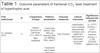
Table 1 shows the main results of studies on changes in hypertrophic scars evaluated using clinical measurement scales. Considering that statistically significant results have a p value of <0.05, a statistically significant difference was observed between the clinical scores measured before and after treatment by El-Zawahry et al.7, Azzam et al.3, Makboul et al.5, Lei et al.13, and Hultman et al.14. Choi et al.12 reported significant improvements in flexibility and scar height.
| First author/year | N | Comparison Groups | Platform parameters | Outcome measures | Clinical outcomes | Significance between the groups |
|---|---|---|---|---|---|---|
| El- Zawahry, 20157 | 11 | Before sessions of fractional CO2 laser X After treatment | 3 sessions, DEKA, 30w, 800-µm spacing, 800-µs dwelling time, stack 1 | VSSa POSASb |
Hypertrophic scars showed improvement in texture and significant decreases in Vancouver score and POSAS. | p = 0.011
(VSS) p = 0.017 (POSAS observer score) p = 0.180 (POSAS patient score |
| Azzam, 20153 | 7 | Half of the scar treated with fractional CO2 laser X Half of the scar left untreated | 4 sessions with 6-week intervals; DEKA; 25w; stack 3; 600-µs dwelling time; 700-800 spacing- hypertrophic keloid: 30w; stack 4; 1000 µs; 800 spacing | VSS | The VSS score was significantly lower in the treated scar halves than in the untreated scar halves. | p = 0.042 (after 3
months) p = 0.027 (after 6 months) |
| Makboul, 20145 | 40 | Before sessions of fractional CO2 laser X After treatment | 4 sessions with 1-month intervals; ATL 250 CO2 medical laser system; 25 w; time on = 1 ms; pixels per inch = 6 | VSS | A statistically significant difference in the VSS score was found between before and after the fractional CO2 laser treatment. | p > 0.001 (VSS) |
| Drooge, 201511 | 12 | Half of the scar treated with fractional CO2 laser X Half of the scar left untreated | 3 sessions with 8-week intervals; UltraPulse Encore - Lumenis Inc, Santa Clara, CA - spot diameter 120 µm, 600 Hz, 30-40 mJ | POSAS GPAc |
GPA showed no statistically significant difference between the treated and untreated sides of the scar.Statistical analysis revealed no significant difference in POSAS score between the two sides of the scar. | p = 0.70 (GPA at 6
months) p = 0.09 (POSAS) |
| Choi, 201312 | 10 | Before sessions of fractional CO2 laser X After treatment | 1-9 sessions with 4- to 8- interval; Lutronic Corp, Korea; 40-60 mJ; 150 spots/cm2 | VSS 5-point rating scale |
Flexibility and scar height significantly improved, while the improvements in vascularization and pigmentation were negligible. | 49.8% (change in VSS) 51% (flexibility) 75% (height) |
| Lei, 201713 | 158 | Before the fractional CO2 laser sessions X After treatment | 3 sessions with 3-month intervals; UltraPulse Encore Lumenis, Yokneam, Israel; 150-175 mJ, 40 Hz; distance between spots 3-5 mm | VSS UNCd patient satisfaction survey |
VSS and UNC scores showed statistically significant differences from before to after fractional CO2 laser treatment. | p < 0.0001
(VSS) p < 0.0001 (UNC) |
| Hultman, 201414 | 147 | Before sessions of fractional CO2 laser X After treatment | 2-6 sessions with 4- to 6-week intervals; Lumenis UltraPulse, Santa Clara, CA | VSS UNC4Pe |
Fractional CO2 laser treatment resulted in significant improvements in the scars. | p < 0.001
(VSS) p < 0.001 (UNC4P) |
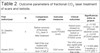
Clinical outcomes in keloids
Only two studies that compared pretreatment and posttreatment keloids were found considering the criteria established for this review. Azzam et al.3 reported a significant difference in the clinical scale scores that they used, with improvement in the treated scar halves (p = 0.006). El-Zawahry et al.7, in turn, reported no improvement in scars, which can be seen in Table 2.
| First author/year | N | Comparison groups | Outcome measures | Clinical outcomes | Significância entre os gruposc |
|---|---|---|---|---|---|
| El- Zawahry, 20157 | 3 | Before sessions of fractional CO2 laser X After treatment | VSS scorea POSAS scoreb |
Keloid scars showed no improvement in texture or Vancouver or POSAS score. | p = 0.102
(VSS) p = 0.180 (POSAS observer score) p = 0.018 (POSAS patient score) |
| Azzam, 20153 | 12 | Half of the scar treated with fractional CO2 laser X Half of the scar left untreated | VSS score | The VSS score was significantly lower in the treated halves than in the untreated halves. | p = 0.006 (after 3
months) p = 0.018 (after 3 months) |
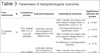
Histopathological outcomes
El-Zawahry et al.7 and Azzam et al.3 reported significant differences in the orientation and density (p = 0.001 and p < 0.05, respectively) of collagen fibers before and after treatment. The latter also reported an increase in the immunohistochemical expression level of MMP9 (p < 0.05). Makboul et al.5 reported greater thickness of the epidermis after treatment (p < 0.001) and decreased immunohistochemical expression of TGF-B1 (p < 0.008). The main histopathological results are shown in Table 3.
| First author/year | N | Comparison groups | Outcome measures | Histological outcomes | Significance between the groupsd |
|---|---|---|---|---|---|
| El-Zawahry, 21057 | 10 | Scara treated with CO2 X Scar left untreated | - Uniformity, density, and orientation of collagen fibers in the ablation areab | - After 3 months: less dense and more aligned collagen fibers in hypertrophic scars (n = 8) | p = 0.001 |
| - Decreased thickness of hypertrophic scars* | p = 0.012 | ||||
| - After 3 months: less dense and more aligned collagen fibers in keloids (n = 2) | p = 0.046 | ||||
| - No difference in average keloid thickness | p = 0.18 | ||||
| Azzam, 20153 | 30 | Scarc treated with CO2 X Scar left untreated | - Uniformity, density, and orientation of collagen fibers in the ablation areab | - After 3 months: less dense and more aligned collagen fibers | p < 0.05 |
| - Immunohistochemical evaluation of MMP9c | - Larger expressionImmunohistochemistry of MMP9 after treatment | p < 0.05 | |||
| Makboul, 20145 | 8 | Scar treatedwith CO2 X Scar left untreated | - Thickness of the epidermis- Presence of TGF-B1 | - Increased thickness after treatment (3 months) | p < 0.001 |
| - Lower immunohistochemical expression (6 months) | p < 0.008 |
a A 2.5-mm punch biopsy (tests: hematoxylin and eosin, Masson trichome for collagen, and Elastica van Gieson for elastic fibers);
b superficial papillary and reticular dermis;
c a 4-mm punch biopsy (tests: hematoxylin and eosin, Masson trichome for collagen, immunohistochemistry for anti-matrix metalloproteinase [MMP9]);
d mean and standard deviation;
* no significant difference between decreased thickness and clinical scores.
DISCUSSION
The treatment of pathological scars is considered to be unpredictable, although it is standardized worldwide. The mechanism of keloid formation and hypertrophic scars, which may help guide treatment, is still under study. The functions of growth factors (TGF-B1) and degradation proteins (MMP9) are still uncertain. Options such as the CO2 laser are important adjuvants in treatment1.
Azzam et al.3 reported clinical improvements in Vancouver scar scale (VSS) scores and histological findings 3 months after treatment with an ablative CO2 laser. They observed more flexible scars and better organized and thinner collagen bundles, with significant increases in MMP9 level after 1 month.
A study that evaluated the effect of fractional ablative CO2 in burn scars reported decreased densities of collagen bundles and changes in the orientation of these fibers through histopathological examination, which clinically contributed to changes in scar texture6.
Another prospective and descriptive study conducted with a sample of 40 scars in a population of 30 patients reported that the use of a combination of Nd:YAG of 1064 nm and fractional CO2 laser at 20 W was significantly effective in improving the vascularization and flexibility of the treated skin, besides reducing itching, only in hypertrophic scars. Moreover, one of the most important effects of the laser on the scar is the generation of heat, which culminates in an inflammatory process that increases vascular permeability, the production of metalloproteinases, and the decomposition of collagen fibers9.
Scar clinical evaluation scales were developed to better understand treatment results, although histopathological analysis of collagen changes and immunohistochemical markers is important for providing scientific evidence15,16.
Although the mechanism of photothermolysis in the treatment of scars is uncertain, the columns produced by thermal injury, characterized by localized epidermal necrosis and denaturation of collagen, initiate a sequence of events that results in a balance between collagenesis and collagenolysis17.
This review selected studies that compared hypertrophic scars and keloids of any nature between before and after treatment with fractional CO2 laser, either from a clinical or histological point of view. Some studies reported a significant improvement in the characteristics and symptoms of scars3,5,6,12-14. Evidence of modification of histopathological characteristics related to collagen, growth factors, and immunohistochemical markers was also reported3,5,6.
The authors argue that fractional CO2 laser should be considered as a promising treatment option for pathological scars. Despite the scarcity of studies with good methodology, this treatment option has been shown to clinically and histologically alter scar tissues, thereby modifying collagen fibers and improving clinical signs and symptoms such as pruritus, color, and thickness.
COLLABORATIONS
|
LEAS |
Analysis and/or data interpretation, conception and design study, conceptualization, data curation, investigation, methodology, project administration, supervision, visualization, writing - original draft preparation, writing - review & editing. |
|
AH |
Analysis and/or data interpretation, conception and design study, conceptualization, final manuscript approval, project administration, supervision. |
|
MF |
Conceptualization, final manuscript approval, project administration, supervision, writing - review & editing. |
|
IC |
Analysis and/or data interpretation, data curation, realization of operations and/or trials, writing - original draft preparation. |
REFERENCES
1. Martin MS, Collawn SS. Combination treatment of CO2 fractional laser, pulsed dye laser, and triamcinolone acetonide injection for refractory keloid scars on the upper back. J Cosmet Laser Ther. 2013;15(3):166-70. DOI: http://dx.doi.org/10.3109/14764172.2013.780448
2. Lorenz P, Sina Bari A. Scar Prevention, treatment, and revision. In: Neligan PC, ed. Plastic Surgery. 3rd ed. Volume 1. Philadelphia: Elsevier Saunders; 2012. p. 297-318.
3. Azzam OA, Bassiouny DA, El-Hawary MS, El Maadawi ZM, Sobhi RM, El-Mesidy MS. Treatment of hypertrophic scars and keloids by fractional carbon dioxide laser: a clinical, histological, and immunohistochemical study. Lasers Med Sci. 2016;31(1):9-18. DOI: http://dx.doi.org/10.1007/s10103-015-1824-4
4. Nowak KC, McCormack M, Koch RJ. The effect of superpulsed carbon dioxide laser energy on keloid and normal dermal fibroblast secretion of growth factors: a serum-free study. Plast Reconstr Surg. 2000;105(6):2039-48. PMID: 10839401 DOI: http://dx.doi.org/10.1097/00006534-200005000-00019
5. Makboul M, Makboul R, Abdelhafez AH, Hassan SS, Youssif SM. Evaluation of the effect of fractional CO2 laser on histopathological picture and TGF-ß1 expression in hypertrophic scar. J Cosmet Dermatol. 2014;13(3):169-79. DOI: http://dx.doi.org/10.1111/jocd.12099
6. Mustoe TA, Cooter RD, Gold MH, Hobbs FD, Ramelet AA, Shakespeare PG, et al.; International Advisory Panel on Scar Management. International clinical recommendations on scar management. Plast Reconstr Surg. 2002;110(2):560-71. PMID: 12142678 DOI: http://dx.doi.org/10.1097/00006534-200208000-00031
7. El-Zawahry BM, Sobhi RM, Bassiouny DA, Tabak SA. Ablative CO2 fractional resurfacing in treatment of thermal burn scars: an open-label controlled clinical and histopathological study. J Cosmet Dermatol. 2015;14(4):324-31. DOI: http://dx.doi.org/10.1111/jocd.12163
8. Campolmi P, Bonan P, Cannarozzo G, Bassi A, Bruscino N, Arunachalam M, et al. Highlights of thirty-year experience of CO2 laser use at the Florence (Italy) department of dermatology. ScientificWorldJournal. 2012;2012:546528. DOI: http://dx.doi.org/10.1100/2012/546528
9. Kaminsky S. Guia Ilustrado - Laser e outras tecnologias em dermatologia. 1ª ed. Rio de Janeiro: Di Livros; 2016.
10. Moher D, Liberati A, Tetzlaff J, Altman DG; PRISMA Group. Preferred reporting items for systematic reviews and meta-analyses: the PRISMA statement. PLoS Med. 2009;6(7):e1000097.
11. van Drooge AM, Vrijman C, van der Veen W, Wolkerstorfer A. A randomized controlled pilot study on ablative fractional CO2 laser for consecutive patients presenting with various scar types. Dermatol Surg. 2015;41(3):371-7. DOI: http://dx.doi.org/10.1097/DSS.0000000000000306
12. Choi JE, Oh GN, Kim JY, Seo SH, Ahn HH, Kye YC. Ablative fractional laser treatment for hypertrophic scars: comparison between Er:YAG and CO2 fractional lasers. J Dermatolog Treat. 2014;25(4):299-303. DOI: http://dx.doi.org/10.3109/09546634.2013.782090
13. Lei Y, Li SF, Yu YL, Tan J, Gold MH. Clinical efficacy of utilizing Ultrapulse CO2 combined with fractional CO2 laser for the treatment of hypertrophic scars in Asians-A prospective clinical evaluation. J Cosmet Dermatol. 2017;16(2):210-6. DOI: http://dx.doi.org/10.1111/jocd.12334
14. Hultman CS, Friedstat JS, Edkins RE, Cairns BA, Meyer AA. Laser resurfacing and remodeling of hypertrophic burn scars: the results of a large, prospective, before-after cohort study, with long-term follow-up. Ann Surg. 2014;260(3):519-29.
15. Sullivan T, Smith J, Kermode J, McIver E, Courtemanche DJ. Rating the burn scar. J Burn Care Rehabil. 1990;11(3):256-60. DOI: http://dx.doi.org/10.1097/00004630-199005000-00014
16. Draaijers LJ, Tempelman FR, Botman YA, Tuinebreijer WE, Middelkoop E, Kreis RW, et al. The patient and observer scar assessment scale: a reliable and feasible tool for scar evaluation. Plast Reconstr Surg. 2004;113(7):1960-5. DOI: http://dx.doi.org/10.1097/01.PRS.0000122207.28773.56
17. Alster T, Zaulyanov L. Laser scar revision: a review. Dermatol Surg. 2007;33(2):131-40.
1. Instituto Israelita de Ensino e Pesquisa Albert
Einstein, São Paulo, SP, Brazil
2. Pontifícia Universidade Católica do Rio Grande
do Sul, Porto Alegre, RS, Brazil.
Corresponding author: Luciana El Halal Schuch Av. Luiz Manoel Gonzaga, nº 187/602 - Porto Alegre, RS, Brazil Zip Code 90470-280 E-mail: luciana_schuch@yahoo.com.br / contato@lucianahalal.com.br
Article received: April 12, 2018.
Article accepted: January 14, 2019.
Conflicts of interest: none.
 Read in Portuguese
 Read in English
 PDF PT
 Print
Print
 Send this article by email
 How to Cite
 Mendeley
 Pocket