


Case Report - Year 2018 - Volume 33 -
The use of digital photography in the follow-up of surgical neoplastic wound: a case report
O uso da fotografia digital na evolução de ferida cirúrgica neoplásica: relato de caso
ABSTRACT
Introduction: The characteristics of cancer wounds are
unusual and are often difficult to describe in writing. Therefore,
digital photography may be used to describe and manage
these wounds.
Case Report: A young female patient with a
recurring lesion measuring 20 × 18 × 13.7 cm in the left thigh
was diagnosed with synovial sarcoma. Digital photography
was used to facilitate evaluation and treatment of her complex
cancer wound. The patient underwent tumor excision,
dressings, and skin grafting; the follow-up was carried out with
photographic records using a cell phone camera, which were
inputted into the electronic medical record and interpreted
by the medical team.
Conclusion: The use of photographs
was important because it allowed rapid wound evaluation,
could be shared with the team to evaluate the treatment plan,
and was an important tool for legal purposes and teaching.
Keywords: Photographs; Surgical oncology; Scar; Surgical wound; Oncology
RESUMO
Introdução: As feridas oncológicas apresentam características peculiares, muitas vezes
difíceis de expressar no texto escrito. A fotografia digital pode contribuir
na descrição e na assistência destas feridas.
Relato do Caso: Relatar o caso de paciente do sexo feminino, jovem, com lesão recidivada
medindo 20x 18x13,7cm na coxa esquerda e diagnóstico de sarcoma sinovial.
Utilizou-se a fotografia digital como facilitador na avaliação e tratamento
de ferida oncológica complexa. A paciente foi submetida à exérese do tumor,
curativos e enxertia de pele; o acompanhamento foi realizado com registros
fotográficos com câmera do telefone celular, inserido no prontuário
eletrônico e proporcionou a fotointerpretação pela equipe de saúde.
Conclusão: O uso da fotografia foi importante, pois permitiu a avaliação rápida da
ferida, pôde ser transmitida à equipe para avaliação do plano de tratamento
e foram importantes ferramentas legais e para o ensino.
Palavras-chave: Fotografias; Oncologia cirúrgica; Cicatriz; Ferida cirúrgica; Oncologia
INTRODUCTION
A total of 600,000 new cancer cases are estimated to be diagnosed in 2018-2019 in Brazil1. Cancer formation results from the carcinogenic process, also known as oncogenesis, which is responsible for uncontrolled cell proliferation. A tumor only becomes visible after several stages, including loss of cutaneous integrity and infiltration of malignant cells into the skin structures, forming neoplastic wounds2. Among the predicted 600,000 new cancer cases, approximately 5–10% of patients will have tumor wounds, a more common condition in patients undergoing palliative care, which also increases due to primary tumors, secondary tumors, or recurring disease3,4.
Therefore, the team must treat these lesions appropriately to maintain patient comfort or, at least, minimize the discomfort resulting from this type of injury. Thus, these professionals seek to devise strategies that allow more accurate record of patient history, aiding in the development of an effective treatment plan5,6.
Therefore, technologies are commonly incorporated in patient care. Among them, digital photography is used for the documentation that facilitates recording the cicatricial evolution of the wounds. This practice positively facilitates proper communication among the team members, affecting the quality of care, providing support for the professional decision-making, and minimizing time and costs of treatment and patient suffering7,8.
In a systematic review on the methods used to evaluate wounds performed by Jørgensen et al.9, digital planimetry and imaging were considered the most accurate and reliable methods in measuring specific areas, especially large and irregular wounds. Until now, three-dimensional technologies have not yet been able to overcome this method due to their low accuracy, high cost, handling complexity, and difficult system interpretation.
OBJECTIVE
To report a case that used digital photography as a facilitator in the evaluation and treatment of a complex cancer wound
METHODS
This case report is conducted by evaluating the data extracted from an electronic medical record. Photographic records obtained during the treatment of a tumor wound between November 2017 and March 2018 were analyzed. The research was approved by the Research Ethics Committee of the Association of Social Pioneers, with the Certificate of Presentation for Ethics Evaluation no. 90589618.6.0000.0022.
The photographic records were obtained using digital cameras of cell phones in the nursing team. The images were taken during dressing procedures, with the patient in the supine position, accommodated in a ward with ambient light. Data were analyzed and correlated to the number of media attached to the electronic patient record; clinical, photographic, and imaging conditions; scenario; and materials used for the assessment and analysis. Qualitative and quantitative data on the affected region, dimensions, appearance, solution of continuity, three-dimensional volume, and presence of necrosis and granulation tissues were obtained using the images.
CASE REPORT
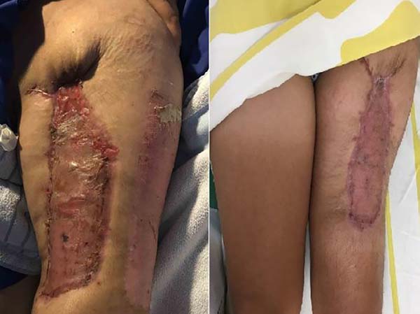
A 31-year-old woman was admitted with a history of left thigh lesion 9 years ago. She has undergone two surgical procedures for tumor excision in another medical service, without confirmation of the diagnosis. She had recurrence and was admitted to our hospital. The physical examination revealed an infiltrative, expansive lesion measuring 20.0 × 18.3 cm on its larger dimensions (Figure 1).
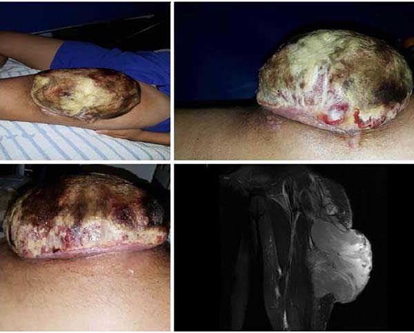
The biopsy and histopathological examination showed a grade 3 biphasic synovial sarcoma classified by the Federation Nationale de Centers de Lutte Contre le Cancer, associated with immunohistochemical and cytogenetic study. Nuclear magnetic resonance imaging in the left thigh at admission (Figure 1) revealed an expansive lesion with aggressive features, and infiltrative appearance, affecting the anterior compartment muscles in the left thigh, with skin ulceration, and bulk tumor component through the cutaneous discontinuity area. The finding has a heterogeneous signal, which possibly delimits areas with hypersignal, probably secondary to necrosis.
No signs of bone involvement were found, the part of the non-compromised muscles was interposed between the femoral cortical and tumor lesion. Two satellite nodular lesions were found near the upper margin of the lesion, with similar signal characteristics. The lesion measured, in its largest diameters, 20.0 × 18.3 × 13.7 cm, and the satellite lesions measured 3.6 × 3.4 × 2.4 cm and 4.3 × 3.3 × 2.9 cm. Small lymph nodes were observed in the left inguinal region and were partially evaluated during the examination.
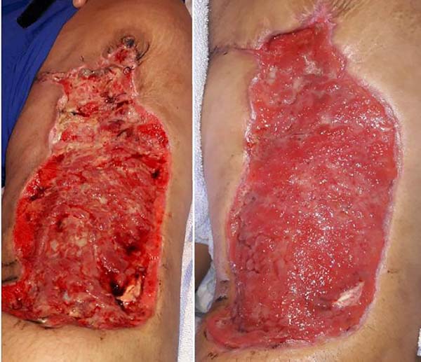
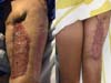
Computed tomography of the chest was normal. The patient underwent wide tumor excision in the left thigh. Follow-up was conducted with daily dressings, and reconstruction with partial skin graft was performed in the follow-up.
A digital photograph was taken upon admission, which serves as the baseline record of the image, identifying the cancer wound, such as the region affected, dimensions, appearance, skin continuity solution, three-dimensional volume, and presence of necrosis and granulation tissue (Chart 1). It also allowed correlation with clinical examination, magnetic resonance imaging, and histopathological examination, providing opportunities for all team members to evaluate the image at any time for descriptive and comparative analysis. Changes in the treatment plan should be guided during the follow-up or dressings (Figures 2–4).
| Quantitative aspects | Size |
| Continuity | |
| Circumference | |
| Color | |
| Qualitative aspects | Improvement |
| Worsening | |
| Stable |


DISCUSSION
Visual communication has been an important tool from the earliest days of human history, even before writing. We have tried to improve the communicating methods through imaging, allowing the observer to understand what they see, absorb the image, associate with their previous knowledge, and process it in order to draw their own conclusions.
The photo interpretation consisted of qualitative and quantitative data analysis of the photographic image considering the criteria presented in Chart 18. The analysis of photographic records facilitated obtaining and evaluating the image, mainly because the object could not be touched and the wound was complex. The acquisition of images according to photographs has been used by plastic surgeons for a long time for pre-, trans-, and postoperative records10.
Many authors propose standardizing photograph procedures to obtain images that improve the evaluation of results11. Tavares et al.12 described the need for photographs and reports in the patient’s medical records in order to document their diagnoses, treatments, and progress. Taking this tool into consideration, besides being a duty, it is a source of scientific knowledge, and a great ally in the potential juridical processes; besides, the authors described a technique in taking transoperative photographs.
The images presented in this study were easily acquired by the professional during the dressing procedure, and the images could be shared with all the team members that managed the patient. Additionally, these images can be used as an important tool in changing the treatment plan, such as the products used during dressings, the time to perform skin grafting, and the wound conditions after skin grafting Moreover, the images were used as a teaching material and as a legal document.
CONCLUSION
The use of photography significantly facilitates the evaluation of a complex wound healing, allows taking images that facilitate the diagnosis, measurements, and surgical programming. It assisted all team members during the follow-up and intervention. Furthermore, data acquisition was rapid, could be shared, and facilitated interdisciplinary communication, which allowed designing strategic plans for the treatment, and are considered legal document tools as well as for teaching purposes.
REFERENCES
1. Brasil. Ministério da Saúde. Instituto Nacional de Câncer José Alencar Gomes da Silva (INCA). Estimativa 2018: Incidência de Câncer no Brasil. Rio de Janeiro: INCA; 2017. [acesso 2018 Mar 23]. Disponível em: http://www.inca.gov.br/estimativa/2018/estimativa-2018.pdf
2. Brasil. Ministério da Saúde. Instituto Nacional de Câncer José Alencar Gomes da Silva (INCA). Tratamento e controle de feridas tumorais e úlceras por pressão no câncer avançado. Rio de Janeiro: INCA; 2009.
3. Silva KRM, Bontempo PSM, Reis PED, Vasques CI, Gomes IP, Simino GPR. Intervenções Terapêuticas em Feridas Tumorais: Relato de Casos. Rev Bras Cancerol. 2015;61(4):373-9.
4. Agra G, Fernandes MA, Platel ICS, Freire MEM. Cuidados Paliativos ao Paciente Portador de Ferida Neoplásica: uma Revisão Integrativa da Literatura. Rev Bras Cancerol. 2013;59(1):95-104.
5. Sacramento CJ, Reis PED, Simino GPR, Vasques CI. Manejo de Sinais e Sintomas em Feridas Tumorais: Revisão Integrativa. R Enferm Cent O Min. 2015;5(1):1514-27.
6. Adderley U, Smith R. Topical agents and dressings for fungating wounds. Cochrane Database Syst Rev. 2007;(2):CD003948.
7. Gardona RGB, Ferracioli MM, Salomé GM, Pereira MTDJ. Avaliação da qualidade dos registros dos curativos em prontuários realizados pela enfermagem. Rev Bras Cir Plást. 2013;28(4):686-92.
8. Faria NGF, Peres HHC. Análise da produção científica sobre documentações fotográficas de feridas em enfermagem. Rev Eletr Enferm. 2009;11(3):704-11.
9. Jørgensen LB, Sørensen JA, Jemec GB, Yderstraede KB. Methods to assess area and volume of wounds - a systematic review. Int Wound J. 2016;13(4):540-53.
10. Stocchero IN, Torres FC. Fotografia digital em cirurgia plástica. São Paulo: LMP Editora; 2005.
11. Coombes AG, Sethi CS, Kirkpatrick WN, Waterhouse N, Kelly MH, Joshi N. A standardized digital photography system with computerized eyelid measurement analysis. Plast Reconstr Surg. 2007;120(3):647-56.
12. Tavares MV, Cotta FB, Corrêa AG, Gomes RCB, Barros VM, Maia MR, et al. Documentação fotográfica intra-operatória. Rev Bras Cir Plást. 2010;25(4):705-7.
1. Rede Sarah de Hospitais de reabilitação,
Brasília, DF, Brazil.
Corresponding author: Barbara Braga Cavalcante, SMHS, 501 Bloco A - Brasília, DF, Brazil, Zip Code 70335-901. E-mail: 13516@sarah.br
Article received: September 21, 2018.
Article accepted: October 4, 2018.
Conflicts of interest: none.
 Read in Portuguese
 Read in English
 PDF PT
 Print
Print
 Send this article by email
 How to Cite
 Mendeley
 Pocket