


Reviw Article - Year 2018 - Volume 33 -
Use of stereophotogrammetry for measuring the volume of external facial anatomy: a systematic review
Uso da estereofotogrametria para mensuração do volume da anatomia externa da face: revisão sistemática
ABSTRACT
Introduction: Photographic documentation is important
in several medical specialties, such as plastic surgery.
Two-dimensional photography has limitations in capturing
structure depth and volume, requiring other instruments
to evaluate these changes. Several technologies have been
developed for three-dimensional analysis of objects, of
which stereophotogrammetry uses computerized analysis
of two or more simultaneous photographs of the object to
produce a three-dimensional geometric model. The advantages
of stereophotogrammetry include lower cost, portability,
absence of radiation, and speed of image capture. The
aim of the present study was to perform a bibliographic
review evaluating the use and accuracy of stereophotogrammetry
for measuring the volume of facial structures.
Methods: Using a combination of MeSH keywords and free
terms, a search was performed in the Cochrane Library
and MEDLINE databases. The search included all articles
published on or before May 2018.
Results: 2,213 studies
were initially retrieved using this search strategy. Of these,
27 studies were selected based on the eligibility criteria,
of which 21 were non-randomized case studies and 6 were
randomized clinical trials. The methodological quality of
the studies varied between 50 and 67%, on a grading scale
from 0 to 100%.
Conclusions: Stereophotogrammetry is
a promising technology that is increasingly being used to
check for facial volume variations with high accuracy and
reproducibility. More studies with higher methodological
quality are needed for evaluating the accuracy and use
of stereophotogrammetry for facial volume evaluations.
Keywords: Three-dimensional image; Photogrammetry; Face; Organ size
RESUMO
Introdução: O registro fotográfico tem sido importante para diversas especialidades
médicas como a Cirurgia Plástica. A fotografia em duas dimensões apresenta
limitações para capturar profundidade e volume de estruturas outros
instrumentos para avaliar essa alteração. Diversas tecnologias foram
desenvolvidas para analisar objeto em três dimensões, sendo a
estereofotogrametria uma tecnologia que utiliza a análise computadorizada de
duas ou mais fotografias simultâneas do objeto para produzir um modelo
geométrico em três dimensões. As vantagens da estereofotogrametria incluem
menor custo, portabilidade, ausência de radiação e rapidez da captura das
imagens. O objetivo deste trabalho foi realizar uma revisão bibliográfica
avaliando o uso e a acurácia da estereofotogrametria para mensuração de
volume de estruturas na face.
Métodos: Foi realizada pesquisa nos bancos de dados Cochrane Library e Medline até
maio de 2018 utilizando uma combinação de descritores Mesh e termos
livres.
Resultados: Foram obtidos inicialmente 2213 estudos observando a estratégia de busca.
Seguindo os critérios de elegibilidade, foram selecionados 27 artigos, sendo
21 relatos de casos não randomizados e 6 ensaios clínicos randomizados. A
qualidade metodológica dos estudos variou de 50 a 67%, segundo uma pontuação
que vai de 0 a 100%.
Conclusões: A estereofotogrametria é uma tecnologia promissora e tem sido cada vez mais
utilizada para verificar variações de volume na face com alta acurácia e
reprodutibilidade. Faltam estudos com melhor qualidade metodológica
avaliando a acurácia e o uso da estereofotogrametria na avaliação de volume
facial.
Palavras-chave: Imagem tridimensional; Fotogrametria; Face; Tamanho do órgão
INTRODUCTION
Photographic documentation has been important for several medical specialties, such as plastic surgery, dermatology, and head and neck surgery, to name a few. Photography is used to document pre- and post-operative events and, through measurements of distances and angles, surgical planning. 1.
Two-dimensional (2D) photography has limitations when used for capturing depth and volume of three-dimensional (3D) structures. Procedures involving volume changes after surgery, therefore, require other instruments to evaluate those changes, through comparison of photographs2,3.
Several technologies have been developed for the analysis of 3D objects. These technologies can be divided into those emitting radiation, such as computerized tomography, and those that do not emit radiation, such as 3D cephalometry, Moire topography, 3D laser scanning, and stereophotogrammetry4-7.
Due to advantages such as lower cost, portability, harmlessness, faster image capturing, and storage and software processing, technologies that do not emit radiation, especially 3D laser scanning and stereophotogrammetry, have increasingly been used to obtain 3D images7,8.
Stereophotogrammetry has advantages over 3D laser scanning in that it is more portable, it has the ability to capture an object’s color and texture, and there is no need to protect the patient’s eyes during its use9. Stereophotogrammetry was first described by Thalmann in 1944, who attempted to capture a 3D image of a face. In 1967, it was improved and simplified by Burke and Beard. In 1995, Ras and collaborators concluded that it was adequate to obtain 3D records of changes in facial morphology. Subsequently, Deacon et al. in 1999 improved the stereophotogrammetry technique by using digitalized images and analysis software5.
Stereophotogrammetry uses two or more simultaneous photographs of an object to produce a 3D geometric model, after software analysis. The software produces 3D images based on the difference between the photographs at a known angle between cameras. Color and texture are subsequently added.
Two types of triangulation algorithms can be used in stereophotogrammetry: Passive and active. The active type involves projecting an unstructured light pattern (visible or infrared) on the object’s surface, whereas the passive uses only the natural ambient light pattern, instead of projected light1,3,5,10.
Active stereophotogrammetry, therefore, has the advantage of depending less on external light to obtain images; however, the surroundings need to be dark when photographs are taken. Despite depending on external light for obtaining images, most available equipment in the market still use passive stereophotogrammetry.
Equipment that use passive stereophotogrammetry technology have been shown to be more flexible for use in doctors’ offices and hospitals, and it has been the technology of choice in many companies using 3D photography. There is also a hybrid equipment that uses both passive and active stereophotogrammetry11-14.
Stereophotogrammetry has great potential to become the standard method for evaluating volumes and distances in facial and body anatomy. Regarding the face, it can be used to evaluate the fill volume necessary for a given wrinkle, post-surgery edemas or estimate volume increases obtained after aesthetic and surgical procedures11,12.
OBJECTIVE
The objective of the present study was to perform a bibliographic review evaluating the use and accuracy of stereophotogrammetry equipment to measure the volume of facial structures.
METHODS
Search strategy for study identification
A search, including studies published on or before May 2018, was performed in the Cochrane Library and MEDLINE virtual databases.
The search in MEDLINE was performed using a combination of free terms and MeSH keywords such as [three-dimensional imaging (MeSH Terms)], [photogrammetry (MeSH Terms)], [face (MeSH Terms)], and the Cochrane Sensitivity Maximizing version strategy (Figure 2, Appendix). The search in the Cochrane Library database was performed using free terms, following the strategy presented in Figure 3 (Appendix). No restrictions were placed on the basis of the study language.
Inclusion and exclusion criteria:
Studies that evaluated the use of stereophotogrammetry to measure the volume of facial structures in humans were included. Studies that used technology unavailable commercially or studies that were reviews or response articles were excluded. The studies retrieved were divided into two groups: Randomized and non-randomized studies.
Two independent researchers read the titles and abstracts and then selected the studies according to the eligibility criteria. A consensus reached by the two researchers was used to solve disagreements on study inclusion. The risk of bias in this study was evaluated using a similar tool to the one used by Cochrane Collaboration.
RESULTS
A total of 2,213 studies were retrieved using the search strategy presented in Figure 1. After reading the titles and/or the abstracts, 2,051 studies were excluded based on the eligibility criteria. A total of 162 studies were selected for full reading and, following selection, 27 studies were included in the study, of which 21 were uncontrolled unblinded non-randomized case studies4,8,12-29 and 6 were randomized clinical trials10,30-34.


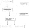
Of the 162 studies, 87 were excluded because facial parameters other than volume were evaluated, 27 studies were excluded because they used technologies other than stereophotogrammetry, 10 studies were excluded because they were reviews, 8 studies were excluded because they were letters to the editor or response letters, and 3 studies were excluded because mannequins or cadavers were used.
Of the 21 case studies, 17 used stereophotogrammetry to evaluate the differences in facial volume before and after an intervention (surgery, fillings, fat graph), 3 used stereophotogrammetry to evaluate the differences in facial volume overtime (aging, monitoring of hemangioma), and 1 aimed at evaluating the reliability of stereophotogrammetry.
A total of 703 individuals were evaluated in the case studies, of which 264 (37.55%) were men and 397 (56.47%) were women, while 42 (5.97%) did not report their gender. The average age was 38.48 years (range; 4 months to 91 years). The average monitoring time was 11.07 months (range; 1 to 24 months).
A total 219 individuals (111 patients and 108 controls) were analyzed in the 6 randomized studies, of which 132 (60.27%) were women, 61 (27.85%) were men, while 26 (11.87%) did not report their gender. The average age was 32.83 (range; 20 to 55 years) and average monitoring time was 6.59 months (range; 7 days to 18 months). These studies aimed at using stereophotogrammetry to evaluate differences in facial volumes between a group subjected to an intervention (such as surgery, filling, or use of devices to decrease post-operative swelling) and control groups.
The methodological quality of the studies varied between 50 and 67%, on a grading scale from 0 to 100% (Table 1).
| Study | A | B | C | D | Absolute | Relative |
|---|---|---|---|---|---|---|
| Hans- Joachim Niekenig 2014 | 2 | 1 | 0 | 1 | 4 | 67% |
| Maieed Rana 2012 | 1 | 1 | 0 | 1 | 3 | 50% |
| M. Rana 2011 | 1 | 1 | 0 | 1 | 3 | 50% |
| Jeff Downie 2009 | 1 | 2 | 0 | 1 | 4 | 67% |
| Kyung Suk Koh 2012 | 1 | 0 | 0 | 1 | 3 | 50% |
| Majeed Rana 2011 | 1 | 1 | 0 | 1 | 3 | 50% |
Items from the metodogical qualification [ score]: (A) allocation [2-randomization with defined metodology ; 1- randomization without defined metodology; 0- partially randomized]; (B) Concealment of allocation [2- double-blind; 1-simple-blind; 0-non-blind or not defined]; (C) previous calculation of the sample size [1- described previously; 0-non-described] and (D) external validation [1- inclusion and exclusion criteria defined; 0- criteria for inclusion and/or exclusion].

DISCUSSION
Although many studies showed the use of stereophotogrammetry in medical practice, relatively few described its usefulness for measuring facial volumes. Moreover, many studies mentioned other 3D photography technologies, evaluated regions other than the face, or measured parameters other than volume.
Most studies used stereophotogrammetry to evaluate volume differences before and after an intervention, follow the evolution of diseases such as changes in hemangioma volume, or evaluate its reliability when compared with direct volume measurements. The scarcity of double-blinded randomized studies should be noted: Only 6 out of the 2,213 initially retrieved studies were double-blinded.
Of the 6 double-blinded randomized trials, three studies31,32,34 compared two methods of cooling the inferior third of the faces of patients subjected to orthognathic or odontological procedures. In these studies, stereophotogrammetry was used to determine which cooling method caused a lesser post-operative volume increase, and, therefore, had a higher efficacy in decreasing post-operative edema.
The methodology was very similar in the 3 studies in that the same facial cooling and stereophotogrammetry equipment (FaceScan3D) was used. Only the type of surgery performed differed. In all the three studies, subject allocation was randomized and the observers were blinded with regards to the use of cooling equipment.
One study used stereophotogrammetry (Di3D) to evaluate the volume gain obtained using four different types of facial fill10. Similar to the other three studies, the subjects were allocated randomly, and subjects and observers were blinded to the type of fill used.
Another study evaluated post-operative edema using stereophotogrammetry (CAM3D), comparing two types of oral and maxillofacial surgical procedures30. Evaluations, done by measuring lower facial volumes using stereophotogrammetry, were used to determine the surgical strategy that caused less post-operative edema. Still, subjects were allocated randomly and observers were blinded.
Finally, one study evaluated survival in subjects who underwent facial fat grafting with or without the use of fat mesenchymal stem cells 33. Fat grafting survival was monitored by measuring the maintenance of volume gain using stereophotogrammetry (Vectra 3D) during post-operative follow-up. Group allocation was randomized but there was no mention as to whether observer or patient blinding was done.
Stereophotogrammetry was observed to be more reliable for volume analysis of inanimate objects than of living beings. It was concluded that this difference could be explained by the effect of muscle contraction and face animation on soft tissues. Despite this difference in reliability, there were no significant intraclass differences in coefficients. This indicated good method reproducibility8.
The accuracy of measurements using the equipment’s software is thought to be dependent on the operator. To avoid this operator bias, appropriate pre-operative volumes were obtained for proper comparison with the volumes measured after surgery. In addition, it is important to avoid facial animation or head rotations19; the patient should have a neutral expression, with closed mouth and lips4,7.
The disadvantages of stereophotogrammetry include the lack of portability of some of the equipment and the need for image analysis using a software that may not be common users. Another disadvantage is that it is difficult to tell whether volume variations in children are due to the intervention or due to growth. Meanwhile, in adults, very small volume variations may be attributed to changes such as edema that could, in reality, be unrelated to the intervention18,22,25.
Another important consideration is whether stereophotogrammetry allows for the calculation of volume in regions with hair, cavities or depressions, such as the sub-nasal and submental regions. Failing to perfectly align the pre- and post-operative photos may also cause errors. Objects that reflect light, such as jewels, may cause photographic artifacts. It is therefore recommended that patients secure their hair and remove jewels and other ornaments. The high cost of the 3D photography equipment, which may vary somewhere between U$15,000 and U$35,0000, is a limiting factor for the availability and use of this technology35.
As validated in previous studies, stereophotogrammetry has good accuracy and reproducibility for the measuring of facial distances and volumes 3,26,29,36,37.
Studies have shown that the most commonly used equipment available in the market have high accuracy. For example, 3dMD, Vectra, and Di3D systems have shown an average error of 2%, 1.2%, and 1%, and a coefficient of reproducibility of 0.80, 1 and 0.13, respectively5,38.
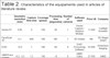
A limitation of the present study is that the studies for analysis were retrieved from only two databases (PubMed and Cochrane) and the majority of studies were of low quality: Only 2 of the 6 studies scored higher than 50% according to the methodological qualification used (Table 1). Also, the present review lacks reliable studies. Of the 6 randomized studies, 3 analyzed the same facial cooling device. Worthy of note is that the stereophotogrammetry equipment differs with regards to software, accuracy, reproducibility, and ease of use (Table 2).
| Geometric resolution (mm) | Capture time (ms) | Coverage (graus) | Processing time (segundos) | Number of cameras | Software used | Price U$ | Company | |
|---|---|---|---|---|---|---|---|---|
| Vectra H1 | 0,95 | 2 | 100 | 20 | 1 | Vectra Capture / Analysis Module | 11.000 | Canfleld Scientific |
| FaceScan 3D | 0,1 | 800 | 180 | ND | 1 | 3D Viewer | NA | 3D-Shape GmbH |
| Di3D | <0,2 | 1 | 180 | 60 | 4 | Di4D Processing Software | 35.000 | Dimension Imaging |
| 3dMDFace System | <0,2 - 0,5 | 1,5 | 190 | <8 | 6 | 3dMD Vultus Software | 27.000 | 3dMD |
CONCLUSION
Stereophotogrammetry is a promising technology that is being increasingly used to evaluate facial volume variations in patients before and after surgery or to monitor the evolution of facial diseases that may involve volume changes.
Measuring facial volume using this technology had high inter- and intra-operator accuracy and reproducibility in the reviewed studies.
Although a good number of studies were retrieved, studies with better methodological quality that evaluate stereophotogrammetry accuracy and use for evaluation of facial volumes are still lacking.
COLLABORATIONS
|
REM |
Data analysis and/or interpretation; final manuscript approval; data collection; conceptualization; conception and design of the study; project management; methodology; writing- preparation of the original manuscript; writing - revision and editing; supervision; visualization. |
|
SM |
Writing- preparation of the original manuscript; writing - revision and editing; supervision; validation. |
|
JLB |
Data analysis and/or interpretation; data collection. |
|
LHM |
Data analysis and/or interpretation; data collection. |
REFERENCES
1. Knoops PG, Beaumont CA, Borghi A, Rodriguez-Florez N, Breakey RW, Rodgers W, et al. Comparison of three-dimensional scanner systems for craniomaxillofacial imaging. J Plast Reconstr Aesthet Surg. 2017;70(4):441-9.
2. Meier JD, Glasgold RA, Glasgold MJ. 3D photography in the objective analysis of volume augmentation including fat augmentation and dermal fillers. Facial Plast Surg Clin North Am. 2011;19(4):725-35.
3. Lane C, Harrell W Jr. Completing the 3-dimensional picture. Am J Orthod Dentofacial Orthop. 2008;133(4):612-20.
4. van Loon B, Maal TJ, Plooij JM, Ingels KJ, Borstlap WA, Kuijpers-Jagtman AM, et al. 3D Stereophotogrammetric assessment of pre- and postoperative volumetric changes in the cleft lip and palate nose. Int J Oral Maxillofac Surg.2010;39(6):534-40.
5. Tzou CH, Frey M. Evolution of 3D surface imaging systems in facial plastic surgery. Facial Plast Surg Clin North Am. 2011;19(4):591-602.
6. Modabber A, Peters F, Kniha K, Goloborodko E, Ghassemi A, Lethaus B, et al. Evaluation of the accuracy of a mobile and a stationary system for three- dimensional facial scanning. J Craniomaxillofac Surg. 2016;44(10):1719-24.
7. Heike CL, Upson K, Stuhaug E, Weinberg SM. 3D digital stereophotogrammetry: a practical guide to facial image acquisition. Head Face Med. 2010;6:18.
8. van der Meer WJ, Dijkstra PU, Visser A, Vissink A, Ren Y. Reliability and validity of measurements of facial swelling with a stereophotogrammetry optical three-dimensional scanner. Br J Oral Maxillofac Surg. 2014;52(10):922-7.
9. Toriumi DM, Dixon TK. Assessment of rhinoplasty techniques by overlay of before-and-after 3D images. Facial Plast Surg Clin North Am. 2011;19(4):711-23.
10. Downie J, Mao Z, Rachel Lo TW, Barry S, Bock M, Siebert JP, et al. A double-blind, clinical evaluation of facial augmentation treatments: a comparison of PRI 1, PRI 2, Zyplast and Perlane. J Plast Reconstr Aesthet Surg. 2009;62(12):1636-43.
11. Tzou CH, Artner NM, Pona I, Hold A, Placheta E, Kropatsch WG, et al. Comparison of three-dimensional surface-imaging systems. J Plast Reconstr Aesthet Surg. 2014;67(4):489-97.
12. Camp MC, Wong WW, Filip Z, Carter CS, Gupta SC. A quantitative analysis of periorbital aging with three-dimensional surface imaging. J Plast Reconstr Aesthet Surg. 2011;64(2):148-54.
13. Meier JD, Glasgold RA, Glasgold MJ. Autologous fat grafting: long-term evidence of its efficacy in midfacial rejuvenation. Arch Facial Plast Surg. 2009;11(1):24-8.
14. Luebberding S, Krueger N, Kerscher M. Comparison of Validated Assessment Scales and 3D digital fringe projection method to assess lifetime development of wrinkles in men. Skin Res Technol. 2014;20(1):30-6.
15. Schendel SA. Enriched autologous facial fat grafts in aesthetic surgery: 3D volumetric results. Aesthet Surg J. 2015;35(8):913-9.
16. Mailey B, Baker JL, Hosseini A, Collins J, Suliman A, Wallace AM, et al. Evaluation of Facial Volume Changes after Rejuvenation Surgery Using a 3- Dimensional Camera. Aesthet Surg J. 2016;36(4):379-87.
17. Nelson L, Stewart KJ. Experience in the treatment of HIV-associated lipodystrophy. J Plast Reconstr Aesthet Surg. 2008;61(4):366-71.
18. Guibert M, Franchi G, Ansari E, Billotet B, Diner PA, Cassier S, et al. Fat graft transfer in children's facial malformations: a prospective three-dimensional evaluation. J Plast Reconstr Aesthet Surg. 2013;66(6):799-804.
19. Gerth DJ, King B, Rabach L, Glasgold RA, Glasgold MJ. Long-term volumetric retention of autologous fat grafting processed with closed-membrane filtration. Aesthet Surg J. 2014;34(7):985-94.
20. Nkenke E, Eitner S, Radespiel-Tröger M, Vairaktaris E, Neukam FW, Fenner M. Patient-centred outcomes comparing transmucosal implant placement with an open approach in the maxilla: a prospective, non-randomized pilot study. Clin Oral Implants Res. 2007;18(2):197-203.
21. van der Vlis M, Dentino KM, Vervloet B, Padwa BL. Postoperative swelling after orthognathic surgery: a prospective volumetric analysis. J Oral Maxillofac Surg. 2014;72(11):2241-7.
22. Donath AS, Glasgold RA, Meier J, Glasgold MJ. Quantitative evaluation of volume augmentation in the tear trough with a hyaluronic Acid-based filler: a three-dimensional analysis. Plast Reconstr Surg. 2010;125(5):1515-22.
23. Chen HH, Javadi P, Daines SM, Williams EF, 3rd. Quantitative assessment of the longevity of poly-L-lactic acid as a volumizing filler using 3-dimensional photography. JAMA Facial Plast Surg. 2015;17(1):39-43.
24. Bernstein EF, Bloom JD. Safety and Efficacy of Bilateral Submental Cryolipolysis With Quantified 3-Dimensional Imaging of Fat Reduction and Skin Tightening. JAMA Facial Plast Surg. 2017;19(5):350-7.
25. Jacono AA, Malone MH, Talei B. Three-Dimensional Analysis of Long-Term Midface Volume Change After Vertical Vector Deep-Plane Rhytidectomy. Aesthet Surg J. 2015;35(5):491-503.
26. van Loon B, van Heerbeek N, Bierenbroodspot F, Verhamme L, Xi T, de Koning MJ, et al. Three-dimensional changes in nose and upper lip volume after orthognathic surgery. Int J Oral Maxillofac Surg. 2015;44(1):83-9.
27. Hermans DJ, Maal TJ, Bergé SJ, van der Vleuten CJ. Three-dimensional stereophotogrammetry: a novel method in volumetric measurement of infantile hemangioma. Pediatr Dermatol. 2014;31(1):118-22.
28. Nada RM, van Loon B, Schols JG, Maal TJ, de Koning MJ, Mostafa YA, et al. Volumetric changes of the nose and nasal airway 2 years after tooth-borne and bone-borne surgically assisted rapid maxillary expansion. Eur J Oral Sci. 2013;121(5):450-6.
29. Yip E, Smith A, Yoshino M. Volumetric evaluation of facial swelling utilizing a 3-D range camera. Int J Oral Maxillofac Surg. 2004;33(2):179-82.
30. Nickenig HJ, Wichmann M, Zöller JE, Eitner S. 3-D based minimally invasive one-stage lateral sinus elevation - a prospective randomized clinical pilot study with blinded assessment of postoperative visible facial soft tissue volume changes. J Craniomaxillofac Surg. 2014;42(6):890-5.
31. Rana M, Gellrich NC, von See C, Weiskopf C, Gerressen M, Ghassemi A, et al. 3D evaluation of postoperative swelling in treatment of bilateral mandibular fractures using 2 different cooling therapy methods: a randomized observer blind prospective study. J Craniomaxillofac Surg. 2013;41(1):e17-23.
32. Rana M, Gellrich NC, Joos U, Piffkó J, Kater W. 3D evaluation of postoperative swelling using two different cooling methods following orthognathic surgery: a randomised observer blind prospective pilot study. Int J Oral Maxillofac Surg. 2011;40(7):690-6.
33. Koh KS, Oh TS, Kim H, Chung IW, Lee KW, Lee HB, et al. Clinical application of human adipose tissue-derived mesenchymal stem cells in progressive hemifacial atrophy (Parry-Romberg disease) with microfat grafting techniques using 3-dimensional computed tomography and 3-dimensional camera. Ann Plast Surg. 2012;69(3):331-7.
34. Rana M, Gellrich NC, Ghassemi A, Gerressen M, Riediger D, Modabber A. Three-dimensional evaluation of postoperative swelling after third molar surgery using 2 different cooling therapy methods: a randomized observer-blind prospective study. J Oral Maxillofac Surg. 2011;69(8):2092-8.
35. Lekakis G, Claes P, Hamilton GS, 3rd, Hellings PW. Three-Dimensional Surface Imaging and the Continuous Evolution of Preoperative and Postoperative Assessment in Rhinoplasty. Facial Plast Surg. 2016;32(1):88-94.
36. Fourie Z, Damstra J, Gerrits PO, Ren Y. Evaluation of anthropometric accuracy and reliability using different three-dimensional scanning systems. Forensic Sci Int. 2011;207(1-3):127-34.
37. Codari M, Pucciarelli V, Pisoni L, Sforza C. Laser scanner compared with stereophotogrammetry for measurements of area on nasal plaster casts. Br J Oral Maxillofac Surg. 2015;53(8):769-70.
38. Winder RJ, Darvann TA, McKnight W, Magee JD, Ramsay-Baggs P. Technical validation of the Di3D stereophotogrammetry surface imaging system. Br J Oral Maxillofac Surg. 2008;46(1):33-7.
1. Hospital das Clínicas, Faculdade de Medicina,
Universidade de São Paulo, São Paulo, SP, Brazil.
Corresponding author: Ricardo Eustachio de Miranda, Rua Bandeira Paulista, nº 530, sala 43 - Itaim Bibi - São Paulo, SP, Brazil, Zip Code 04532-001. E-mail: ricardomiranda@hotmail.com
Article received: June 24, 2018.
Article accepted: October 4, 2018.
Conflicts of interest: none.
 Read in Portuguese
 Read in English
 PDF PT
 Print
Print
 Send this article by email
 How to Cite
 Mendeley
 Pocket