


Original Article - Year 2018 - Volume 33 -
Breast measurement using Adobe Photoshop®
Medição das mamas com o Adobe Photoshop®
ABSTRACT
INTRODUCTION: With the globalization and improvement of digital media and, consequently, the increasing use of digital photography, it is necessary to create reliable measurement methods. In clinical practice, direct measurement using tape measure, ruler, and compass, is most commonly used. However, there are intrinsic (body contour) and extrinsic (patient collaboration or respiratory movements) factors that may influence the data obtained. Photography and graphics software can be an accurate, comfortable, and reproducible alternative to direct measurement. The objective is to demonstrate how to perform breast measurement using Adobe Photoshop software.
METHODS: Participants were described the position to assume. Imaging capturing techniques and software calibration were performed in order to obtain the final breast measurement. Markers were placed on the breast and arms of 40 women volunteers aged 18 to 60 years using an anterior view. The union of these markers in each hemibody resulted in seven linear segments, one angle and one transverse segment common to both hemibodies. These photographs were evaluated as a RAW file, by 3 evaluators, assisted by Adobe Photoshop CS6® software.
RESULTS: Photoshop CS6® proved effective in the breast measurement using RAW files.
CONCLUSION: Photoshop® showed precision and effectiveness for breast measurement.
Keywords: Photogrammetry; Anthropometry; Software; Breast; Body weights and measures.
RESUMO
INTRODUÇÃO: Com a globalização e melhora dos meios digitais e, consequentemente, a crescente utilização de fotografias digitais, faz-se necessária a criação de métodos de mensuração confiáveis. Na prática clínica, a medida direta utilizando-se fita métrica, régua e compasso é mais comumente utilizada. Porém, existem fatores intrínsecos (relevos corporais) e extrínsecos (colaboração do paciente ou movimentos respiratórios) que podem influenciar nos dados obtidos. A fotografia e os softwares gráficos podem ser uma alternativa precisa, mais cômoda e reprodutível à medida direta. O objetivo é demonstrar a forma de medir as mamas pelo software Adobe Photoshop.
MÉTODOS: Foram descritas a posição das voluntárias, a captação das imagens, calibração do software até a obtenção mensuração final das mamas. Em 40 mulheres voluntárias, com idade entre 18 e 60 anos, foram demarcados pontos sobre a região mamária e braços em posição frontal. A união destes pontos em cada hemicorpo formou 7 segmentos lineares, 1 angular e 1 segmento mediano comum a ambos os hemicorpos. Essas fotografias foram avaliadas no arquivo RAW, por 3 observadores, com auxílio do software Adobe Photoshop CS6®.
RESULTADOS: O Photoshop CS6® demonstrou ser eficaz na mensuração das mamas no arquivo RAW.
CONCLUSÃO: O Photoshop® apresentou precisão nas medidas e ser factível para mensuração das mamas.
Palavras-chave: Fotogrametria; Antropometria; Software; Mama; Pesos e medidas corporais.
It is important to perform a thorough breast examination for the diagnosis of any deformity as part of the surgical preparation for procedures in that region. Changes in contour, volume, and prominences in female breasts make them difficult to be properly measured1.
Currently, pre-operative evaluation and documentation using digital photography is performed in nearly all plastic surgery procedures, being an important step to gain awareness of existing body shape and contours and, as a consequence, to provide information about the limitations of the surgical procedure, so that unrealistic expectations or postoperative doubts do not arise.
Sometimes, each individual's natural asymmetry is not perceptible by itself until it is presented in a clear manner2. In addition to the physical examination, a standardized photographic analysis is required mainly due to the achieved reproducibility, an essential aspect to the scientific rigor required in academia3.
Breast evaluation can be performed by direct measurement using a ruler, tape measure, or compass, or indirectly, using measurements performed on an image, which can be printed or in digital form, the latter being performed using graphics software.
Comparison studies between direct and indirect anthropometry began with the face and nose. Ward4, in 1979, compared nose measurements and found that there was no difference between those obtained by photogrammetry and direct anthropometry. In this context, Nechala et al.5 also found that both angles and measurements between two points in the same sagittal plane are not changed in photogrammetry.
Sivagnanavel6 and Assunção7 proposed software validation studies with comparisons between them, since despite using the same digital tool and sharing the same theoretical basis, they could generate different results.
Indirect mammary region measurements made using a digital image are of great value to avoid patient constraints and contribute to the objective analysis of postoperative results, mainly after mastoplasty.
Quieregatto et al.8 demonstrated that there may be differences in mammary region measurements obtained through different software, and so the standardization of the method used to obtain digital photography is of great value, as is the way in which the measurements will be made by the chosen software.
OBJECTIVE
To demonstrate how to perform breast measurements using graphics software.
METHODS
This study was approved by the Research Ethics Committee of the São Paulo Federal University (UNIFESP), in São Paulo, SP, protocol number 430.239/ 2013. It was conducted at UNIFESP/EPM from June 2014 to January 2016.
The study included 40 female volunteers aged 18 to 60 years (mean 29,1 ± 10,3). Women who underwent mastectomy, with a history of breast surgery, thoracic deformities, or severe breast ptosis in which the nipples crossed a transverse line at the lower limit of the umbilicus, were not included in the study.
Self-adhesive labels (Primaco BIC®, OP-4433 model, 0.6 cm diameter) were used to mark eight points per hemibody, of which 5 were anthropometric points: the center of the jugular notch (IJ) and the base of the xiphoid process (X); the bilateral points were the center of the nipple (PAP), acromion (Ac) and the anterior projection of the lateral epicondyle (EpL).
Three other anatomical points were used in both hemibodies: the point corresponding to half the distance between the center of the jugular notch and the acromion, denominated clavicle "x" (xCL), the projection of the point most proximal to the anterior axillary line (Ax) and a point corresponding to half the distance between the acromion and the lateral epicondyle, denominated "humerus midpoint" (½ Um) (Figure 1).
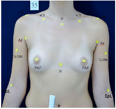
Figure 1. Measurement of anthropometric and anatomical references. Counter-clockwise. IJ: Center of the jugular notch; xCl: 50% of the distance between the IJ and the acromion; Ac: Lateral prominence of the acromion; Ax: Proximal point of the anterior axillary line; ½ Um: Mean distance between the Ac and EpL; EpL: Anterior projection of the lateral epicondyle; PAP: Center of the nipple; Xi: Base of the xiphoid process.
The union of one point to another formed 15 segment lines; one of these was common to both hemibodies and 7 were bilateral. The common segment was formed by joining the jugular notch center to the base of the xiphoid process (IJ-Xi). The seven bilateral segments were as follows: from the center point of the jugular notch to nipple center (IJ-PAP), from the clavicle "x" to nipple center (xCl-PAP), from the acromion to the nipple center (Ac-PAP), from the acromion to the anterior projection of the lateral epicondyle (Ac-EpL), from the proximal point of the axillary line anterior to the center of the mammary papilla (Ax-PAP), from the mammary papilla center to the median line and from the acromion and the midpoint of the humerus (Ac-½ Um) (Figure 2).
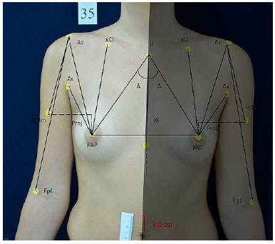
Figure 2. Schematic representation of segments and angular measure for each hemibody. Description of segments: IJ-Xi: Distance from the center of jugular notch to the base of the xiphoid process; IJ-PAP: Distance from the center of the jugular notch to the center of the nipple; xCl-PAP: 50% of the distance between the center of the jugular notch and the acromion to the center of the nipple; Ac-PAP: Distance from the lateral prominence of the acromion to the center of the nipple; Ax-PAP: Distance from the proximal point of the anterior axillary line to the center of the nipple; LM-PAP: Distance from the anterior medial line to the center of the nipple; Ac-EpL: Distance from the lateral prominence of the acromion to the anterior projection of the lateral epicondyle; Ac-½ Um: 50% the distance from the lateral prominence of the acromion to the anterior projection of the lateral epicondyle; Â: Angle formed by the confluence of segments IJ-Xi and IJ-PAP; Proj: Projection of ½ Um in the line of the homolateral nipple.
The confluence of the (IJ - Xi) and (IJ - PAP) segments formed an angle for each hemisphere (RA and LA) denominated "sternal angle". These angles were measured with a protractor.
Indirect anthropometry was performed by three independent researchers to verify the inter-rater reliability. The main researcher conducted a second evaluation in graphic software for evaluation of intra-rater reliability. All raters obtained specific prior training on Photoshop CS6® measuring tools.
A photography setting was prepared for obtaining the photographs, with the volunteers placed in the same standard spot marked using vinyl foam placed on the floor. Light sources and camera were arranged so that photos were obtained at the same position and with the same light exposure. After obtaining digital photography, software tools were calibrated using a 3-cm measurement taken from a numerical scale from a ruler taped to the right mesogastric region of the volunteers in order to obtain real measurements.
Direct anthropometry of the linear segments was performed by opening a compass and then transferring the measurement to a ruler; angular measurements were performed with a protractor.
After obtaining the standardized photos, defining the measurement points and ruler placement to be used in the software calibration, measurement was initiated using Photoshop CS6® software. The software can be obtained from the Adobe website.
The images were obtained in RAW format.
To start the measurements, the pictures must be stored on the same computer in which Adobe Photoshop CS6® is installed.
When opening the software, click File, and then Open.
Choose a picture to be analyzed and the file type, which can be NEF (if picture is obtained by an Nikon® machine) or JPEG. Once the photo appears, click Open. In the present study, we chose to measure in the NEF format.
Click on the icon to use range of measures - alongside L2 in the second line from the top.
After the picture is open, increase zoom 4 times (Ctrl ++++). This way, the ruler for software calibration will become more evident.
To calibrate the software, one must click on the Image item, then Analysis, then choose Define Scale of Measure, and finally Personalize.
This will open a window showing pixel length, logical length, and logical units. In this framework, we must define the measuring scale. For this study, we chose the scale with 3.0 cm. Therefore, put the mouse and drag the scale from zero to 3 cm on the ruler. This will show the pixel length which corresponds to the 3.0 cm of the strip. Write 3 on the logical length and cm in logical units. Click ok and the software will now be calibrated (Figure 3).
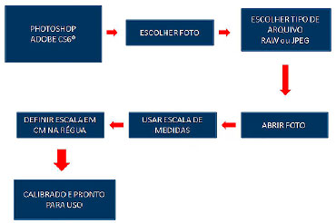
Figure 3. Step-by-step use of the software.
The figure corresponding to the measurements in centimeters will appear in Table L1. To facilitate the measurements, we suggest reducing the zoom once (Ctrl -) to perform measurements in a more comfortable way.
The angle measurements will appear in the Angle a: x degrees box.
We performed the measurements of the previously described segments through the graphics software and with direct measurement.
All analyzes were made with a significance level of 5%.
Variable concordance/reproducibility was assessed by the intra- and inter-observer ICC (Intraclass Coefficient of Correlation) for each method among the 3 evaluators.
Concordance/reproducibility by ICC among methods for measurements of the main evaluator, compared with direct measurement (compass).
The T test was used as a basis for assessing the significance (p) of the main evaluator's measurement results compared with the direct measurements.
RESULTS
Comparisons were made between measurements taken at 2 different occasions by the same examiner and between three different evaluators on the same graphics software.
The results were:
1. Inter-examiner ICC
Concordance/reproducibility between examiners 1, 2 and 3
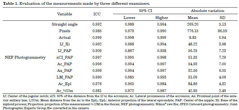
All variables had an ICC value larger than 0.8, meaning that there is a high correlation between measurements (Table 1).
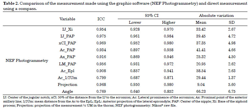
2. ICC obtained by NEF photogrammetry compared with direct measurement (compass)
Except for "Ac_1/2um" and "angle" in the NEF photogrammetry method, all remaining variables had an ICC>0.8, which means there was high correlation between the measurements. Lower values of ICC indicate a lower correlation, but still within an acceptable range (Table 2).
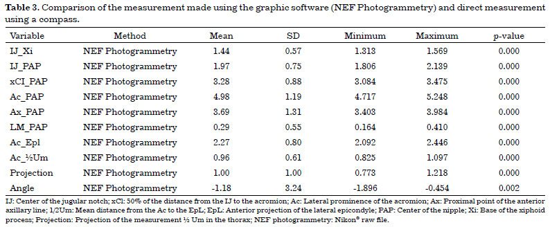
3. Description of the absolute differences betweensoftwareand direct measurements in each evaluated segment and results of the comparisons of between-methods differences.
All variables presented mean differences (p < 0.05) when compared to the direct measurement obtained using a compass (Table 3).
DISCUSSION
Since the 18th century, there have been reports of physical anthropometry on Marco Polo's travel experiments, in which some significant differences in body size and height were found in people from different ethnic groups9.
Penn10 was the precursor of breast evaluation by direct anthropometry, publishing an article that described the measurements of 20 women with breasts considered to be aesthetically perfect, with the objective of finding a normal breast pattern. After this article was published, different authors conducted other studies on this topic in order to develop breast measurement protocols by direct anthropometry5,11-15.
Odo et al.16 analyzed pre- and post-operative mammary asymmetry outcomes and verified that this asymmetry can be evaluated by direct anthropometry. In comparative studies of mammary asymmetry correction surgery, Pozzobon et al.17 used both breast MRI and linear measurements. These authors found that MRI associated with linear measurements is a good method for performing breast measurements.
Direct anthropometry allows for the quantification of breast measurements from predetermined points in linear and angular measurements using, for example, a ruler, measuring tape, and compass12,13,14,18.
Direct thoracic region measurements may have only relative accuracy due to the number of curves, depressions, and contours. Moreover, chest wall mobility during breathing can oscillate in the same individual at a given time and also at different times5,13.
These issues should warrant caution because they may create differences when measurements are carried out by different researchers and in different measurement collection19. To minimize changes resulting from breathing, Agbenorku et al.20 proposed to choose the smallest value of two consecutive mammary region measurements performed with a tape measure. In this way, the values obtained are closer to the actual measurements.
Quieregatto et al.21 described anthropometric and anatomical points to be used for breast measurement in the frontal plane. These points were selected in order to standardize breast measurement and increase methodological reproducibility. It has been shown that there are limitations on points that could possibly be used for photographic measurement in the oblique and profile views21.
Obtaining photographic linear measurements instead of directly from the individual has been an efficient way to evaluate the breasts22. According to Nechala et al.5, indirect anthropometry has advantages in comparison to direct anthropometry such as minimizing measurement errors, millimetric precision, possibility of taking measurements over time, possibility of pre- and post-operative comparison, reduction of discomfort to which the patient is exposed, and less exposure time during measurements.
Indirect anthropometry is faster and more efficient because it allows for immediate or later viewing of the photos after being you've got the or after, without the need for specific technical training and reducing costs when compared to the 3D scanners.
Some previous studies by the aforementioned author show there is no reliable pattern in breast measurement regarding methodological reproducibility and accuracy. Several factors can interfere in these measurements, such as the type of camera used, photographic setting standardization, file type used, and processing software8,21,23.
Quieregatto et al.8 analyzed the comparison of direct anthropometry using a compass and ruler with 3 different softwares and found no correlation among them. In addition, these authors suggest that after choosing the software for breast measurement, comparisons should only be made to the same software and not to others, even when using the same digital measurement tool. They also demonstrated that direct anthropometry measurements were always different from those found by graphics software and suggested further studies to elucidate these differences.
Mallucci & Branford24 analyzed the proportion of upper and lower breast poles, as well as nipple position and angle, in order to identify a relationship between quadrant proportion and breast aesthetic standards. These authors observed that these parameters can be identified in a simple and objective way, not taking into account only the papilla as the main part of breast evaluation. The software used in this study (Adobe Photoshop CS4®) was suitable for the measurement of breast angles and proportions.
According to Kouchi et al.25, measurements smaller than 10 cm tend to be more reliable and do not show significant differences in comparison to larger measurements. This suggests that studies on breast classification obtained by direct anthropometry should have higher values of standard deviation.
The present study shows that indirect anthropometry evaluation performed using Adobe Photoshop CS6 software® proved feasible using RAW image files. It is a readily available tool and is proving important for the evaluation of digital breast photography. It has also proved to be the only graphics software that allows measurements using RAW files, among the 3 other softwares previously studied8.
CONCLUSION
Adobe Photoshop CS6® was effective for breast measurement using a computer and RAW files, with a specific software, without the need for specific training.
The direct breast measurements were different from the ones obtained using Adobe Photoshop CS6®.
COLLABORATIONS
PRQES Analysis and/or interpretation of data; statistical analyses; final approval of the manuscript; conception and design of the study; completion of surgeries and/or experiments; writing the manuscript or critical review of its contents.
MSN Analysis and/or interpretation of data; statistical analyses; final approval of the manuscript; conception and design of the study; completion of surgeries and/or experiments; writing the manuscript or critical review of its contents.
FF Analysis and/or interpretation of data; statistical analyses; final approval of the manuscript; conception and design of the study; completion of surgeries and/or experiments; writing the manuscript or critical review of its contents.
TWTJ Analysis and/or interpretation of data; statistical analyses; final approval of the manuscript; conception and design of the study; completion of surgeries and/or experiments; writing the manuscript or critical review of its contents.
AAQES Analysis and/or interpretation of data; conception and design of the study; completion of surgeries and/or experiments.
FLN Analysis and/or interpretation of data; conception and design of the study; completion of surgeries and/or experiments.
RAT Conception and design of the study; completion of surgeries and/or experiments.
LMF Analysis and/or interpretation of data; statistical analyses; final approval of the manuscript; conception and design of the study; completion of surgeries and/or experiments; writing the manuscript or critical review of its contents.
REFERENCES
1. Christie D, Sharpley C, Curtis T. Improving the accuracy of a photographic assessment system for breast cosmesis. Clin Oncol (R Coll Radiol). 2005;17(1):27-31. DOI: http://dx.doi.org/10.1016/j.clon.2004.09.009
2. Ellis H, Colborn GL, Skandalakis JE. Surgical embryology and anatomy of the breast and its related anatomic structures. Surg Clin North Am. 1993;73(4):611-32. PMID: 8378813 DOI: http://dx.doi.org/10.1016/S0039-6109(16)46077-9
3. Hochman B, Nahas FX, Ferreira LM. Fotografia aplicada na pesquisa clínico-cirúrgica. Acta Cir Bras. 2005;20(Suppl 2):19-25. DOI: http://dx.doi.org/10.1590/S0102-86502005000800006
4. Ward CM. An analysis, from photographs, of the results of four approaches to elongating the columella after repair of bilateral cleft lip. Plast Reconstr Surg. 1979;64(1):68-75. PMID: 377332 DOI: http://dx.doi.org/10.1097/00006534-197907000-00013
5. Nechala P, Mahoney J, Farkas LG. Digital two-dimensional photogrammetry: a comparison of three techniques of obtaining digital photographs. Plast Reconstr Surg. 1999;103(7):1819-25. PMID: 10359240 DOI: http://dx.doi.org/10.1097/00006534-199906000-00002
6. Sivagnanavel V, Smith RT, Lau GB, Chan J, Donaldson C, Chong NV. An interinstitutional comparative study and validation of computer aided drusen quantification. Br J Ophthalmol. 2005;89(5):554-7. DOI: http://dx.doi.org/10.1136/bjo.2004.046813
7. Assunção WG, Gomes EA, Tabata LF, Gennari-Filho H. A comparison of profilometer and AutoCAD software techniques in evaluation of implant angulation in vitro. Int J Oral Maxillofac Implants. 2008;23(4):618-22.
8. Quieregatto PR, Hochman B, Furtado F, Machado AF, Sabino Neto M, Ferreira LM. Image analysis software versus direct anthropometry for breast measurements. Acta Cir Bras. 2014;29(10):688-95. DOI: http://dx.doi.org/10.1590/S0102-8650201400160010
9. Roebuck JA Jr, Kroemer KHE, Thomson WG. Engineering anthropometry methods. New York: John Wiley & Sons Inc; 1975.
10. Penn J. Breast reduction. Br J Plast Surg. 1955;7(4):357-71. DOI: http://dx.doi.org/10.1097/00006534-195507000-00028
11. Smith DJ Jr, Palin WE Jr, Katch V, Bennett JE. Surgical treatment of congenital breast asymmetry. Ann Plast Surg. 1986;17(2):92-101. PMID: 3273092 DOI: http://dx.doi.org/10.1097/00000637-198608000-00002
12. Malata CM, Boot JC, Bradbury ET, Ramli AR, Sharpe DT. Congenital breast asymmetry: subjective and objective assessment. Br J Plast Surg. 1994;47(2):95-102. PMID: 8149066 DOI: http://dx.doi.org/10.1016/0007-1226(94)90166-X
13. Westreich M. Anthropomorphic breast measurement: protocol and results in 50 women with aesthetically perfect breasts and clinical application. Plast Reconstr Surg. 1997;100(2):468-79. PMID: 9252618 DOI: http://dx.doi.org/10.1097/00006534-199708000-00032
14. Brown TP, Ringrose C, Hyland RE, Cole AA, Brotherston TM. A method of assessing female breast morphometry and its clinical application. Br J Plast Surg. 1999;52(5):355-9. DOI: http://dx.doi.org/10.1054/bjps.1999.3110
15. Farkas LG, Bryson W, Klotz J. Is photogrammetry of the face reliable? Plast Reconstr Surg. 1980;66(3):346-55. PMID: 7422721
16. Odo LM, Guimarães PA, Silva ALAL, Sabino Neto M, Ferreira LM. Avaliação do tratamento cirérgico da assimetria mamária por meio de medidas lineares. Arq Catarinenses Med. 2009;38(Supl 1):43-5.
17. Pozzobon AV, Sabino Neto M, Veiga DF, Abla LE, Pereira JB, Biasi TL, et al. Magnetic resonance images and linear measurements in the surgical treatment of breast asymmetry. Aesthetic Plast Surg. 2009;33(2):196-203. PMID: 18709409 DOI: http://dx.doi.org/10.1007/s00266-008-9224-9
18. Smith DJ Jr, Palin WE Jr, Katch VL, Bennett JE. Breast volume and anthropomorphic measurements: normal values. Plast Reconstr Surg. 1986;78(3):331-5. DOI: http://dx.doi.org/10.1097/00006534-198609000-00008
19. Quieregatto PR, Hochman B, Ferrara SF, Furtado F, Liebano RE, Sabino Neto M, et al. Anthropometry of the breast region: how to measure? Aesthetic Plast Surg. 2014;38(2):344-9. PMID: 24610111
20. Agbenorku P, Agbenorku M, Iddi A, Amevor E, Sefenu R, Osei D. Measurements of breasts of young West African females: a guideline in anatomical landmarks for adolescent breast surgery. Aesthetic Plast Surg. 2011;35(1):49-54. DOI: http://dx.doi.org/10.1007/s00266-010-9555-1
21. Quieregatto PR, Hochman B, Furtado F, Ferrara SF, Machado AF, Sabino Neto M. Photographs for anthropometric measurements of the breast region. Are there limitations? Acta Cir Bras. 2015;30(7):509-16. DOI: http://dx.doi.org/10.1590/S0102-8650201500700000010
22. Sacchini V, Luini A, Tana S, Lozza L, Galimberti V, Merson M, et al. Quantitative and qualitative cosmetic evaluation after conservative treatment for breast cancer. Eur J Cancer. 1991;27(11):1395-400. PMID: 1835855 DOI: http://dx.doi.org/10.1016/0277-5379(91)90019-A
23. Trigo T. Equipamento Fotográfico - Teoria e Prática. São Paulo: Editora Senac; 2014.
24. Mallucci P, Branford OA. Concepts in aesthetic breast dimensions: analysis of the ideal breast. J Plast Reconstr Aesthet Surg. 2012;65(1):8-16. DOI: http://dx.doi.org/10.1016/j.bjps.2011.08.006
25. Kouchi M, Mochimaru M, Tsuzuki K, Yokoi T. Random errors in anthropometry. J Hum Ergol (Tokyo). 1996;25(2):155-66.
1. Sociedade Brasileira de Cirurgia Plástica, São Paulo, SP, Brazil
2. Colégio Brasileiro de Cirurgiões, Rio de Janeiro, RJ, Brazil
3. Escola Paulista de Medicina, Universidade Federal de São Paulo, São Paulo, SP, Brazil
Institution: Universidade Federal de São Paulo, São Paulo, SP, Brazil
Corresponding author:
Miguel Sabino Neto
Rua Napoleão de Barros, 715, 4º andar - Vila Clementino
São Paulo, SP, Brazil - Zip Code 04023-002
E-mail: msabino@uol.com.br
Article received: October 16, 2017.
Article accepted: January 24, 2018.
Conflicts of interest: none.
 Read in Portuguese
 Read in English
 PDF PT
 Print
Print
 Send this article by email
 How to Cite
 Mendeley
 Pocket