


Original Article - Year 2018 - Volume 33 -
Getting to know the types of JPEG and RAW photo files used in research
Conhecendo os tipos de arquivo de fotografia JPEG e RAW utilizados em pesquisa
ABSTRACT
INTRODUCTION: Indirect anthropometric measurements are important in research but may also be significant in the legal area as a quantitative instrument for pre- and post-operative evaluation. The type of file used determines variations in the manipulation of the images. The objective is to study the types of image files used in digital photography that will find utility in research.
METHODS: The breasts of 40 women volunteers were measured in 80 hemithoraces, and the mammary region and arms were marked in the frontal position. The union of these points in each hemithorax formed seven linear segments, an angular measure, and a median segment common to both hemithoraces. These photographs were evaluated as high definition RAW and JPEG files by three observers using Adobe Photoshop CS6® software.
RESULTS: RAW and JPEG files were shown to be effective in breast measurement.
CONCLUSION: RAW and JPEG files presented similar results in the measurement of female breasts.
Keywords: Anthropometry; Breast; Body weights and measures; Mammoplasty; Thorax; Photography; Software.
RESUMO
INTRODUÇÃO: Mensurações por antropometria indireta são importantes na área de pesquisa, mas também podem se tornar significativas na área jurídica, como instrumento quantitativo de avaliação pré e pós-operatória. O tipo de arquivo utilizado determina variações na manipulação das imagens. O objetivo é evidenciar tipos de arquivos de imagens utilizados na fotografia digital que será utilizada na pesquisa.
MÉTODOS: Foram realizadas mensurações das mamas em 80 hemitórax de 40 mulheres voluntárias. Foram demarcados pontos sobre a região mamária e braços em posição frontal. A união destes pontos em cada hemicorpo formou sete segmentos lineares, uma medida angular e um segmento mediano comum a ambos os hemicorpos. Essas fotografias foram avaliadas em arquivos RAW e JPEG de alta definição, por 3 observadores, com auxílio do softwareAdobePhotoshop CS6>®.
RESULTADOS: Os arquivos RAW e JPEG demonstraram serem eficazes na mensuração das mamas.
CONCLUSÃO: Os arquivos RAW e JPEG apresentaram medidas semelhantes na mensuração das mamas femininas.
Palavras-chave: Antropometria; Mama; Pesos e medidas corporais; Mamoplastia; Tórax; Fotografia; Software.
The importance of anthropometric studies using photos as research has an increasingly vital role, as it is essential to know the types of files obtained using the digital camera1. Photography has become an important analytical tool, as it is performed in a standardized manner, which determines the scientific rigor necessary for any analysis2.
Image analysis can be performed using graphic software, and that is necessary a previously calibration, and this calibration would be determined by linear markers or pixels. It is a form of measurement that can be used in the absence of the patient, as the measurements are performed on a previously-obtained image3.
Photography, besides allowing a preoperative record, presents advantages in terms of evaluation of the subject, such as reduced inconvenience to the patient, the possibility of making several measurements that can be made by several researchers at any time, and approximation of centesimal order (up to 0.01 centimeters) offered by the tools of the graphic software3.
Computerized photogrammetry, when used on patients, can be of great value in avoiding patient constraints and contributing to the objective analysis of postoperative results.
Sivagnanavel et al.4 and Assunção et al.5 proposed software validation studies with comparisons between them, because although they used the same digital tools and had the same theoretical basis, they generated different results.
Quieregatto et al.6 demonstrated that there may be differences in the measurements obtained for the mammary region using different softwares.
A digital image comprises elements represented by a long series of numbers. These elements are the pixels (Picture elements). Each pixel forms a digital image, and its color and position are represented by a series of numbers. These numbers are identified by two coordinates (x and y), and divided into red (Red), green (Green), and blue (Blue) tones (Figure 1).
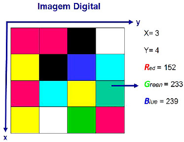
Figure 1. Schematic of image distribution mathematically in the X and Y coordinates. Each number corresponds to a color with variations in tones of red, green, and blue.
The raw image (RAW) obtained by the camera is unique and does not allow modifications, considering the image matrix. It is captured in shades of black, gray, and white (gray scale) and is represented in a matrix by numbers from 0 to 255, 0 representing black color; 255, white; and the intermediate values, grayscale (Figure 2). When this image that is originally in black and white becomes colored, the camera or graphic software uses these numbers and groups of pixels to determine the color of the image. The way these pixels are used vary with the file format of the image 7.
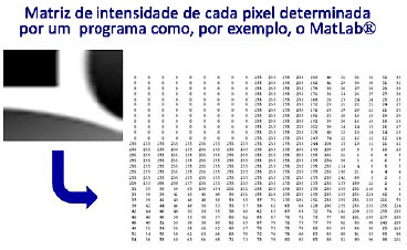
Figure 2. Example of mathematical representation of a RAW image in white, gray, and black tones. Zero corresponds to pure black, 255 corresponds to white, and intermediate numbers correspond to grey-scale color tones.
A file format is a method that uses a computer to store information, images, and texts. For images, we highlight the most common formats, including RAW (raw file) and JPEG (Joint Photographic Experts Group).
The digital image obtained by a photographic machine is always in the RAW format and can be converted to other types of image files by a camera (on board) or using graphical software (off board).On board conversion will depend on the photographic equipment used as well as the adjustment made by the machine operator.7
For off board conversion, a graphic software is required, which offers different levels of image quality for different types of image files selected by the operator.
To give color to the image, pixel groups are used, varying based on the type of file used (JPEG, TIFF) as well as the image quality output of the software7.
In pictures with a higher image quality, to obtain a better image definition, the software uses a determined number of pixel groups. The smaller the number of pixels used in the group, the greater the color resolution of the image obtained, because a group with a smaller number of pixels will be used to color a given segment and may cause changes in image definition (Figure 3).
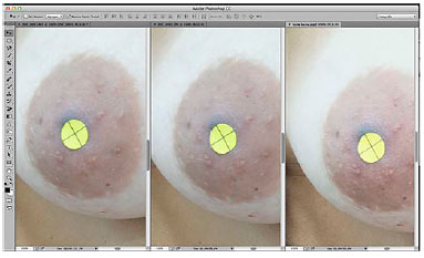
Figure 3. Example of different types of image files for the same image. Left to right, RAW file, high-definition JPEG, and low-definition JPEG. Note the blurring of the image at the top of the areolas.
OBJECTIVE
The objective of this study is to reveal the types of image files used in digital photography that will be used in research.
METHODS
This study was approved by the Research Ethics Committee of the Universidade Federal de São Paulo (UNIFESP) 430.239/ 2013. It was conducted at UNIFESP from February 2014 to October 2016.
How the image is obtained using the camera, the different types of RAW and JPEG image files used, and the factors in the conversion of this obtained image will be demonstrated.
The study included 40 female volunteers aged 18-60 years (mean 29.1 ± 10.3). The exclusion criteria were women who underwent mastectomy, volunteers with a history of breast surgery, thoracic deformities, or severe breast ptosis in which the nipples crossed a transverse line at the lower limit of the umbilicus.
Self-adhesive labels Primaco BIC® brand OP-4433 model of 0.6 cm in diameter were used to demarcate eight points per hemibody, of which 5 were anthropometric points: the center of the jugular notch (IJ) and the base of the xiphoid process (X); bilaterally, the center of the nipple (PAP), acromion (Ac) and the anterior projection of the lateral epicondyle (EpL).
Three other anatomical points were used in both hemibodies: the point corresponding to half the distance between the center of the jugular notch and the acromion, called "x" of the clavicle (xCL), the projected point more proximal to the anterior axillary line (Ax), and a point corresponding to half the distance between the acromion and the lateral epicondyle, called the "mid-point of the humerus" (a 1/2), according to the study by Quieregatto et al.6,8,9 (Figure 4).
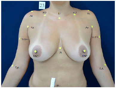
Figure 4. Measurement of anthropometric and anatomical points. Counter-clockwise. IJ: Center of the jugular notch; xCl: 50% of the distance between the IJ and acromion; Ac: Lateral prominence of the acromion; Ax: Proximal point of the anterior axillary line; 1/2Um: Mean distance between the Ac and EpL; EpL: Anterior projection of the lateral epicondyle; PAP: Center of the nipple; Xi: Base of the xiphoid process.
The union of one point to another formed 15 segment lines; of these, one was common to hemibodies and 7 were bilateral. The common segment was formed by joining the jugular notch center of the base of the xiphoid process (IJ- Xi). The other seven bilateral points were formed by the segment center point of the jugular notch to the center of the nipple (IJ- PAP), the distance between the "x" point of the clavicle to the center of the nipple (xCl- PAP), the acromion to the center of the nipple (Ac-PAP), the anterior projection of the lateral epicondyle (Ac-EpL), the proximal point of the axillary line anterior to the center of the mammary papilla (Ax- PAP), the mammary papilla center to the median line and the distance between the acromion and the midpoint of the humerus (Ac ½ a) (Figure 5).
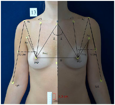
Figure 5. Schematic representation of the segments and angular measures in each hemibody. Description of segments: IJ-Xi: Distance from the center of jugular notch to the base of the xiphoid process; IJ-PAP: Distance from the center of the jugular notch to the center of the nipple; xCl-PAP: 50% of the distance from the center of the jugular notch and acromion to the center of the nipple; Ac-PAP: Distance from the lateral prominence of the acromion to the center of the nipple; Ax-PAP: Distance from the proximal point of the anterior axillary line to the center of the nipple; LM-PAP: Distance from the anterior medial line to the center of the nipple; Ac-EpL: Distance from the lateral prominence of the acromion to the anterior projection of the lateral epicondyle; Ac - ½ Um: 50% of the distance from the lateral prominence of the acromion to the anterior projection of the lateral epicondyle; Angle formed by the confluence of segments IJ-Xi and IJ-PAP; Proj: Projection of ½ Um in the line of the homolateral nipple.
The confluence of the segments (IJ - Xi) and (IJ - PAP) formed an angle for each hemisphere (RA and LA) called the "sternal angle." These angles were measured with a protractor.
Indirect anthropometry was performed by three independent researchers to verify inter-reproducibility. The principal investigator conducted a second evaluation using graphic software for evaluation of intra-rater reproducibility. All raters obtained specific prior training with the tools for measurement using software Photoshop CS6®.
A photographic setting was set for obtaining photographs, with the volunteers placed in the same standard point through vinyl foam on the floor. The light sources and camera were arranged so that the photos were taken in the same position and with same light exposure. After obtaining digital photographs, the software tools were calibrated with the measurement of 3 cm, obtained from a numerical scale the pasted rule in mesogastric right region of voluntary to obtain actual measurements.
Direct anthropometry of the linear measurements was performed by opening a compass and then transferring to a ruler and the angular measurements performed with a protractor.
By obtaining the photos in a standardized way, the settings of the measurement points and ruler placement were used in the software calibration. The measurement was initiated through Photoshop CS6® software. The same can be obtained from the Adobe website.
All analyses considered a significance level of 5%.
Concordance/reproducibility of the variables was determined by the application of intra- and inter-observer ICC (Intraclass Coefficient of Correlation) for each method among the three evaluators.
RESULTS
The results were:
1. Intraclass correlation coefficient (ICC) inter-examiner
Concordance / reproducibility between examiners 1, 2, and 3
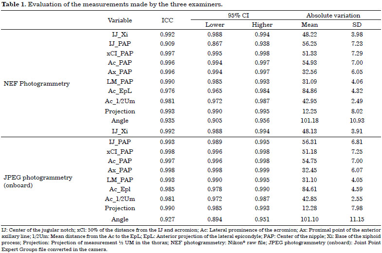
All variables have intraclass correlation coefficients (ICCs) larger than 0.8 (ICC> 0.8), indicating a high correlation between measurements (Table 1).
2. Description of absolute differences between each method on each reported segment and results of comparisons of these differences between the methods
On average, all variables in all methods are similar (p < 0.05) compared with each other (Table 2).

DISCUSSION
Since the eighteenth century, there have been reports of physical anthropometry that have noted significant differences in body size and height of people from different ethnic groups10.
The best understand the mammary region, can provide improved health11. For evaluation of the breasts, a pioneer in breast measurements was Penn12, who evaluated aesthetically perfect breasts to create an aesthetic standard. This was the motivation of several authors for the development of protocols for the measurement of female breasts3,13-16.
Direct anthropometry was previously used by Odo et al.17, who verified mammary asymmetry in a comparative manner pre- and post-surgically. In comparative studies of operations for correction of mammary asymmetry, Pozzobon et al.11 used MRI of breasts and linear measurements. These authors recommended that MRI associated with linear measurements is a good method to evaluate breast measures11.
Direct anthropometric measurements enable quantification of the breasts from predetermined points in Linares and angular measurements using, for example, a ruler, measuring tape, and compass2,18-21.
Measurement of the breasts by direct anthropometric measurements has been well studied and, as it is a measurement obtained through a fixed image, it allows reduction in the time of exposure of and constraint to the individual being analyzed3,15. This was the determining factor in using breast photogrammetry to evaluate different types of image files.
Quieregatto et al.9 described anthropometric and anatomical points to be used for measurements using breast photography. These points were selected to standardize the measurement of breasts and to increase the reproducibility of the methods. It has been shown that there are limitations to the points that could possibly be used for measurements on photographs in the oblique position and profile8.
These points were defined by the authors, and were based on previous studies by Penn12, Smith et al.13 and Westreich15. The points analyzed were based on breast evaluations as a reference to beauty, breast symmetry, and the ideal point of the mammary papilla. Quieregatto et al., based on these studies, defined the points and the best position (frontal) that would allow study of the breasts through photographs6,8,9.
By obtaining the photograph in a standardized way and taking the distance of the object and height of the photographic camera, and lighting into consideration, the necessary conditions for statistical analysis are met, maintaining the scientific rigor20. Sacchini et al.22 were the first to consider linear measurements per photograph rather than directly from the individual as a more efficient way to evaluate breasts.
Photogrammetry provides some advantages over direct anthropometry, such as a way to minimize measurement errors, the precision tool graphics software offers, the possibility of performing measurements over time by being able to compare quantitatively the differences between preoperative and postoperative results, and by reducing the exposure time and discomfort to the patient during3 measurements. In addition to being faster and more efficient, it enables viewing of the photos immediately after they were taken, not requiring specific technical training and its low cost compared to 3-dimensional scanners (3D)3.
The attention given to lighting is important to preserve the technical rigor of the clinical picture. Over- or under-exposure can cause shadows or interfere with the definition of images23. In this study, we used illumination that would allow for the measurement of the breasts without interference.
In the study by Quieregatto et al.6 different softwares were evaluated for breast assessment. The authors concluded that different software, with different complexity, can interfere in the final result of the measurement of the breasts by direct anthropometry. They used Adobe Photoshop CS4®, AUTOCAD® and Image Tool®. They also described differences in the measurements obtained by the software, with measurements obtained directly from the patients. A JPEG (Joint Point Expert Groups) file was used6.
The analysis made using the interclass correlation coefficient between the same evaluator twice and between three other evaluators allowed us to define the reproducibility and accuracy of the method, proving to be reproducible and with high accuracy.
This study shows that the measurement of the breasts using different files can be performed using the same graphic software. The choice of Photoshop CS6® was made because the software allowed images to be analyzed in both file types chosen (JPEG and RAW) among the three software previously researched by the authors.
JPEG and RAW archives were analyzed in the present study because of the tendency to use a RAW file in lawsuits and because no study in literature has evaluated the differences between JPEG and RAW files in breast measurements. We observed a disadvantage to viewing the RAW file, that is, it required a specific software for the image to be viewed.
The authors emphasize that studies involving files of different complexities, with different file sizes, are important to determine an ideal method of analyzing images obtained in the digital form, with less distortion, smaller file sizes, occupying lesser computer memory, as well as making it possible to exchange smaller images via the internet.
CONCLUSION
- Both files could be used to evaluate the mammary region;
- There was no statistical difference between the measurements of the RAW file and high-definition JPEG, but the measurements were not equal.
COLLABORATIONS
PRQES Analysis and/or interpretation of data; statistical analyses; final approval of the manuscript; conception and design of the study; completion of surgeries and/or experiments; writing the manuscript or critical review of its contents.
MSN Analysis and/or interpretation of data; statistical analyses; final approval of the manuscript; conception and design of the study; completion of surgeries and/or experiments; writing the manuscript or critical review of its contents.
FF Analysis and/or interpretation of data; statistical analyses; final approval of the manuscript; conception and design of the study; completion of surgeries and/or experiments; writing the manuscript or critical review of its contents.
TWTJ Analysis and/or interpretation of data; final approval of the manuscript; conception and design of the study; completion of surgeries and/or experiments; writing the manuscript or critical review of its contents.
AAQES Analysis and/or interpretation of data; completion of surgeries and/or experiments.
FLN Analysis and/or interpretation of data; completion of surgeries and/or experiments.
RAT Analysis and/or interpretation of data; writing the manuscript or critical review of its contents.
LMF Analysis and/or interpretation of data; statistical analyses; final approval of the manuscript; conception and design of the study; completion of surgeries and/or experiments; writing the manuscript or critical review of its contents.
REFERENCES
1. Ellis H, Colborn GL, Skandalakis JE. Surgical embryology and anatomy of the breast and its related anatomic structures. Surg Clin North Am. 1993;73(4):611-32. PMID: 8378813 DOI: http://dx.doi.org/10.1016/S0039-6109(16)46077-9
2. Hochman B, Nahas FX, Ferreira LM. Photography in medical research. Acta Cir Bras. 2005;20 Suppl 2:19-25. DOI: http://dx.doi.org/10.1590/S0102-86502005000800006
3. Nechala P, Mahoney J, Farkas LG. Digital two-dimensional photogrammetry: a comparison of three techniques of obtaining digital photographs. Plast Reconstr Surg. 1999;103(7):1819-25. PMID: 10359240 DOI: http://dx.doi.org/10.1097/00006534-199906000-00002
4. Sivagnanavel V, Smith RT, Lau GB, Chan J, Donaldson C, Chong NV. An interinstitutional comparative study and validation of computer aided drusen quantification. Br J Ophthalmol. 2005;89(5):554-7. DOI: http://dx.doi.org/10.1136/bjo.2004.046813
5. Assunção WG, Gomes EA, Tabata LF, Gennari-Filho H. A comparison of profilometer and AutoCAD software techniques in evaluation of implant angulation in vitro. Int J Oral Maxillofac Implants. 2008;23(4):618-22.
6. Quieregatto PR, Hochman B, Furtado F, Machado AF, Sabino Neto M, Ferreira LM. Image analysis software versus direct anthropometry for breast measurements. Acta Cir Bras. 2014;29(10):688-95. DOI: http://dx.doi.org/10.1590/S0102-8650201400160010
7. Trigo T. Equipamento Fotográfico - Teoria e Prática. São Paulo: Editora Senac; 2014.
8. Quieregatto PR, Hochman B, Ferrara SF, Furtado F, Liebano RE, Sabino Neto M, et al. Anthropometry of the breast region: how to measure? Aesthetic Plast Surg. 2014;38(2):344-9. PMID: 24610111
9. Quieregatto PR, Hochman B, Furtado F, Ferrara SF, Machado AF, Sabino Neto M, et al. Photographs for anthropometric measurements of the breast region. Are there limitations? Acta Cir Bras. 2015;30(7):509-16. DOI: http://dx.doi.org/10.1590/S0102-8650201500700000010
10. Roebuck JA Jr, Kroemer KH, Thomson WG. Engineering anthropometry methods. New York: John Wiley & Sons; 1975.
11. Pozzobon AV, Sabino Neto M, Veiga DF, Abla LE, Pereira JB, Biasi TL, et al. Magnetic resonance images and linear measurements in the surgical treatment of breast asymmetry. Aesthetic Plast Surg. 2009;33(2):196-203. PMID: 18709409 DOI: http://dx.doi.org/10.1007/s00266-008-9224-9
12. Penn J. Breast reduction. Br J Plast Surg. 1955;7(4):357-71. DOI: http://dx.doi.org/10.1097/00006534-195507000-00028
13. Smith DJ Jr, Palin WE Jr, Katch V, Bennett JE. Surgical treatment of congenital breast asymmetry. Ann Plast Surg. 1986;17(2):92-101. PMID: 3273092 DOI: http://dx.doi.org/10.1097/00000637-198608000-00002
14. Malata CM, Boot JC, Bradbury ET, Ramli AR, Sharpe DT. Congenital breast asymmetry: subjective and objective assessment. Br J Plast Surg. 1994;47(2):95-102. PMID: 8149066 DOI: http://dx.doi.org/10.1016/0007-1226(94)90166-X
15. Westreich M. Anthropomorphic breast measurement: protocol and results in 50 women with aesthetically perfect breasts and clinical application. Plast Reconstr Surg. 1997;100(2):468-79. PMID: 9252618 DOI: http://dx.doi.org/10.1097/00006534-199708000-00032
16. Brown TP, Ringrose C, Hyland RE, Cole AA, Brotherston TM. A method of assessing female breast morphometry and its clinical application. Br J Plast Surg. 1999;52(5):355-9. DOI: http://dx.doi.org/10.1054/bjps.1999.3110
17. Odo LM, Guimarães PA, Silva ALAL, Sabino Neto M, Ferreira LM. Avaliação do tratamento cirúrgico da assimetria mamária por meio de medidas lineares. Arq Catarinenses Med. 2009;38(Supl 1):43-5.
18. Farkas LG, Bryson W, Klotz J. Is photogrammetry of the face reliable? Plast Reconstr Surg. 1980;66(3):346-55. PMID: 7422721
19. McCausland TM. A method of standardization of photographic viewpoints for clinical photography. J Audiov Media Med. 1980;3(3):109-11. DOI: http://dx.doi.org/10.3109/17453058009167132
20. Rodrigues OR, Geraldelli S, Minamoto H, Schmidt AF Jr. A Fotografia em ciências biológicas: uso no ensino e na documentação científica. Acta Cir Bras. 1995;10(4):173-82.
21. Gherardini G, Matarasso A, Serure AS, Toledo LS, DiBernardo BE. Standardization in photography for body contour surgery and suction-assisted lipectomy. Plast Reconstr Surg. 1997;100(1):227-37. PMID: 9207680 DOI: http://dx.doi.org/10.1097/00006534-199707000-00034
22. Sacchini V, Luini A, Tana S, Lozza L, Galimberti V, Merson M, et al. Quantitative and qualitative cosmetic evaluation after conservative treatment for breast cancer. Eur J Cancer. 1991;27(11):1395-400. PMID: 1835855 DOI: http://dx.doi.org/10.1016/0277-5379(91)90019-A
23. Jakowenko J. Clinical photography. J Telemed Telecare. 2009;15(1):7-22. DOI: http://dx.doi.org/10.1258/jtt.2008.008006
1. Sociedade Brasileira de Cirurgia Plástica, São Paulo, SP, Brazil
2. Colégio Brasileiro de Cirurgiões, Rio de Janeiro, RJ, Brazil
3. Escola Paulista de Medicina, Universidade Federal de São Paulo, São Paulo, SP, Brazil
Institution: Universidade Federal de São Paulo, São Paulo, SP, Brazil.
Corresponding author:
Miguel Sabino Neto
Rua Napoleão de Barros, 715, 4º andar - Vila Clementino
São Paulo, SP, Brazil - Zip Code 04023-002
E-mail: msabino@uol.com.br
Article received: September 23, 2017.
Article accepted: January 24, 2018.
Conflicts of interest: none.
 Read in Portuguese
 Read in English
 PDF PT
 Print
Print
 Send this article by email
 How to Cite
 Mendeley
 Pocket