
Case Report - Year 2016 - Volume 31 -
Surgical treatment of complications resulting from adjuvant radiotherapy: a case report
Tratamento cirúrgico das complicações resultantes da radioterapia adjuvante: relato de caso
ABSTRACT
INTRODUCTION: Breast cancer is the leading cause of death among malignancies affecting women. Treatment options range from surgical treatment, radiotherapy, chemotherapy and hormone therapy. The immediate breast reconstruction helps to benefit the psychosocial aspects of patients, however, depend on the technique used a number of complications can appear, especially after adjuvant radiotherapy.
CASE REPORT: We report a case of a 65-year-old woman, underwent quadrantectomy and radiotherapy in 1988. In 2010, the patient presented an injury on the scar and she was referred to mastectomy with reconstruction of retail large dorsal and adjuvant radiotherapy. Upon examination, we observed flap necrosis with infection on the axillary region, lymphedema, radiodermatitis, fibroses and joint limitation. In 2014, the patient sought our service to perform a new restorative approach. A bipedicled transverse abdominal flap was decided to be adequate to her case. After surgery, the patient reported mild pain in the upper pole that managed with debridement and dressing. Seven months after surgery there was a complete healing of the flap and patient was satisfied with the surgery.
CONCLUSION: The knowledge of surgical techniques associated with the correct scaling of steps are essential for surgical success and management of complications that may appear in breast reconstruction of patients undergoing radiotherapy.
Keywords: Mammaplasty; Radiotherapy; Breast neoplasms; Postoperative complications.
RESUMO
INTRODUÇÃO: O câncer de mama é a principal causa de morte entre as mulheres por doenças malignas. As opções de tratamento variam entre a abordagem cirúrgica, radioterápica, quimioterápica e hormonal. A reconstrução mamária imediata atua em aspectos psicossociais das pacientes, contudo, a depender da técnica realizada, diversas complicações podem surgir, principalmente pós-radioterapia adjuvante.
RELATO DO CASO: Paciente sexo feminino, 65 anos, submetida à quadrantectomia e radioterapia em 1988. Evoluiu com aparecimento de ferimento em local de cicatriz em 2010, sendo submetida à mastectomia com reconstrução com retalho grande dorsal imediata e radioterapia adjuvante. Apresentou necrose do retalho, com infecção em região axilar, linfedema, radiodermite, retração cicatricial intensa e limitação articular. Em 2014, procurou nosso serviço para nova abordagem reparadora. Indicado retalho transverso do abdome bipediculado. No seguimento pós-operatório houve pequeno sofrimento em polo superior manejado com desbridamentos e curativos. Após sete meses de pós-operatório, houve cicatrização completa do retalho e satisfação da paciente com procedimento cirúrgico.
CONCLUSÃO: O conhecimento das técnicas cirúrgicas, associado ao correto escalonamento, é fundamental para um bom sucesso cirúrgico e manejo das complicações que venham a acontecer nas reconstruções mamárias em pacientes submetidas à radioterapia.
Palavras-chave: Mamoplastia; Radioterapia; Câncer de mama; Complicações pós-operatórias.
Breast cancer is the cancer that has the highest incidence among female population. In Brazil it is the leading cause of death from malignant disease in this population1.
Surgical treatment includes partial mastectomy with axillary dissection and radiotherapy, or radical mastectomy2.
Radiation therapy may act as adjuvant or neoadjuvant therapy to surgery, which is paramount in locoregional control. However, radiation also affects normal tissue regions that can lead to soreness, fatigue, and cutaneous sensory changes such as radiodermatitis and infections. Patients undergoing primary reconstruction with alloplastic materials are most likely to present capsular contracture in postoperative period due to this factor3.
In advanced stages in which prospective use of radiotherapy as adjuvant therapy become necessary, the reconstruction with autologous tissue is desirable, if available. However, despite the current high rates of capsular contracture, in the need of alloplastic materials combination, these materials should be used as reconstructive techniques4,5.
Currently, reconstructive techniques have a broad spectrum ranging from the use of dermoglandular local falas and alloplastic materials to the use of autologous and microsurgical flaps. The knowledge about the variety of surgical approaches techniques for breast reconstruction is fundamental to plastic surgeons because of complications that caused by radiation therapy that can compromise prior reconstruction in some cases, 3,6,7.
OBJECTIVE
To report a case of breast reconstruction that presented complications after adjuvant radiotherapy and highlight the importance of technical knowledge of plastic surgeons on correct scaling of steps for breast reconstruction.
CASE REPORT
A 65-year-old woman underwent quadrantectomy and axillary dissection with adjuvant chemotherapy and radiotherapy in 1988 for cancer in her right breast. In 2010 her clinical profile evolved with injury on the scar site. The biopsy, which was negative for malignancy, presented positivity for frozen biopsy therefore resulting in radical right mastectomy with reconstruction with latissimus dorsi flap.
The patient underwent again the adjuvant radiotherapy. In postoperative period she had necrosis of latissimus dorsi flap, and infection in the axillary region, also involving the lung and bones (ribs, acromion-clavicular joint and spine).
The various surgical debridements, use of antibiotics and long-term maintenance of the exposed wound caused major local scarring with retractions. The therapy with moxifloxacin, based on advised of infection advisors, generated intermittent clinical improvements, maintaining the persistent secretion drainage in her right armpit.
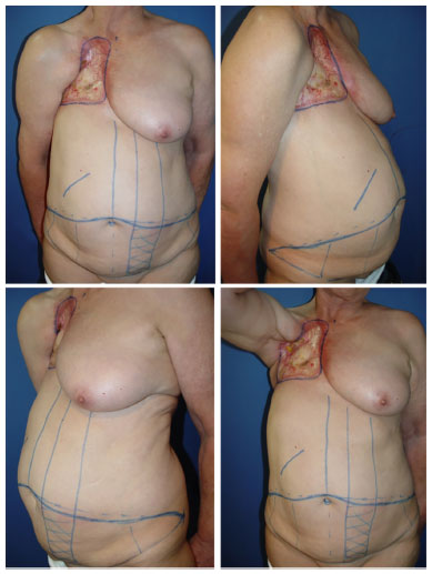
In 2014, she sought our service to undergo a new reconstruction. Upon physical examination we observed intense area of fibrosis in her right axilla with two points of drainages, severe radiodermatitis, striking lymphedema in the upper right limb and great functional limitation of right shoulder. The patient's weight was 63 kg, height 1.55 cm, and her body mass index was 26.22 BMI, in addition she had an abdominal "apron" (Figure 1).

Figure 1. Images showing pre-operative marking for flap creation.
A multidisciplinary approach was performed with prior assessment of infectious diseases, orthopedics, neurosurgery and mastology.
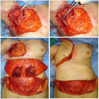
After clinical, laboratory and imaging examination by appropriate professionals, there were no contraindications regarding a new reconstructive approach. Infectious diseases evaluation requested armpit fragments for culture. Because of the large scar with retraction in the axillary region, the team of neurosurgery opted to perform the dissection from the axillary region and isolate brachial nerve plexus to avoid local injuries.
A transversal abdominal flap bipedicled for breast reconstruction was done, which proceeded uneventfully.
The armpit fragments culture showed Serratia marcescens. The antimicrobial therapy with ertapenem and clindamycin was administered for 21 days (Figure 2).

Figure 2. Images show axillary dissection and flap creation (intraoperatively).
The patient developed pain of the upper portion of the transversal abdominal flap and hematoma on the 11th after the surgery which were managed with the use of 70% alcohol and Rifocina spray on the site, and local care.
After 23 days in postoperative period the wound progressed with suffering of upper medial edges, middle and bottom of the flap, and 42 days after surgery, we used a dressing with calcium alginate because of dehiscence of lower medial edge. The conservative treatment was performed with mechanical and chemical debridement. Healing process was slow and gradual, the scar improved 4 months after surgery with the alginate suspension.
In postoperative follow-up a local small debridement was done weekly to remove excessive fibrin and also daily topical applications of essential fatty acids up to 6 months after the surgery.
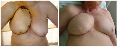
The complete wound healing occurred 7 months after the surgery (Figure 3).

Figure 3. Images showing progress of the scaring process of the flap.
Currently the patient has a discreet lymphedema in the right arm that after physical therapy improved the movement of shoulder joint. She is satisfied with the surgical outcome of the reconstructed breast.
DISCUSSION
The benefits of immediate breast reconstruction after removal of a tumor are unquestionable today. The cancer biology is not changed by reconstruction and it does not compromise the proper treatment of disease8.
Reconstructions are increasingly taking a central role for the treatment of breast cancer, especially in terms of psycho-emotional aspects generated for patients9.
A variety of studies in the literature report that immediate surgery shows improvement in patients' quality of life. The preservation of the breast contouring leads to high satisfaction with follow-up of treatment of disease1,10.
The breast reconstruction technique with tissue expander and implant involves a simple procedure than the reconstruction with autologous tissue,and it is often indicated based on preference of the patient and clinical or anatomical conditions that may limit more complex surgeries7.
Radiation therapy is a key component of conservative treatment of breast cancer. Although consensus exists on the use of breast reconstruction techniques with autologous tissues in patients with referral or suspension of radiotherapy in the future after the surgery, there are situations inherent to the patient that lead to indication of implants associated or not with flaps6.
In our study we observed that the patient had complications associated with radiotherapy, i.e., necrosis of the latissimus dorsi flap. Surgical technique used followed the principles defined for breast reconstruction, however, the patient presented complications.
The occurrence of these complications was due to vascular micro-injuries, modification of proteins and cell nucleus, therefore causing ineffective healing, dermal and subcutaneousatrophy7.
The use of radiation on myocutaneous flap is also associated to capsular contracture, radiodermatitis and burns, more complex surgical treatment and possibility of high unsatisfactory results. In addition to these complications, hypoesthesia, pain and lymphedema are described3.
Our patient had lymphedema, restriction in the range of movement of shoulder joint, severe scar retraction and radiodermatitis - complications described in the literature. The therapeutic approach with multidisciplinary teams was essential to the outcome of the case.
The presence of hypoesthesia is observed in the course of intercostobrachial nerve of the upper limb ipsilateral to the irradiated area. Because adjuvant radiation therapy should be used within five working days during five to six weeks, it is common the presence of skin reactions in the irradiated area associated with neuropathies resulting from fibrosis and ischemia of the nervous tissue site. This may explain the decreased in sensitivity immediately after the period of treatment3,4.
Furthermore, the radiation acts in onset and persistence of pain causing an increase in disability, showing worsening in the upper limb function3,6.
These factors were highlighted in a Canadian study11 that explored the occurrence of morbidity observed in the upper limb ipsilateral to the irradiated breast, between six and 12 months after surgical treatment in 347 patients. Of these patients, about 12% of them had lymphedema, 39% reported pain and 50% had restrictions on the range of movement in the arm ipsilateral to the irradiated region.
The knowledge of different breast reconstruction techniques as well as their correct indication, are essential for the management of complications. In our study, we observed that the transverse myocutaneous flap indication for the correction of breast reconstruction was adequate for success in the surgery9.
The transversal abdominal flap is not a procedure without complications, even if performed by experienced professionals. It is important to highlight the considerable incidence of flap fat necrosis (27%)9,10.
The necrosis and partial necrosis occur in a small number of patients and they often resolve with conservative measures. The partial skin necrosis of the recipient area can occur in almost 10% of cases and it is related with wide resection of the subcutaneous tissue that nourishes the skin site, in addition to excessive time due to breast reconstruction9.
Our study showed a compromising of upper pole, upper medial edges, middle and bottom of transverse myocutaneous flap. However, good response was seen to chemical and mechanical debridement, and by the use of dressings with calcium alginate and essential fatty acid, which generated a satisfactory result to the patient.
The knowledge of surgical techniques associated with the correct scaling of steps are fundamental to surgical success.
COLLABORATIONS
OMC Analysis and/or interpretation of data, experiments and/or activities of the study, drafting the manuscript or critical review of the content.
DASS Analysis and/or interpretation of data, drafting the manuscript or critical review of the content.
LMCD Analysis and/or interpretation of data.
IRJ Analysis and/or interpretation of data.
LGM Analysis and/or interpretation of data.
MCAG Analysis and/or interpretation of data.
JCD Conception of the project and design of the study.
REFERENCES
1. Saliba GAM, Carvalho EES, Silva Filho AF, Alves JCRR, Tavares MV, Costa SM, et al. Reconstrução mamária: análise de novas tendências e suas complicações maiores. Rev Bras Cir Plást. 2013;28(4):619-26.
2. Gregório TCR, Sbalchiero JC, Leal PRA. Exame histopatológico das cicatrizes de mastectomia nas reconstruções tardias de mama: existe relevância oncológica? Rev Bras Cancerol. 2007;53(4):421-4.
3. Cordeiro PG, Pusic AL, Disa JJ, McCormick B, VanZee K. Irradiation after immediate tissue expander/implant breast reconstruction: outcomes, complications, aesthetic results, and satisfaction among 156 patients. Plast Reconstr Surg. 2004;113(3):877-81.
4. Carnevale A, Scaringi C, Scalabrino G, Campanella B, Osti MF, De Sanctis V, et al. Radiation therapy after breast reconstruction: outcomes, complications, and patient satisfaction. Radiol Med. 2013;118(7):1240-50.
5. Bezerra TS, Rett MT, Mendonça ACR, Santos DED, Prado VM, DeSantana JM. Hipoestesia, dor e incapacidade no membro superior após radioterapia adjuvante no tratamento para câncer de mama. Rev Dor. 2012;13(4):320-6.
6. Cordeiro PG. Discussion: Current status of implant-based breast reconstruction in patients receiving postmastectomy radiation therapy. Plast Reconstr Surg. 2012;130(4):524e-525e.
7. Cowen D, Gross E, Rouannet P, Teissier E, Ellis S, Resbeut M, et al. Immediate post-mastectomy breast reconstruction followed by radiotherapy: risk factors for complications. Breast Cancer Res Treat. 2010;121(3):627-34.
8. Lamartine JD, Ino Júnior JáG, Daher JC, Guimarães GS, Camara Filho JPP, Borgatto MS, et al. Reconstrução mamária com retalho do músculo grande dorsal e materiais aloplásticos: análise de resultados e proposta de nova tática para cobertura do implante. Rev Bras Cir Plást. 2012;27(1):58-66.
9. Paredes CG, Pessoa SGP, Amorim DN, Peixoto DTT, Araújo JS. Complications in breast reconstruction using a transverse rectus abdominis myocutaneous flap. Rev Bras Cir Plast. 2012;27(4):552-5.
10. Taylor CW, Horgan K, Dodwell D. Oncological aspects of breast reconstruction. Breast. 2005;14(2):118-30.
11. Thomas-Maclean RL, Hack T, Kwan W, Towers A, Miedema B, Tilley A. Arm morbidity and disability after breast cancer: new directions for care. Oncol Nurs Forum. 2008;35(1):65-71.
1. Sociedade Brasileira de Cirurgia Plástica, São Paulo, SP, Brazil
2. Hospital Daher Lago Sul, Brasília, DF, Brazil
Institution: Hospital Daher Lago Sul, Brasília, DF, Brazil.
Corresponding author:
Daniel Augusto dos Santos Soares
CCSW 02, Lote 03, Ed. Unique Duplex, Apt 105 - Sudoeste
Brasília, DF, Brazil Zip Code 70680-250
E-mail: daniel.soares@globo.com
Article received: April 12, 2016.
Article accepted: October 13, 2016.
Conflicts of interest: none.


 Read in Portuguese
 Read in English
 PDF PT
 Print
Print
 Send this article by email
 How to Cite
 Mendeley
 Pocket
 Twitter