
Ideas and Innovation - Year 2015 - Volume 30 -
The bundle of twigs cartilage graft for augmentation of the nasal dorsum
Enxerto de cartilagem em "feixe de gravetos" no aumento do dorso nasal
ABSTRACT
INTRODUCTION: Autologous grafts play an important role in rhinoplasty, they provide support with improved functional and aesthetic results. Fragmented cartilaginous grafts are an alternative that help avoid the perceptible deformities that occur with sculpted grafts. Grafts in the form of unbound strips have also shown good results. This study demonstrated the effectiveness of a new technique, which involves filling of the nasal dorsum using cartilaginous strips bound in the form of a bundle of twigs.
METHOD: A total of 28 open structure rhinoplasties were performed from January to June 2015 at the Plastic Surgery Clinic of the Felício Rocho Hospital. V-shaped infracolumellar incisions were made by dissection of the deep planes. The nasal septum was the main donor area of the cartilage grafts. The grafts were prepared with a #11 scalpel blade into thin strips with an average thickness of 1 mm. Using simple 5-0 catgut wire, the grafts were bound into a self-contained bundle.
RESULTS: Thirteen patients were female and 15 male, with a mean age of 34.8 years. The "bundle of twigs" technique displayed good postoperative results, with maintenance of the filling and elevation of the nasal dorsum. Follow-up assessments confirmed the augmentation of the root and dorsum in 100% of the cases, without local complications or need for surgical revision.
CONCLUSION: The bundle of twigs technique was efficient and easily performed. It offers an attractive alternative for treatment of the nasal dorsum and root in aesthetic, functional, and traumatic cases.
Keywords: Plastic surgery; Rhinoplasty; Nose; Nasal cartilages; Autologous transplantation.
RESUMO
INTRODUÇÃO: Os enxertos autólogos conquistaram importante papel na rinoplastia gerando sustentação com melhor resultado funcional e estético. Os enxertos cartilaginosos fragmentados surgiram como alternativa para evitar deformidades perceptíveis que ocorrem com enxertos esculpidos. O uso de enxertos em forma de filetes não fixados entre si demonstrou bons resultados. Este estudo objetiva divulgar nova técnica de preenchimento de dorso nasal utilizando filetes cartilaginosos amarrados em forma de "feixe de gravetos".
MÉTODO: Foram realizadas 28 rinoplastias abertas estruturadas de janeiro a junho de 2015 na Clínica de Cirurgia Plástica do Hospital Felício Rocho. Realizou-se incisão infracolumelar em V e dissecção em planos profundos. A principal área doadora de enxerto cartilaginoso foi o septo nasal. Os enxertos foram preparados com lâmina 11, em longas e finos filetes com espessura média de 1 mm. Com fio catgut simples 5-0, os enxertos foram agrupados em um feixe autossustentável.
RESULTADOS: Treze pacientes eram do gênero feminino e 15 do masculino. A idade média foi 34,8 anos. A técnica do feixe de gravetos demonstrou bons resultados no per e pósoperatório com manutenção do preenchimento e elevação do dorso nasal. O aumento da raiz e do dorso foi obtido em 100% dos casos, sem complicação local ou necessidade de revisão cirúrgica no seguimento atual.
CONCLUSÃO: O enxerto de cartilagem em feixe de gravetos mostrou-se eficiente e de fácil execução. Oferece boa forma no tratamento do dorso e raiz nasal em casos estéticos, funcionais e traumáticos.
Palavras-chave: Cirurgia plástica; Rinoplastia; Nariz; Cartilagens nasais; Transplante autólogo.
The functional importance of the nose and its prominent position on the face makes rhinoplasty a challenging procedure in plastic surgery1,2. This procedure has undergone a major transformation since its creation; in particular, there has been a change in principles in the last 15 years to abandon aggressive procedures with excessive reduction, in favor of augmentation and nasal structuring3-5. Plastic surgeons must monitor evolving techniques to offer patients better aesthetic and functional results.
Open structure rhinoplasty has played an increasingly important role2, and grafts are often used to provide support and functionality2-4. In recent years, the use of autologous grafts has become more widespread; they are considered one of the best options for treatment of nasal deformities because of their high biocompatibility and low risk of infection and extrusion3. Today, these grafts are considered an essential part of rhinoplasty3,6-8.
While septal, conchal, and costal cartilages are all sources of autologous cartilage7, septal cartilage is favored because it is easy to obtain and manipulate; it also provides good support and a more rectilinear structure3.
While sculpted single-block or layered cartilage grafts have been in widespread use since many years, the natural thinning of the skin can result in tangible and perceptible deformities in the medium and long term, a frequent complaint that often requires surgical revision1,6,8-10.
Two groups have described fragmented grafts as an alternative method2, in which the cartilage is diced and crushed and may be wrapped with autologous tissue such as the temporal fascia7,8 or synthetic material such as an absorbable mesh of oxidized cellulose3,6.
However, diced grafts have also been shown to be perceptible in the long term1,2,5,6 and are associated with higher incidences of fibrosis and local absorption. However, crushed grafts offer greater stability and better preserve volume2.
The senior author previously preferred unwrapped diced grafts because of their versatility and generally good results. However, some nasal dorsum and root procedures resulted in a change in the latero-lateral volume, which reduced the initial gain in height and increased the diameter undesirably2. We therefore sought to improve the technique in order to obtain better results.
Souza et al.2 described their search for a particulate graft that was slightly perceptible or palpable, but with a regular and more predictable contour suitable for increasing the longitudinal axis of the nasal dorsum without excessive increase in length of the transverse axis. They proposed using cartilage grafts in the form of strips, used in several units, and reported good results regarding the augmentation of the nasal dorsum and root.
OBJECTIVE
The objective of this study was to describe and demonstrate a new technique for filling of the nasal dorsum, which uses strips of cartilage bonded by catgut to form a self-contained structure similar to that of a bundle of wood twigs. This technique was developed to improve results and reduce long-term distortion.
METHODS
A total of 28 open structure rhinoplasties were performed from January to June 2015 at the Felício Rocho Hospital in Belo Horizonte, MG. Thirteen patients were female and 15, male. Patient age ranged from 16 to 64 years, with a mean of 34.8 years. There were 12 and 16 primary and secondary cases, respectively.
Functional disturbances were also present in 24 patients; surgical treatment was therefore performed in conjunction with the otorhinolaryngology procedure.
Surgical Technique
The rhinoplasty surgeries were performed under general anesthesia. The technique described here can be performed with exo- or endorhinoplasty. The authors opted for open rhinoplasty with V-shaped incisions in all patients. Dissection was performed in a deep plane, with thick flaps to avoid ischemic complications. The dissection of the nasal dorsum was restricted in all patients, aiming for better graft positioning to avoid graft migration.
The nasal septum received donor cartilage in all patients; however, 12 patients also received finely diced cartilage of the ear concha to enhance and define the nasal tip.
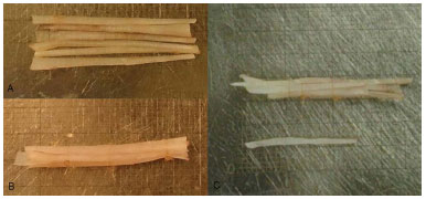
A #11 scalpel blade was used to form cartilage twigs (long strips on the larger diameter of the quadrangular cartilage of 1.0 mm thickness). Six to eight of these units were grouped by tying them with a 5-0 catgut wire to simulate a bundle of twigs (Figures 1A, 1B, and 1C).

Figure 1. A: Septal cartilage grafts in unbound strips as proposed by Souza et al.2. B: Cartilaginous strips bound with 5-0 catgut, forming a graft in the shape of a bundle of twigs. C: Demonstration that this self-contained bundle can be used to increase the graft length, compared to use of isolated strips. In this patient, there was a 10-mm gain in the final length of the graft.
In all patients, columellar struts of septal cartilage measuring approximately 0.3 × 2.5 cm were also used. A fixation was made between the medial crura of the alar cartilages using absorbable monofilament 5-0 sutures.
Then, the height deficit of the nasal dorsum and root to be filled were verified and the bundle of twigs was inserted under the superficial musculoaponeurotic system from the nasal dorsum up to the root for filling, support, and profile improvement (Figures 2A and 2B).

Figure 2. A: Bundle of twigs graft placed on the nasal dorsum to simulate the filling of the defect. B: The dorsum and nasal root aspect after grafting of the "bundle of twigs." Note the rectification of the dorsal line.
After structuring, the skin was sutured with 6-0 mononylon. In cases in which finely diced cartilage was also used to fill the nasal tip, the cartilage was injected through an incision in the nasal wing, before the complete suture.
Bilateral septum splints were affixed in all patients and maintained for 7-15 days.
RESULTS
The bundle of twigs technique showed good pre- and short-term postoperative results. Augmentation of the root and dorsum was achieved in 100% of the cases, without any local complications. Diced grafts were also used, mainly for definition of the nasal tip, in 23 patients.
The conchal and septal cartilage donor regions healed without complications.
The filling and elevation of the nasal dorsum was maintained satisfactorily in patients with 6 months or more of follow-up (Figures 3-5). No grafts were visible under the skin, and no surgical revisions were necessary in the most recent follow-ups.

Figure 3. Pre (A) and 6-month (B) postoperative follow-up showing the height gain at the nasal root, removal of the dorsal hump, and correction resulting from the bundle of twigs graft.

Figure 4. Pre (A) and 6-month (B) postoperative follow-up showing correction of the nasal dorsum hump, maintenance of the root and nasal dorsum height, and smooth nasal profile contour.

Figure 5. Pre (A and B) and 6-month postoperative follow-up (C and D) demonstrating the root height gain, removal of the dorsal hump, and correction obtained by use of a bundle of twigs graft and lateral osteotomy for treatment of laterorhinia.
DISCUSSION
Rhinoplasty is an important procedure in plastic surgery due to the central position and functional importance of the nose on the face2. The search for long-term, well-structured nasal root and dorsum remains a challenge for plastic surgeons1,10.
Technical developments to improve results must be encouraged and monitored by surgeons. Open structure rhinoplasty has become the technique of choice for several authors1,2,5; however, regardless of the access route, grafts are essential tools for nasal structuring3,6,10.
Diced cartilage was previously underused; however, its use was revived after Erol reported good results using this type of graft6. Several studies have shown excellent results using finely diced cartilage, whether or not wrapped in Surgicel3,4,6-8. However, postoperative complications, such as grafts being visible or palpable under thin skin, and unwanted gains in the transverse dimension of the nasal dorsum, led us to seek methods to minimize these effects2.
Grafts in the form of strips, with a dominant longitudinal dimension, are better suited for filling the nasal root and dorsum. They can be used to define the dorsum because of their natural form and are nearly imperceptible to palpation, as they are fine particulates, and are flexible, without memory or elastic force, the characteristics that overcome fibrosis and pressure from the surrounding cutaneous covering. It is also believed that they undergo less absorption and loss, compared to diced grafts, because they preserve more chondrocytes within the long axis of the cartilaginous matrix2.
By binding the longitudinal grafts (strips) in a bundle of twigs structure similar to a thin and irregular wooden beam, this study observed similar outcomes to those offered by the separated strip graft proposed by Souza et al.2, with the additional benefits of extending the graft and increasing its resistance. This potentially increases the long-term success of the results. However, additional long-term studies are necessary to test this hypothesis.
The bundle of twigs graft can be manipulated to perform minor adjustments in the first 15 days after the procedure. This manipulation of fragments has been demonstrated in other rhinoplasty grafting techniques6,8. The bundle of twigs graft also has the advantage that it can be structured, unlike diced cartilage grafts8.
The bundle of twigs grafts showed better results, that is, maintaining the desired shape, with less potential for long-term distortion. The possible dimensions of the graft can be augmented using smaller cartilage fragments. The only disadvantage of septal cartilage is the limited quantity available in some patients3, mainly in secondary approaches. In all our patients, the authors used septal cartilage; however, the principles of this technique may be extended to the use of twigs made from conchal and costal cartilage, thus increasing the reproducibility of this technique.
CONCLUSION
The bundle of twigs cartilage is was efficient and easy to perform. It offers good results for treatment of the nasal dorsum and root in primary or secondary aesthetic, functional, and traumatic cases. Thus, this promising approach in rhinoplasty grafts combines the advantages of strip grafts with the advantage of high resistance offered by the bundle conformation.
REFERENCES
1. Richardson S, Agni NA, Pasha Z. Modified Turkish delight: morcellized polyethylene dorsal graft for rhinoplasty. Int J Oral Maxillofac Surg. 2011;40(9):979-82. PMID: 21514116 DOI: http://dx.doi.org/10.1016/j.ijom.2011.03.013
2. Souza GMC, Costa SM, Afiune RG, Oliveira KR, Miolo TL. Enxertos de cartilagem em rinoplastia: técnica de enxerto em forma de gravetos (stick graft). Arq Catarin Med. 2015;44( Supl 1):166-71.
3. Sajjadian A, Rubinstein R, Naghshineh N. Current status of grafts and implants in rhinoplasty: part I. Autologous grafts. Plast Reconstr Surg. 2010;125(2):40e-49e. PMID: 19910845 DOI: http://dx.doi.org/10.1097/PRS.0b013e3181c82f12
4. Sajjadian A, Naghshineh N, Rubinstein R. Current status of grafts and implants in rhinoplasty: Part II. Homologous grafts and allogenic implants. Plast Reconstr Surg. 2010;125(3):99e-109e PMID: 20195087 DOI: http://dx.doi.org/10.1097/PRS.0b013e3181cb662f
5. Souza GMC, Costa SM, Penna WCNB. Enxerto de cartilagem picada injetável para rinoplastia: método e experiência do Hospital Felício Rocho. Rev Bras Cir Craniomaxilofac. 2012;15(1):17-20.
6. Erol OO. The Turkish delight: a pliable graft for rhinoplasty. Plast Reconstr Surg. 2000;105(6):2229-41. DOI: http://dx.doi.org/10.1097/00006534-200005000-00051
7. Daniel RK, Calvert JW. Diced cartilage grafts in rhinoplasty surgery. Plast Reconstr Surg. 2004;113(7):2156-71. PMID: 15253210 DOI: http://dx.doi.org/10.1097/01.PRS.0000122544.87086.B9
8. Daniel RK. Diced cartilage grafts in rhinoplasty surgery: current techniques and applications. Plast Reconstr Surg. 2008;122(6):1883-91. PMID: 19050542 DOI: http://dx.doi.org/10.1097/PRS.0b013e31818d2104
9. Kelly MH, Bulstrode NW, Waterhouse N. Versatility of diced cartilage-fascia grafts in dorsal nasal augmentation. Plast Reconstr Surg. 2007;120(6):1654-9. DOI: http://dx.doi.org/10.1097/01.prs.0000285185.77491.ab
10. Gunter JP, Landecker A, Cochran CS. Frequently used grafts in rhinoplasty: nomenclature and analysis. Plast Reconstr Surg. 2006;118(1):14e-29e. PMID: 16816668 DOI: http://dx.doi.org/10.1097/01.prs.0000221222.15451.fc
1. Hospital Felício Rocho, Belo Horizonte, MG, Brazil
2. Sociedade Brasileira de Cirurgia Plástica, São Paulo, SP, Brazil
3. Associação Brasileira de Cirurgia de Crânio-Maxilo-Facial, São Paulo, SP, Brazil
Institution: Clínica de Cirurgia Plástica do Hospital Felício Rocho, Belo Horizonte, MG, Brazil.
Corresponding author:
Sérgio Moreira da Costa
Rua Timbiras, 3642, Barro Preto
Belo Horizonte, MG, Brazil Zip Code 30140-062
E-mail: sergio.plastica@bol.com.br
Article received: August 1, 2015.
Article accepted: November 17, 2015.


 Read in Portuguese
 Read in English
 PDF PT
 Print
Print
 Send this article by email
 How to Cite
 Mendeley
 Pocket
 Twitter