


Original Article - Year 2015 - Volume 30 -
Surgical correction of prominent ears: association of the Furnas and Mustardé techniques
Correção cirúrgica de orelhas em abano: associação das técnicas de Furnas e Mustardè
ABSTRACT
INTRODUCTION: Prominent ears, popularly known as bat ears, are the most common deformity of the head and neck, occurring in approximately 5% of the general population. This study aims to demonstrate that, with the use of simple surgical techniques, it is possible to correct the deformity and achieve optimal patient satisfaction.
METHOD: A total of 60 patients of both sexes, aged between 11 and 40 years, underwent bilateral otoplasty between February 2009 and December 2010.
RESULTS: In all cases, bilateral otoplasty was performed. There were no cases of hematomas, surgical site infection, or hypertrophic scars. In 5 cases (8.3%) visible scars or foreign body granulomas were found in the posterior surface of the ear. Chondritis occurred in one patient. Bilateral residual deformity occurred in 3 cases at 1 year post-surgery. Total recurrence was observed in 1 case (1.7%). After one year of surgery, 56 patients (93.3%) considered the result as good and were satisfied, 3 patients underwent reintervention for residual deformity, and total recurrence occurred in 1 case, which the patient did not wish to correct.
CONCLUSION: The present study demonstrates that concurrent use of the Mustardé and Furnas otoplasty techniques results in a high degree of satisfaction and a low rate of complications. The procedure can easily be performed at the outpatient level and at a low cost.
Keywords: Ear/surgery; Surgical procedures; Acquired ear defects.
RESUMO
INTRODUÇÃO: Orelha proeminente, popularmente conhecida como orelha em abano, é uma afecção muito frequente em nosso meio. Constitui a deformidade mais comum da cabeça e pescoço, prevalecendo em aproximadamente 5% da população em geral. O trabalho tem como objetivo demonstrar que, com a junção de técnicas cirúrgicas simples, é possível corrigir as deformidades e obter um ótimo índice de satisfação dos pacientes.
MÉTODO: Um total de 60 pacientes de ambos os sexos, entre 11 e 40 anos foram submetidos à otoplastia bilateral entre fevereiro de 2009 e dezembro de 2010.
RESULTADOS: Em todos os casos se realizou otoplastia bilateral. Não houve casos de hematomas, infecção de sítio cirúrgico ou cicatriz hipertrófica. Cicatrizes visíveis ou granulomas de corpo estranho na face posterior da orelha foram constatados em 5 casos (8,3%). Houve 1 caso de condrite. O índice de deformidade residual com 1 ano de pós-operatório ocorreu em 3 casos, sendo todos bilaterais. Recidiva total foi presenciada em 1 caso (1,7%). Após um ano da cirurgia, 56 pacientes (93,3%) consideraram o resultado como bom e estavam satisfeitos, 3 pacientes sofreram reintervenção por deformidade residual e em 1 caso ocorreu a recidiva total, em que o mesmo não desejou correção.
CONCLUSÃO: O presente estudo vem demonstrar que a associação das técnicas de Mustardè com a de Furnas traz alto grau de satisfação, baixo índice de complicações, podendo ser realizada com tranquilidade em nível ambulatorial e com baixo custo.
Palavras-chave: Orelha/cirurgia; Procedimentos cirúrgicos operatórios; Deformidades adquiridas da orelhas.
Popularly known as "bat ears" the prominent ear is the most common deformity of the head and neck, and is present in about 5% of the general population. 8% of cases have a family history. Children, adolescents and adults with this condition may have psychiatric disorders related to social interaction.
Prominent ears are characterized by the following changes:
1) Deletion or absence of antihelix, with scapholunate-conchal angle of > 90 º;The ear has two stages of growth: one that is completed around 6-7 years of age, which coincides with school life, and one that begins in the last three decades of life.
2) Excessively deep or hyperdeveloped shell with increased cephalo-auricular angle of > 40 º;
3) Combination of the deformities of item 1 and 2, the most common finding;
4) Protrusion of the earlobe.
The correction for this anatomical defect should be performed from the age of 7 years, when the auricular development is complete. At preschool age, children are not aware of the deformity and therefore do not suffer psychological trauma, which is frequently observed in older school children.
In the correction of prominent ear, in order to obtain the best results, one must observe and comply with the following:
1) Resect an ellipse of skin in post-auricular region where the final scar should be hidden in the retroauricular sulcus.The history of protruding ear correction surgery (otoplasty) commenced in 1848 with Dieffenbach1 proposing excision of retroauricular skin along the posterior sulcus with primary suture of the borders. In 1881, Ely2 reduced the cephalo-auricular angle, removing a strip of the lateral and medial cartilage with the skin.
2) Create an antihelix, with rounded contours, avoiding borders with sharp edges.
3) Avoid overcorrection of the antihelix which causes effacement of the helix.
4) Reduction of conchal hypertrophy in selected cases, when indicated.
5) Control the position of the earlobe, which often remains anterior when the ear is positioned posteriorly.
6) Attachment of the concha to the fascia of the mastoid when required.
In 1963 Stenstrom3 modified the Pierce4 technique performing cartilage scarification instead of incising it. The area of scapholunate-conchal angle was narrowed by rasping and posterior access was made through a tunnel to reach the anterior surface of the cartilage.
After using several techniques, in 1963, Mustardè5 , proposed avoiding making excisions, incisions or weakening the cartilage, because he considered that a simple suture was sufficient to maintain the shape of the ear during healing.
Furnas6 in 1968, advocated the posterior detachment and sectioning of the posterior auricular muscle, with the fixation of the cartilage to the periosteum of the mastoid.
In 1977, Brent B.7 performed the resection of a strip of conchal cartilage with repositioning of the upper third of the ear by incorporating the technique of Stenstrom and Mustardé.
Psillakis et al.8 (1979) used the Mustardè method associated with the Furnas procedure.
Although there are over 200 described techniques, the best surgery for prominent ear correction that would be that which was easily reproducible, versatile, simple, and which produced good results and few complications.
This study aims to demonstrate that with simple surgical techniques, it is possible to correct the deformity and achieve an optimal patient satisfaction index.
METHODS
A total of 60 patients of both sexes, between 11 and 40 years underwent bilateral otoplasty in the Agamenon Magalhães Hospital between February 2009 and December 2010.
All patients were evaluated in preoperative consultations and postoperative consultations up to 1 year to obtain the patient's medical history, to ascertain the real desire to be submitted to the deformity correction, to explain the proposed treatment, the technique, its limitations and risks, and postoperative consultations up to 1 year for postoperative care and to assess the level of satisfaction with the outcome. The procedures were performed on an outpatient basis in the operating room. Patients were discharged on the same day.
Conchal hypertrophy associated with an obliterated antihelix was present in 25 patients (41.7%), obliterated antihelix alone was present in 20 cases (33.3%) and conchal hypertrophy alone was found in 15 patients (25%).
Surgical Technique (Figures 1 to 5)

Figure 1. Demarcation of the antihelix.

Figure 2. Cartilage Weakening.

Figure 3. Demarcation of the area of the concha being resected.

Figure 4. Stitches joining the concha and mastoid.

Figure 5. Final Appearance.
1. With the patient in the supine position, after previous demarcation of the zone of skin to be resected, and with the posterior auricular sulcus as reference, antisepsis of the whole face of the ears and neck was carried out. This allowed visualisation of the whole face in relation to the ears. Prophylactic antibiotics were not used due to lack of data supporting its use for this type of procedure.The patient was discharged with instructions to return to the hospital on the first postoperative day to remove the dressing and to start using an elastic band or cap to protect against injury, for 7 days continuously, followed by 7 days only while sleeping. The stitches were removed 21 days after surgery (Figure 6 and 7).
2. Local anesthesia with 0.5% lidocaine and epinephrine in a proportion of 1: 100,000. First, the greater auricular nerve was blocked, followed by subdermal infiltration of the entire ellipse that was to be resected, covering the entire posterior surface of the ear, to facilitate resection of the skin and release the antihelix, as well as to reduce the risk of inadvertent cartilage injury. Subsequently, blocking of the auriculotemporal nerve and subdermal infiltration of the concha was carried out to facilitate its resection when removal was required.
3. Resection of the zone of skin, release of the concha and subsequent antihelix exposure. The auricular muscle was sectioned routinely to view the concha more clearly.
4. Marking with dye, using 25 x 7 mm needles, for further demarcation of the new anti-helix (Figure 1).
5. Scoring and weakening of the posterior region of the cartilage was performed for better modelling and preparation of stitches in the antihelix (Figure 2).
6. Making the stitches with 4-0 nylon in the form of a "U" (usually 03 stitches), as recommended by Mustardè, to construct the new antihelix.
7. Economic resection of the concha in cases of severe hypertrophy, where the resection would deform and obstruct the auditory canal ("kidney"-shaped resection) (Figure 3).
8. Fixation and rotation of concha with 4-0 nylon sutures (three stitches) between the concha and the periosteum of the mastoid as recommended by Furnas (Figure 4).
9. Closure of skin with continuous intradermal suture with 4-0 nylon suture. Appearance at the end of surgery (Figure 5).
10. Dressing with cotton soaked with saline in the projections and recesses of the helix and antihelix.

Figure 6. (A and B) Pre-operative female patient, 24 years-old. (C and D) Post-operative female patient, 24 years-old.
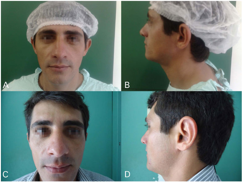
Figure 7. (A and B) Pre-operative male patient, 30 years-old. (C and D) Post-operative male patient, 30 years-old.
RESULTS
The average age of the sample was 25.7 years, ranging between 11 and 40 years. Of the 60 operated patients, 26 (43.3%) were female and 34 (56.7%) were male.
All patients were operated in ambulatory surgery, 55 patients (91.7%) with only local anesthesia and 5 patients (8.3%) operated under local anesthesia and sedation.
In all cases, bilateral otoplasty was performed. There was no hematoma or surgical site infection. Hypertrophic scar did not occur in any case.
Visible scars or foreign body granuloma in the posterior aspect of the ear were found in 5 cases (8.3%). There was one case of chondritis in the fourth month after surgery.
The residual deformity rate at 1 year post surgery occurred in 3 cases, and all were bilateral. Complete recurrence was observed in 1 case (1.7%).
One year after surgery, 56 patients (93.3%) considered the outcome as good and were satisfied. Three patients underwent unilateral reintervention to correct the small deformity. The patient with complete relapse chose not undergo further surgery.
DISCUSSION
Mustardé5 reports a series of 391 cases operated with his own technique over a period of 10 years and found 17 unsatisfactory cases with residual deformity (4.3%). Other authors, using the same technique were not able to achieve the same results.
Tan et. al.9 reported a comparative study with 146 patients where 45 were operated by Mustardé technique with 24% residual deformity and 91% satisfaction. 101 patients were operated by the "anterior scoring" technique (Stenstrom), obtaining 10% residual deformity and 85% patient satisfaction.
Guyuron and Deluca10 operated on a series of 44 patients under local anesthesia with the Mustardé technique, and obtained 100% patient satisfaction and no cases of residual deformity. However, there were 13 cases of slight imperfections observed on follow-up.
Grabb11, in a series of 135 patients, operated by the Mustardé technique, obtained 12.3% residual deformity.
With the Stenstrom technique, in a series of 562 cases, there was 8% residual deformity12 and Caouette-Laberge et al.13 found deformities in 5.7% of 500 operated cases.
The cartilage weakening technique is based on observations by Gibson and Davis14, in which the cartilage tends to bend in the direction opposite to the affected area. Fry15 later confirmed this principle and Stenstrom12 applied this theory in otoplasty. It is important to note that it is possible to change the level of the curvature according to the degree of cartilage weakening. Although it is quite well established that the cartilage tends to bend in the direction opposite to the weakened area, by scoring the posterior surface to thin the cartilage, it was possible to make an appropriate antihelix without using a posterior approach.
In this study, 5% residual deformity and 93.3% satisfaction was observed. These data are approximate to what is in the literature. The zero rate of infection and hematoma formation demonstrate the safety of the technique. Complications such as visible and palpable scarring (8.3%) were also low. However, visible sutures are higher by the Mustardé technique due to the use of permanent internal sutures. Such problems usually occur later and the sutures can be removed without problems.
When relapses occur in Mustardé technique, they usually occur upto 12 months after surgery. Some degree of loss of correction can occur in up to 2/3rds of patients, mainly in the upper pole of the ear, as seen in the work McCarthy16. These authors advocated a slight overcorrection, especially at the upper pole, anticipating this probable loss of correction. In this study, there was one total recurrence. The complications observed were due to:
A) Incorrect placement or insufficient number of sutures. Mustardé5 reports that four is the minimum number of sutures between the concha and the scapha, and at least two sutures between the concha and the mastoid.In cases of very firm cartilage which is difficult to mould with sutures, it is preferable to use cartilage weakening techniques either by the anterior approach, advocated by most, or by posterior approach to achieve better results, since in this group of patients relapse with the Mustardé technique is more likely.
B) Excessive removal of skin on the posterior surface of the ear, leaving the skin suture taut, with a greater chance of recurrence of ear protrusion to relieve the tension.
C) Local postoperative trauma.
D) "Memory" of the cartilage, which tends to return to its original position.
CONCLUSION
Otoplasty is a procedure that has existed for over a hundred years, and in most cases brings great satisfaction to patients, improving their self-image and social life.
The objectives of performing an otoplasty are widely known and may be achieved through a careful physical examination, observation of asymmetry, analysis of the consistency of the cartilage, affected angles, the position of the lobe, and principally through adequate surgical planning. The latter includes conservative excision of skin, non-absorbable sutures for both antihelix modelling and attachment of the concha to the mastoid, concha resection when indicated, and preventing the effacement of the helix due to overcorrection of the antihelix. The identification and implementation of these manoeuvres, when performed, reduce the need for a reoperation.
This study demonstrates that the association of the Mustardé with Furnas techniques brings high satisfaction and a low rate of complications. It is a simple procedure that can be performed with ease on an outpatient basis at a low cost.
REFERENCES
1. Dieffenbach JE. Die ohrbildung otoplastik. In: Die operative Chirugie. Leipzig: Brockhauss; 1848. P.395-7.
2. Ely ET. An Operation for prominence of the auricle. Arch Otolaryngol. 1881;10:97.
3. Stenstrom SJ. A "natural" technique for correction of congenitally prominent ears. Plast Reconstr Surg. 1963;32:509-18.
4. Pierce GW, Klabunde EH, Bergeron VL. Useful procedures in plastic surgery. Plast Reconstr Surg (1946).1947;2(4)358-61. DOI: http://dx.doi.org/10.1097/00006534-194707000-00009
5. Mustardé JC. The correction of prominent ears. Using simple mattress sutures. Br J Plast Surg. 1963;16:170-176.
6. Furnas D. Correction of prominent ears by concha mastoid sutures, Plast Reconstr Surg. 1968;42:189.
7. Brent B. The acquired auricular deformity. Plast Reconstr Surg. 1977;59:475-485. PMID: 322165 DOI: http://dx.doi.org/10.1097/00006534-197759040-00001
8. Psillakis JM. Prominent ears: evolution of a surgical tecnique. Aesthetic Plst Surg. 1979;3:147-152. DOI: http://dx.doi.org/10.1007/BF01577849
9. Tan ST, Gault DT. When do ears become prominent? Br J Plast Surg. 1994;47(8):573-4. PMID: 7697287
10. Guyuron B, DeLuca L. Ear projection and the posterior auricular muscle insertion. Plast Reconstr Surg. 1997;100(2):457-60. PMID: 9252616 DOI: http://dx.doi.org/10.1097/00006534199708000-00030
11. Grabb CW. Cirurgia Plastica. São Paulo: Salvat; 1984.
12. Stenstrom SJ, Heftner J. The stenstrom otoplasty. Clin Plast Surg. 1978;(3)491-5.
13. Caouette-Laberge L, Guay N, Bortoluzzi P, Belleville C. Otoplasty: anterior scoring technique and results in 500 cases. Plastic and Reconstructive Surgery 105(2):504-15, 2000. PMID: 10697153
14. Gibson T and Davis WD. The distortion of auto-genous cartilage grafts, its cause and prevention. Br J Plast surg 10:257,1958.
15. Fry HJH. Interlocked stresses in human nasal septal cartilage. Br J Plast Surg 19:276,1966. DOI: http://dx.doi.org/10.1016/S0007-1226(66)80055-3
16. McCarthy JG, ed. Plastic Surgery vol 3. Philadelphia: W.B. Saunders; 1990.
1. Sociedade Brasileira de Cirurgia Plástica, Recife, PE, Brazil
2. Hospital Universitário da Univasf-Petrolina, PE, Brazil
3. Hospital Agamenon Magalhães, Recife, PE, Brazil
Institution: Hospital Agamenon Magalhães, Recife, PE, Brazil.
Corresponding author:
Ernani Coelho Alencar
Rua Isaac Salazar, 189, Tamarineira
Recife, PE, Brazil Zip Code 52050-160
E-mail: ernaniplastica@hotmail.com/ecalencar@hotmail.com
Article received February 27, 2012.
Article accepted July 01, 2012.
 Read in Portuguese
 Read in English
 PDF PT
 Print
Print
 Send this article by email
 How to Cite
 Mendeley
 Pocket