


Special Article - Year 2015 - Volume 30 -
Fournier gangrene: Reconstruction of the scrotal sac with a fasciocutaneous flap from the internal thigh region
Síndrome de Fournier: Reconstrução de bolsa testicular com retalho fasciocutâneo de região interna de coxa
ABSTRACT
INTRODUCTION: Fournier gangrene is a rapidly progressing multi-bacterial infection in the perineal region. The treatment of this condition includes debridement, broad-spectrum antibiotic therapy, and oxygen therapy in a hyperbaric chamber. Aggressive debridement typically results in the loss of skin coverage of the entire scrotal sac, and the exposure of both testes. During treatment, it is essential to use well-vascularized flaps to ensure the recovery of function.
METHOD: We describe the application of a fasciocutaneous flap-which takes advantage of the rich arterial network of the internal region of the thigh-in the perineal reconstruction method proposed by Ferreira et al. that allows for the treatment of large defects.
CONCLUSION: The flap is quite versatile. Its advantages include its utility in various clinical situations, low risk of gangrene in the donor area, single-stage reconstruction, and adequate flap thickness for reconstruction.
Keywords: Fournier gangrene; Necrotizing fasciitis; Surgical flaps.
RESUMO
INTRODUÇÃO: A síndrome de Fournier é uma infecção multibacteriana de rápida progressão em região perineal. Seu tratamento inclui desbridamento, antibioticoterapia de amplo espectro e terapia com oxigênio em câmara hiperbárica. O desbridamento agressivo tipicamente resulta em perda da cobertura cutânea de toda bolsa escrotal, expondo ambos os testículos. No tratamento, é necessária a utilização de retalhos bem vascularizados para o reestabelecimento das funções.
MÉTODO: Apresentamos a aplicação de um retalho fasciocutâneo, aproveitando a rica rede arterial da região interna da coxa para a reconstrução perineal, proposto por Ferreira et al., o qual permite o tratamento de amplos defeitos.
CONCLUSÃO: O retalho descrito para reconstrução perineal é bastante versátil. Suas vantagens incluem a possibilidade de ser utilizado em diversas situações clínicas, baixo acometimento de gangrena na região doadora, reconstrução em único estágio e a espessura do retalho adequada para reconstrução desta região.
Palavras-chave: Gangrena de Fournier; Fasciite necrotizante; Retalhos cirúrgicos.
Fournier syndrome was described in 1883 by Jean Alfred Fournier-a French dermatologist specializing in venereal diseases-as a rapidly progressing infection of the perineum and the abdominal wall that extends to the scrotum and penis through the dartos fascia. Fournier stated that there were three characteristic findings of this syndrome: sudden onset in young and healthy males, rapid progression, and absence of a specific etiologic agent1. The etiology of this disease is controversial. Fournier originally reported cases with no known etiology. Currently, approximately 25% of the cases still remain idiopathic, although there are many predisposing factors, such as diabetes mellitus and paraphimosis2.
However, in the majority of the cases, this condition is thought to result from common urologic or colorectal procedures or diseases, including surgical complications of hemorrhoidectomy, orchiectomy, hernia, vasectomy and circumcision. The pathological characteristics of the disease and the port of entry of micro-organisms are also well defined. There are three possible pathophysiological mechanisms: local trauma, which would facilitate the entry of micro-organisms through injured skin; infection of the genitourinary tract, with extension to deep planes; and/or infection of the perineal or retroperitoneal space3.
The bacteria that reach the subcutaneous tissue cause obliterative endarteritis in the vessels that supply blood to the scrotal and perineal region, thus causing necrosis. The combination of edema, inflammation, and infection decreases the local blood supply and the resulting hypoxia serves as a predisposition to the growth of obligate and facultative anaerobic micro-organisms. The anaerobic metabolism of these bacteria results in the production of gas, which accumulates in the subcutaneous tissues and causes the crackling palpation4,5.
Fournier syndrome affects not only men, as proposed by the author. The low incidence in women is attributed to better hygiene and local physiological drainage, through vaginal secretions. In elderly, immunocompromised, bedridden, and dependent patients, this disease may be asymptomatic, whereas it more commonly manifests as sepsis in young adults. The high number of surgical procedures and longer hospitalization time may be factors that impede an early diagnosis, which may be associated with a high mortality rate6.
The necrotizing fasciitis is psychologically devastating and requires treatment for a good esthetic and functional outcome. The treatment includes extensive debridement, broad-spectrum antibiotic therapy, and hyperbaric oxygenotherapy to eliminate the infection, as well as protection of the exposed testicles through reconstruction of the perineal and penile region7. The coverage defect resulting from the debridement of the affected tissues can be extensive, and may result in the exposure of the testicles, penis, abdominal wall, and spermatic funicle.
There are several methods for the closure of the defect resulting from the initial surgical treatment. We present our experience with the use of a versatile flap, which takes advantage of the rich arterial network in this region, for the reconstruction of the perineal region based on the method described by Ferreira et al. in 20068.
METHOD
Surgical Technique
The flap presented here is based on a dense network of perforating vessels of the perineal region and proximal portion of the thigh, as well as the deep branches of the internal pudendal and external pudendal arteries. It is extracted from the medial portion of the root of the thigh, with a longitudinal axis located between the ischial tuberosity and internal femoral condyle; moreover, it has an adequate size to cover the defect and allow for primary closure of the donor area (Figure 1). The patient is maintained in the lithotomy position, with the legs apart, thus exposing the perineal defect area to the maximum extent possible. After debridement of the affected area and determining the appropriate size of the flap, which is usually weeks after resolution of the infection, an incision is made up to the superficial fascia while maintaining the base of the flap in the anterior region. The flap is lifted and transposed to the defect (Figure 1 and 2). The donor area is primarily closed and drained with a penrose or drain.
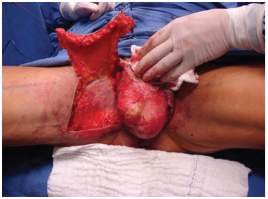
Figure 1. Flap from the internal portion of the raised thigh. Note that the length of the flap can be decided based on the defect.
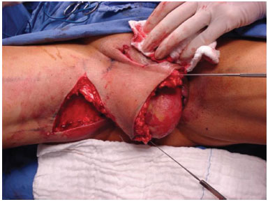
Figure 2. Transposition of the flap and coverage of the debrided area.
CASE REPORT
Case 1
A 64-year-old man had a history of hypertension, and presented with necrotizing fasciitis without any predisposing factor. He initially underwent surgical debridement and antibiotic therapy. Scrotal reconstruction was performed 20 days after the onset of symptoms. Under spinal anesthesia, in the lithotomy position, a local flap was constructed. The surgery time was 80 minutes. He was discharged on the first postoperative day, and was treated with oral analgesics and antibiotics. At the follow-up, the patient showed excellent evolution and proper healing of the wound (Figures 3 and 4).
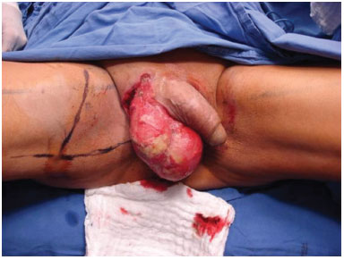
Figure 3. Flap from the internal portion of thigh, designed to cover the defect.
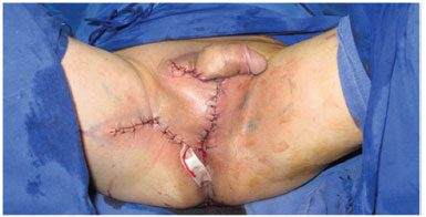
Figure 4. Immediate postoperative period, with complete coverage of the defect and without any tension.
Case 2
A 53-year-old man with diabetes presented with a 3-day history of necrotizing fasciitis (Figure 5). He underwent several debridements prior to perineal reconstruction, which was performed 3 weeks after establishing the clinical framework. The surgery time was 90 minutes, and the patient's course progressed satisfactorily (Figures 6 and 7).
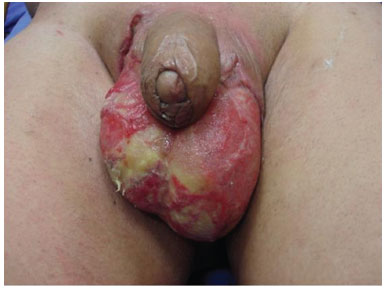
Figure 5. Necrotizing fasciitis, after debridement.
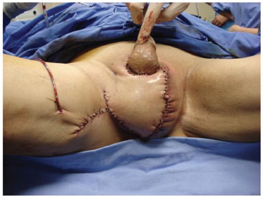
Figure 6. Immediate postoperative period, with complete coverage of the defect and without tension.
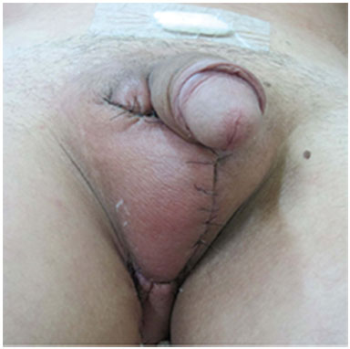
Figure 7. 15th Postoperative day, with good evolution of the wound
DISCUSSION
Necrotizing fasciitis in the perineal and abdominal region is a soft tissue infection that rapidly spreads to the adjacent tissues. This potentially fatal disease is associated with mortality rates between 3% and 67 %8.
In 1889, Manchot described in detail the arterial supply of the perineal region. He divided the genital region in 2 vascularization areas-the anterior and posterior portions. The anterior region is primarily vascularized by the superficial external pudendal artery and the deep pudendal external artery. The posterior region is supplied by the internal pudendal artery, which branches medially and laterally. The lateral branches reach the lower medial portion of the gluteus maximus muscle and supply this region. The medial branches vascularize the skin in the genital region and infero-medial region of the gluteus maximus muscle. The inferior gluteal artery also contributes to the blood supply of the skin in this region, and is anastomosed with the branches of the internal pudendal artery9,10.
In 1973, Horton described for the first time the principles of random flaps from the perineal region11. However, the versatility of these flaps was limited. After a better knowledge of the vascular network and its anastomoses was obtained, safer fasciocutaneous flaps were developed for the reconstruction of the perineum.
For the treatment of raw areas resulting from the necrotizing fasciitis of the scrotum, three aspects should be considered: wound healing, maintenance of function, and aesthetic recovery. A few days after the resection of the necrotic tegument, granulation tissue will cover the raw areas. This period varies according to the local and general conditions of the patient that may affect tissue regeneration. Its formation indicates that the infection that caused the condition is under control. Reconstruction should not be attempted in such cases, as it would represent a new attack on the yet vulnerable tissues. On the other hand, the contraction that follows the formation of granulation may compress the testicles, which may also lead to bending, that may affect spermatogenesis in the future. However, testicular function may have already been impaired by the disease itself. There are several ways to cover the wound, and the ideal method would allow appropriate functionality and a more aesthetic appearance. The correct time for initiating surgery is important in the treatment of these patients 12.
Skin grafting is a simple method that can be used for penile-scrotal reconstruction, and can provide a thin, rough, easily adaptable coverage that closely resembles the natural aspect of the region. It is a good method for the coverage of the penis, but has higher morbidity rates for the treatment of the scrotal sac, due to its relief. It is usually safe and simple. However, the graft undergoes intense contraction over time, may encounter difficulty in attaching to the receptor bed, may experience maceration by urine, may be exposed to contamination by feces, and may be associated with a risk of loss due to local infection13,14.
Hence, local flaps are excellent alternatives for reconstruction. The perineum presents an ample anastomotic network of blood vessels, similar to the face, which allows for the establishment of several flaps. The fasciocutaneous flaps provide good resistance, with coverage of an extensive area of fat and fascia, thus yielding superior results as compared to grafting with split-thickness skin grafts15.
We present our experience with the use of the transposition flap for perineal reconstruction in cases of necrotizing fasciitis, as described by Ferreira et al. It is very versatile, and its advantages include its utility in various clinical situations, low risk of gangrene in the donor area, the single-stage reconstruction method, a suitable flap thickness for reconstruction. In addition, the flaps are relatively simple, can be established quickly, and leave scars in non-visible locations. The major disadvantage of the flap is the limited transversal dimension due to a low elasticity in the thigh region. No cases of wound dehiscence or other postoperative complications were reported.
There are other options of fasciocutaneous flaps. The fleur-de-lis flap-a fasciocutaneous flap based on the internal pudendal artery-is frequently used for vulvo-vaginal and neoescrotum reconstruction.4 Its anatomic configuration and vascularization are ideal for single-stage procedures, and it also allows for the recreation of scrotum volume and coverage of the testicles. Another advantage of this flap is that the donor area is hidden in the medial portion of the thighs; thus, excellent cosmetic results are obtained, with little difference between the color of the skin of the donor and receiver area, and the functional outcome is good as well. This flap can also be used with dermal matrix to improve the final aesthetic aspect 16.
The inguinal flap, which is another option for defect coverage in the region, should be contraindicated if the inguinal region has previously been used for flaps.
The Singapore flap is based on the cutaneous vessels and results in good coverage of the scrotum. However, it has a short pedicle and does not reach the dorsal region of the penis. In this case, it is necessary to add a skin graft to the penis.
The anterolateral thigh flap is relatively simple, presents enormous local vascularization and minimal morbidity of the donor site, and is thinner than the myocutaneous gracilis flap. It has been widely used since it was first described by Song in 1984. Its long pedicle allows for a large arc of rotation and is the key to success in the implementation of this flap, by allowing the reconstruction of adjacent areas17.
The fasciocutaneous supero-medial thigh flap was described by Hirshowitz (1980) for scrotum and vulva reconstruction. Its extensive vascularization, based on deep external pudendal arteries, offers better safety. Another advantage is the maintenance of sensory innervation by the genital branch of the genito-femoral nerve and ileo-inguinal nerve. Its major limitation is that the wide transverse dimension prevents the primary closure of the donor area18.
When the defect resulting from debridement is extensive and deep, a muscle flap is necessary to reduce the dead space. The gracilis muscle flap, as a muscle flap and in the combined myocutaneous form, can be used for the reconstruction of the scrotum or inguinal region. It is a safe flap, is easily executable, and presents good vascularization, which protects against new local infections. On the other hand, it utilizes a functional muscle and is thus associated with greater morbidity.
The advantages of fasciocutaneous flaps are numerous: they spare the muscle, allow multiple components to be included, and decrease the pain and functional deficit in the donor area. With regard to the disadvantages, the fasciocutaneous flaps based on perforating vessels have a lesser thickness and, therefore, are limited to reconstructions that require more filling. The myocutaneous flaps, in comparison, have good thickness and are very well vascularized. However, the muscle tissue is more sensitive to ischemia and the removal of muscles in the donor area has higher functional morbidity. In the patients in this series, there was no need for more volume, beyond that provided by the fasciocutaneous flap.
CONCLUSION
The fasciocutaneous flap proposed by Ferreira et al. provides easy and quick reconstruction of the cases that present with necrotizing fasciitis and that require reconstruction.
REFERENCES
1. Kılıç A, Aksoy Y, Kılıç A. Fournier's gangrene: etiology, treatment, and complications. Ann Plast Surg, 2001 PMID: 11716264
2. Chen SY, Fu JP, Chen TM, Chen SG. Reconstruction of Scrotal and Perineal Defects in Fournier's gangrene. J Plast Rec Aest Surg, 2010
3. Ellabban MG, Townsend PL. Single-stage Muscle Flap Reconstruction of Major Scrotal Defects: Report pf Two Cases. B J Plast Surg 2003 DOI: http://dx.doi.org/10.1016/S0007-1226(03)00178-4
4. Jones RB, Hirschmann JV, Brown GS, Tremann JA. Fournier's syndrome. Necrotizing subcutaneous infection of the male genitalia. J Urol, 1979 PMID: 381687
5. Fahal AH, Hassan MA. Fournier's gangrene in Khartoum. Br J Urol, 1988 PMID: 3293689
6. Moses, A. E. Necrotizing fasciitis: flesh-eating microbes. Israel J Medical Sciences, 1996
7. Riseman JA, Zamboni WA, Curtis A. Hyperbaric oxygen therapy for necrotizing fasciitis reduces mortality and the need for debridements. Surgery 1990 PMID: 2237764
8. Ferreira PC, Reis JC, Amarante JM, Silva AC, Pinho CJ, Oliveira IC, Silva PN. Fournier's Gangrene: A Review of 43 Reconstructive Cases. Plast Rec Surg 2007 DOI: http://dx.doi.org/10.1097/01.prs.0000244925.80290.57
9. Ye X, Wang C, Yu Y, Zheng S. Pedicled Deep Inferior Epigastric Perforator Flap An Alternative Method to Repair Groin and Scrotal Defects. Ann Plast Surg 2011
10. Yii NW, Niranjan NS. Lotus petal flaps in vulvo-vaginal reconstruction. Br J Plast Surg 1996 PMID: 8976747 DOI: http://dx.doi.org/10.1016/S0007-1226(96)90132-0
11. Mccraw, J. B., Massey, F. M., Shanklin, K. D., & Horton, C. E. Vaginal reconstruction with gracilis myocutaneous flaps. Plast Rec Surg 1976. DOI: http://dx.doi.org/10.1097/00006534-197608000-00006
12. Montoya Chinchilla R, Izquierdo Morejon E, Nicolae Pietricicâ B, Pellicer Franco E, Aguayo Albasini JL, Miñana López B. Fournier's gangrene. Descriptive analysis of 20 cases and literature review. Actas urologicas españolas, 2011
13. Morris SF. Inveted Discussion: Pedicled Deep Inferior Epigastric Perforator Flap: An Alternative Method to Repair Groin and Scrotal Defects. Ann Plast Surg 2006 DOI: http://dx.doi.org/10.1097/01.sap.0000239822.42841.fb
14. Chen, S. Y., Fu, J. P., Chen, T. M., & Chen, S. G. Reconstruction of scrotal and perineal defects in Fournier's gangrene. J Plast, Reconst & Aesthetic Surg, 2011 DOI: http://dx.doi.org/10.1016/j.bjps.2010.07.018
15. Yu P, Sanger, JR, Matloub HS, Gosain A, Larson D. Anterolateral Thigh Fasciocutaneous Island Flaps in Perineoscrotal Reconstruction. Plast Rec Surg 2001
16. Payne CE, Williams AM, Hart NB. Lotus petal flaps for scrotal reconstruction combined with Integra_ resurfacing of the penis and anterior abdominal wall following necrotising fasciitis. J Plast Rec Aest Surg, 2009 DOI: http://dx.doi.org/10.1016/j.bjps.2007.09.015
17. Song, Y., Chen G, Song YL. The free thigh flap: a new free flap concept based on the septocutaneous artery.. Br J Plast Surg, 1984 DOI: http://dx.doi.org/10.1016/0007-1226(84)90002-X
18. Hirshowitz B., Moscona R,Kaufman T, Pnini A. One-stage reconstruction of the scrotum following Fournier's syndrome using a probable arterial flap. Plast Reconst surg ,1980 PMID: 7208675 DOI: http://dx.doi.org/10.1097/00006534-198010000-00021
1. Universidade Federal do Paraná. Curitiba, PR, Brazil
2. Sociedade Brasileira de Cirurgia Plástica. São Paulo, SP, Brazil
3. Universidade de Yale, USA
4. Universidade de São Paulo, São Paulo, SP, Brazil
Institution: Universidade Federal do Paraná, Curitiba, PR, Brazil.
Corresponding author:
Priscilla Balbinot
Rua General Carneiro, 181, Alto da Glória
Curitiba, PR, Brazil Zip code 80060-900
E-mail: pribalbinot@hotmail.com
Article received: December 23, 2013.
Article accepted: March 15, 2015.
 Read in Portuguese
 Read in English
 PDF PT
 Print
Print
 Send this article by email
 How to Cite
 Mendeley
 Pocket