


Original Article - Year 2015 - Volume 30 -
Unique surgery for the upper body segment in patients with massive weight loss: thoracobrachio-mammoplasty
Abordagem do segmento superior do corpo em pacientes ex-obesos com cirurgia única: toracobraquio-mamoplastia
ABSTRACT
INTRODUCTION Ex-obese patients require a complex surgical approach because of the large amount of excess skin due to their massive weight loss. In some cases, several plastic surgeries are needed, and there is no existing standard in the coordination of these surgeries. In the upper segment of the body, the arms, side of the thorax, and breasts are usually affected, mainly in women. Several techniques have been developed with the aim of achieving better results with better hidden scars. Some techniques may be associated, being carried out in a single surgical procedure. A well-structured surgical team leads to a reduced surgical time, which means higher safety for the patient.
METHOD: We present a technique for standardization in the treatment of ex-obese patients that is performed in a single step, comprising mammoplasty (according to Pitanguy's technique or with placement of breast prosthesis), thoracoplasty (with the removal of excess skin on the side of the chest), and brachioplasty (performed with a rectilinear drawing at the lowest part of the arms).
RESULTS: Seven cases were evaluated in terms of surgical time, location of the scars, and final shape and symmetry. The complications included partial dehiscence (14%) and hypertrophic scars (14%). The aesthetic result was satisfactory for 84% of the patients; on the other hand, the quality of cicatrization, keloid, and hypertrophic scars were the major causes of dissatisfaction.
CONCLUSION: The use of the thoracobrachio-mammoplasty technique in a single surgical time was effective in the treatment of ex-obese patients, offering yet another option among other surgeries that these patients usually need.
Keywords: Mammoplasty; Brachioplasty; Thoracoplasty; Plastic surgery in ex-obese.
RESUMO
INTRODUÇÃO Os pacientes ex-obesos necessitam de uma abordagem complexa, diante do grande excesso de pele decorrente da perda ponderal. Em alguns casos, muitas cirurgias plásticas são necessárias, não havendo uma padronização na associação destas cirurgias. No segmento superior do corpo, a região dos braços, a lateral do tórax e as mamas normalmente são acometidas, principalmente nas mulheres. Diversas técnicas foram desenvolvidas com o objetivo de alcançar resultados melhores e com cicatrizes mais escondidas. Algumas técnicas podem ser associadas, sendo realizadas em um único tempo cirúrgico. Quando a equipe é bem estruturada, o tempo cirúrgico é reduzido, significando mais segurança para o paciente.
MÉTODO: Os autores apresentam uma técnica que oferece padronização no tratamento do ex-obeso, que é realizada em tempo único: a Mamoplastia (pela técnica de Pitanguy ou com aposição de prótese mamária), a Toracoplastia (com a retirada do excesso de pele na lateral do tórax) e a Braquioplastia (realizada com um desenho retilíneo na parte mais inferior dos braços).
RESULTADOS: Os sete casos foram avaliados quanto ao tempo cirúrgico, à localização das cicatrizes, à forma final e à simetria. Entre as complicações, houve deiscências parciais (14%) e cicatrizes hipertróficas (14%). O resultado estético foi satisfatório para os pacientes em 84% dos casos, sendo que a qualidade da cicatrização do paciente, queloide ou cicatrizes hipercrômicas, foi a maior causa de insatisfação.
CONCLUSÃO: A utilização da técnica de Toracobraquio-mamoplastia em um único tempo se mostrou efetiva no tratamento do ex-obeso, oferecendo mais uma opção, diante das outras cirurgias que estes pacientes normalmente necessitam.
Palavras-chave: Mamoplastia; Braquioplastia; Toracoplastia; Cirurgia plástica em ex-obeso.
Currently, a large number of patients with significant weight loss after a reduction gastroplasty have become part of a new segment of plastic surgery constituting a wide field of activity, in view of the multiple surgeries required to remove the excess skin in these patients. A standardization of the surgeries to be performed is required, especially in the abdomen, breasts, arms, and thighs. For a better approach to these patients, the body is divided into four segments, from the upper to the lower portion (Figure 1). Segment 1 represents the face and neck, being treated with rhytidoplasty. Segment 2 is the upper segment of the body, including the upper limbs, chest, and breasts, treated with thoracobrachio-mammoplasty, which is the procedure addressed in this report. Segment 3 is circumscribed by the region of the abdomen, flanks, and gluteal region, and is treated with circumferential abdominoplasty. Finally, segment 4 includes the lower limbs and is treated with a crural lift.
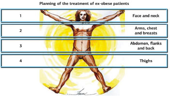
Figure 1. Planning of the treatment of ex-obese patients.
The treatment of the flank of the chest associated with arm dermolipectomy has been performed for some time in patients who are obese or have had a major weight loss1,2, well before the advent of reduction gastroplasty. Normally, mammoplasty (or mastopexy) was performed in a separate surgical stage, usually with the requirement of silicone prostheses because of large tissue atrophy of the mammary gland and adipose tissue. Other techniques are performed in a single surgical step, but with separate incisions3, in which the lateral breast scar subsequently follows the chest (by the bra mark), and brachioplasty does not continue on the chest, being restricted in the armpit, with an "L"-shaped skin incision within the axila4,5. We add markings on the chest and arms similar to the manner proposed by Pitanguy in 19751. Pitanguy's technique of mammoplasties is also followed, and, if necessary, silicone prostheses are positioned, all at the same surgical stage, in which the resulting scar follows the medial epicondyle of the elbow to the sternal region (Figure 2). Note that, when necessary, the scars are joined medially with a reverse abdominoplasty (Figure 3).
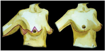
Figure 2. Thoracobrachio-mammoplasty.
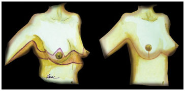
Figure 3. Thoracobrachio-mammoplasty + Reverse abdominoplasty.
In 2005, the Pittsburgh rating scale was developed to standardize ex-obese patients according to the degrees of ptosis in all regions of the body6. Brachioplasty is indicated for grades 2 and 3 brachial flaccidity (Figure 4), associated with grade 3 breasts, in which there is more accentuated deformity, with significant ptosis, large flaccid skin, severe atrophy of the parenchyma, and medial deviation of the nipple-areola complex, due to the large lateral thoracic excess (Figure 5) and on the back (Figure 6).
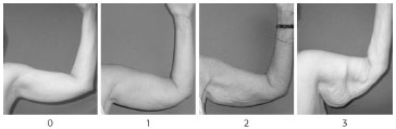
Figure 4. Pittsburg scale - Arms.
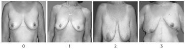
Figure 5. Pittsburg scale - Breasts.
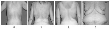
Figure 6. Pittsburg scale - Back.
The aim of this work is to standardize the treatment of the segment including the breasts, lateral chest, and arms, in a single surgical stage and with only one scar on each side.
METHOD
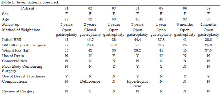
This work was carried out in seven female patients, age from 27 to 65 years (mean, 47 years), from 2011 to 2014. In a single surgery, mammoplasty, thoracoplasties, and brachioplasties were performed.
The markings on the patient were made in the orthostatic position, which is one of the most important aspects of the technique. The arms are open at 90º and the forearms are in a vertical position. The marking begins in the breast according to Pitanguy's technique, but with points AB and AC higher than normal, and point AC always 1 cm higher than AB. In the lateral region of the breast, at the level of the anterior axillary line, the ascending marking begins, and with manual clamping, the amount of skin that can be removed is marked. The marking is curvilinear and toward the posterior axillary fold, with the lateral portion always much larger than the medial portion; perpendicular reference points, around three, are necessary to avoid compensation mistakes at the end of surgery. In the region of the axilla, we reduce the marking of the resected skin, to 1-2 cm, to avoid axillary adhesions in the late postoperative period. From the posterior axillary line, we draw a straight line up to the region of the medial epicondyle of the elbow, through the anterior and posterior regions of the arm, always performing digital clamping to check the possibility of closure of the two sides. All of the skin removed from the lateral wall of the chest to the elbow can be approached as a large lateral "ear" of the mammoplasty.
The surgery is performed under general anesthesia, with the breast injected with 120 mL saline solution, 40 mL xylocaine, and a vial of adrenaline, to reduce the bleeding and the amount of venous anesthetic agents. In cases where there is still abundant adipose tissue, a previous liposuction can be performed in the flap to be removed from the thorax and upper arm.
The surgical times follow an order. First, mammoplasty is done. After mounting the breast, thoracoplasty is carried out, as well as dermolipectomy and flap compensation. Finally, as one surgeon finalizes the breast sutures, brachioplasty is performed (Figure 7).
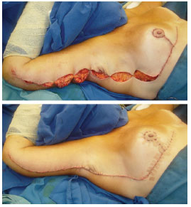
Figure 7. Intra- and immediate postoperative period.
Skin closure is performed with 3-0 monocryl in the higher tension sutures and with 4-0 monocryl in the subdermal and intradermal sutures. Finally, thin strips of micropore surgical tape, and simple and padded gauze are applied. A surgical bra is placed in the breasts, and bandages with moderate compression are placed in the arms.
RESULTS
The seven cases were evaluated in terms of the surgical stage, location of the scars, and final shape and symmetry. The complications included partial dehiscence (14%) and hypertrophic scars (14%) (Figures 8-12). In 86% of cases, the aesthetic result was satisfactory for the patients; on the other hand, the quality of the cicatrization, keloid, and hypertrophic scars were the major causes of dissatisfaction (Table 1).
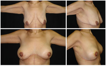
Figure 8. Patient 01.

Figure 9. Patient 02.
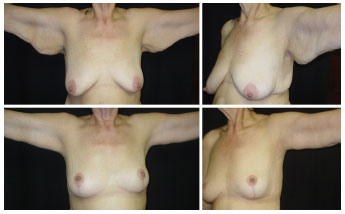
Figure 10. Patient 03.
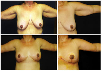
Figure 11. Patient 04a.
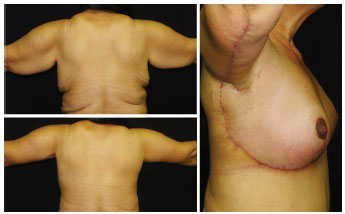
Figure 12. Patient 04b.
DISCUSSION
The techniques were employed for each region (breast, chest, and arms) and associated in a single surgical procedure. The Pittsburg scale6 was used for the indication of these surgeries after an accurate diagnosis, focusing mainly on excess skin in each region. In the breasts, the presence or absence of the gland and fat will determine the need for placement of breast implants.
Mastopexy is indicated according to the degree of mammary ptosis. In the case of pseudoptosis, prostheses can be placed without removing the skin, which can be done only in the chest and arms. High-profile textured prostheses are used.
When there is breast hypertrophy or when the volume is adequate, Pitanguy's technique is employed, for ease of implementation and a shorter surgical time. In such cases, the breast pillars should always be fixed, medially as much as possible, because the traction of thoracoplasty tends to lateralize the breast.
With respect to the thoracoplasty, Hurwitz performs an incision as a continuation of mammoplasty, following horizontally to the back, in the region of the bra mark, with a separate brachioplasty4. This technique involves moving to the supine position when finalizing the thoracoplasty.
The technique presented in this report has a similar design to those of Geoffrey3 and Ruth Graf7, uniting all surgeries in a single incision. However, the drawing of the authors has a much more acute angle in the axilla, unlike in the current report, with a sinuous design and a decrease in the resection of skin in the armpit, in order to reduce the necrosis at the tip of the flap and the formation of axillary adhesions in the late postoperative period.
With respect to the brachioplasty, currently all the techniques for brachial flaccidity involve an inferior incision and no breaks in the scar, avoiding interrupted incisions, as in W-plasty, which feature many bands and retractions in the arm.
The standardization of combined surgeries in ex-obese patients is important for the completion of treatment in all affected areas. Some authors combine mammoplasty with circumferential abdominoplasty4,5, leaving the arms for a second surgical stage. In a continuation of the work published in 20048, we chose to combine mammoplasty, thoracoplasty, and brachioplasty, for being more proximate and possible to perform with a single incision, which facilitates the postoperative period.
CONCLUSION
The mean surgical time and the results of these seven cases leave us optimistic about the single-stage approach of thoracobrachio-mammoplasty, as a single incision and a more segmented operated region in the upper body (segment 2) are good options for the treatment of ex-obese patients.
REFERENCES
1. Pianguy I. Correction of lipodystrophy of the lateral thoracic aspect and inner side of the arm and elbow dermosenescence. Clin Plast Surg. 1975;2(3):477-83. PMid:1097165.
2. Baroudi R. Dermolipectomy of the upper arms. Clin Plast Surg. 1975;3:485-91.
3. Hallock GG, Altobelli JA. Simultaneous brachioplasty, thoracoplasty, and mammoplasty. Aesthetic Plast Surg. 1985;9(3):233-5. http://dx.doi.org/10.1007/BF01570856. PMid:4072818.
4. Hurwitz DJ. Single-staged total body lift after massive weight loss. Ann Plast Surg. 2004;52(5):435-41. http://dx.doi.org/10.1097/01.sap.0000123361.14654.a5. PMid:15096919.
5. Hurwitz DJ, Jerrod K. L-brachioplasty: an adaptable technique for moderate to severe excess skin and fat of the arms. Aesthet Surg J. 2010;30(4):620-9. http://dx.doi.org/10.1177/1090820X10380857. PMid:20829261.
6. Song AY, Jean RD, Hurwitz DJ, Fernstrom MH, Scott JA, Rubin JP. A classification of contour deformities after bariatric weight loss: the Pittsburgh Rating Scale. Plast Reconstr Surg. 2005;116(5):1535-44. http://dx.doi.org/10.1097/01.prs.0000182606.92069.13. PMid:16217505.
7. Colpo PG, Bark AA JR, Oliveira e Cruz GA, Freitas RS, Graf RM. Mama-tóraco-braquioplastia: tratando o tronco superior como unidade estética. Arq Catarin Med. 2009;38(Supl 1):238-40.
8. Furtado RI, Nogueira HC, Lima EM JR. Cirurgia plástica após gastroplastia redutora: planejamento das cirurgias e técnicas. Rev Soc Bras Cir Plast. 2004;19(2):29-40.
1. Pontíficia Universidade Católica do Rio de Janeiro (PUC-Rio), Rio de Janeiro, RJ, Brazil
2. Serviço de Cirurgia Plástica do Professor Ivo Pitanguy, Rio de Janeiro, RJ, Brazil
3. Sociedade Internacional de Cirurgia Plástica Estética, Fortaleza, CE, Brazil
4. Departamento de Dermatologia, Universidade Federal do Ceará (UFC), Fortaleza, CE, Brazil
5. Instituto Doutor José Frota, Fortaleza, CE, Brazil
Institution: Study performed at the Hospital Monte Klinikum, Fortaleza, CE, Brazil.
Corresponding author:
Isaac Rocha Furtado
Harmony Medical Center
Avenida Dom Luís, 1233, Sala 606 - Aldeota
Fortaleza, CE, Brazil Zip Code 60160-230
E-mail: dr.isaacfurtado@gmail.com
Article received: October 12, 2013.
Article accepted: February 8, 2015.
 Read in Portuguese
 Read in English
 PDF PT
 Print
Print
 Send this article by email
 How to Cite
 Mendeley
 Pocket