
Original Article - Year 2013 - Volume 28 -
A surgical technique for nail mimicry
Mimetização Ungueal
ABSTRACT
BACKGROUND: Nails play important roles in helping individuals pick up small objects and protecting the fingertips. However, since nails are located at the distal portion of the fingers, they are susceptible to injuries that can require their amputation. Here we present our technique for nail mimicry that aimed to provide a better aesthetic result after amputation.
METHODS: A total of 14 surgical procedures were performed in 10 patients over 5 years.
RESULTS: The goal of the surgery was achieved in all cases and no complications were observed. The surgery was repeated in three cases due to unsatisfactory results.
CONCLUSIONS: Our technique is easy to perform. Patients were highly satisfied with the results since they no longer had the stigma of an amputated finger with a stump, which improved their quality of life.
Keywords: Nail; Mimicry
RESUMO
INTRODUÇÃO: As unhas cumprem importante papel de auxílio na pinça e proteção das extremidades digitais. Por seu posicionamento distal nos dedos, estão sujeitas a traumas que podem levar a sua amputação. Apresentamos nossa técnica de mimetização ungueal com o objetivo de melhora estética do dedo tratado.
MÉTODO: 14 procedimentos cirúrgicos foram realizados em 5 anos, beneficiando 10 pacientes.
RESULTADO: em todos os casos o objetivo da cirurgia foi atingido, sem nenhuma complicação observada. Em 3 casos a cirurgia foi repetida, devido ao resultado inicial ser insatisfatório.
CONCLUSÃO: é uma técnica de fácil execução e que traz ao paciente um grau de satisfação elevado por retirar o estigma de um coto de amputação com melhor aparência para um convívio social.
Palavras-chave: Unha; Ungueal; Mimetização;
Nails improve our ability to pick up small objects, are useful for scratching, and play an important role in protecting the fingertips. However, due to their location, they are susceptible to injury. Trauma to the distal portion of the fingers may lead to amputation and nail loss, and have a significant impact on the appearance of the affected area.
Our proposed technique is indicated for patients who suffer trauma to the distal phalanx with total or partial amputation and complete nail loss. The surgery is performed for aesthetic reasons and aims to mimic the nail to obtain a better appearance and enable a normal social life.
OBJECTIVE
In this study, we present an easy-to-perform surgical technique that minimizes the stigma caused by nail absence in amputated fingers with stumps after complete nail loss.
MATERIALS AND METHODS
A total of 14 procedures were performed in 10 patients between 2008 and 2012.
The nail mimicking procedure requires that the amputation stumps be rounded off to display a similar shape to that of the remaining fingers of the hand. This procedure can be performed during a single surgery or in different surgeries, the latter of which is our preference.
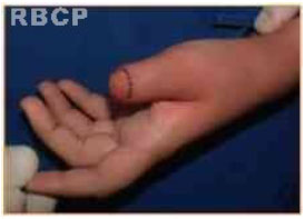
The technique is initiated with a digital nerve block followed by delineation of the incision line in an attempt to obtain a similar shape to those of the remaining non-injured fingers. Subsequently, a scalpel blade No. 15 is used to make a 1-2-mm-deep incision and create a vascularized flap. Next, the borders are cauterized using an electrocauter to achieve hemostasis and deliberate lesion borders. At the end of the procedure, a light compression bandage is placed over the area for 7 days.
RESULTS
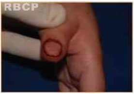
The goal of the surgery was achieved in all cases. The surgery had to be repeated in three cases because the appearance of the scar was inadequate. No cases of infection, flap necrosis, or scar pain or hypersensitivity occurred.
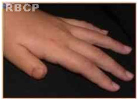
All patients were satisfied with the results obtained using the proposed surgical technique and cited a decreased perception of mutilation by others and were educated about the possibility of having a fake nail placed over the mimicked nail to provide a more natural look.

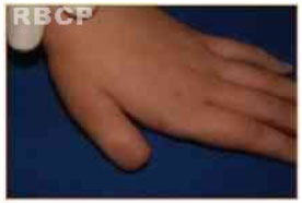
Figure 1: Amputation stump before the surgery

Figure 2: Delineation of the mimicked area

Figure 3: Result immediately after surgery

Figure 4: Result 3 months after surgery
DISCUSSION
The nail should protrude by at least 2 mm from the eponychium to allow for a precise grasp of objects and a better aesthetic look. Several surgical nail bed reconstruction techniques have been described in the literature.
The procedure described by Bakhach is simple and fast and can be used in cases of incomplete nail bed loss. It can also be used in emergency situations. This procedure allows the eponychium to be close together and the nearly complete externalization of the nail matrix, resulting in a 3-mm-long nail.
Treatment is more difficult when the lesion involves loss of most of the distal phalanx and two-thirds of the nail bed. In these cases, microsurgery with an onycho-osteo-cutaneous free flap of the hallux or second finger, as proposed by Koshima & Endo, can be used to reconstruct the nail bed and then support it with bone and the finger pulp in a single surgical procedure. However, this technique is complex and the patient does not always accept it.
The technique proposed in this study is indicated for cases of amputation of the distal phalanx with total nail bed loss. In these cases, the Bakhach technique cannot be used. Our proposed technique represents a simple and easy-to-perform alternative to the onycho-osteo-cutaneous free flap (Koshima, 1991, 1992, and 2000) and the short-pedicle vascularized nail flap (Endo, 1996).
A literature research for a technique similar to that proposed here returned no results. This could be due to the fact that in cases of trauma with fingertip amputation, the goal is always to reestablish function. In this study, finger function was preserved and the patients were concerned about the appearance of the amputation stump, which impacted his/her social life. The use of a prosthesis that replaces the distal phalanx offers no sense of touch since it covers the finger. These prostheses are also impractical.
Unlike the majority of cases in which the plastic surgeon focuses on creating imperceptible scars, here we aimed to produce an obvious scar to mimic the missing nail bed.
Once again, it is important that the amputation stump displays a similar shape to that of the patient's remaining fingers. The patient also has the choice of having fake nails placed over the mimicked area to provide an even better aesthetic result.
CONCLUSIONS
Nail mimicking is a technique that is safe and easy to perform and has low morbidity rates. Patient satisfaction was high since the procedure minimized the stigma associated with an amputation stump, nearly restored the normal appearance of the distal portion of the finger, and eliminated the social discomfort caused by the deformity.
REFERENCES
1)Lemperle G, Schwarz M, Lemperle M. Nail regeneration by elongation of the partially destroyed nail bed. Plast Reconstr Surg. 2003;111(1):167-172.
2) Meals RA. The Nail. Plast Reconstr Surg. 1983;71 (4):579.
3) Endo T, Nakayama Y, Soeda S. Nail transfer: evolution of the reconstructive procedure. Plast Reconstr Surg. 1997;100(4):907-913.
4) Adani R, Marcoccio I, Tarallo L. Nail Lengthening and Fingertip Amputations. Plast Reconstr Surg. 2003;112(5):1287-1294.
5) Koshima, I., Moriguchi, T., Soeda, S., Hamanaka, T., and Umeda, N. Free second toe transfer for reconstruction of the distal phalanx of the fingers. Br. J. Plast.Surg. 44: 456, 1991.
6) Koshima, I., Inagawa, K., Urishibara, K., Okumoto, K., and Moriguchi, T. Fingertip reconstruction using partial-toe transfers. Plast. Reconstr. Surg. 105: 1666, 2000.
7) Bakhach, J. Le lambeau d'eponychium. Ann. Chir. Plast.Estet. 43: 259, 1998.
1. Full Member of the SBCP - Assistant Professor and Private Clinic
2. General Surgeon - Third-year plastic surgery graduate student, University of Nova Iguaçu, - Hospital da Plástica - Rio de Janeiro, RJ, Brazil
3. General Surgeon - Third-year plastic surgery graduate student, University of Nova Iguaçu, Hospital da Plástica - Rio de Janeiro, RJ, Brazil
Correspondence:
Luiz Mário Bonfatti Ribeiro
Hospital da Plástica
Rua Sorocaba, 552 - Botafogo
Rio de Janeiro
Article received on: 07/11/2013
Article accepted: 08/11/2013
Work performed at Hospital da Plástica - INIG


 Read in Portuguese
 Read in English
 PDF PT
 Print
Print
 Send this article by email
 How to Cite
 Mendeley
 Pocket
 Twitter