


Original Article - Year 2012 - Volume 27 -
Rhytidoplasties: cervicofacial SMAS-plasty according to vector suturing
Ritidoplastias: smasplastia cervicofacial mediante sutura de vetores
ABSTRACT
BACKGROUND: Treatment of the superficial muscular aponeurotic system (SMAS) with rhytidoplasty has been continually improving since the pioneering works of the 1970s. The procedures in current usage and described in the literature differ with regard to results as well as to what constitutes an efficient operation. This article describes a surgical procedure that has been applied for SMAS treatment for the last 5 years and which provides satisfactory mid-and long-term results.
METHODS: During the past 5 years, we operated on 274 patients (age range, 35-84; 233 female and 41 male) and monitored them thereafter. In all cases, the procedure followed the same strategy, which involved suturing the SMAS according to 5 vectors, 3 in the face and 2 in the neck, instead of dissecting it.
RESULTS: Most cases were clinically and photographically evaluated yearly. In all age groups, this treatment yielded better results than the traditional methods involving SMAS dissection, as no additional surgeries were required.
CONCLUSIONS: SMAS suturing according to 5 specifically oriented vectors in the face and neck provides better long-lasting results in patients of various ages submitted to rhytidoplasty. Moreover, this procedure avoids the complications described in the literature.
Keywords: Rhytidoplasty/methods. Face/surgery. Plastic surgery/methods. Rejuvenation.
RESUMO
INTRODUÇÃO: O tratamento do SMAS (do inglês, superficial muscular aponeurotic system) nas ritidoplastias faciais tem apresentado contínua evolução desde sua aplicação nos trabalhos pioneiros, na década de 1970. As várias publicações sobre os diferentes tipos de procedimento registram a diversidade do tempo de eficácia e dos efeitos dessa cirurgia. Neste artigo é apresentada mais uma tática cirúrgica que vem sendo aplicada ao SMAS nos últimos 5 anos, cujos resultados a médio e longo prazos têm sido satisfatórios.
MÉTODO: Nos últimos 5 anos, 274 pacientes foram operados e acompanhados, sendo 41 do sexo masculino e 233 do sexo feminino, com idades variando de 35 anos a 84 anos. Em todos os pacientes, a conduta seguiu a mesma sistematização de não dissecar o SMAS, mas realizar sua sutura segundo a direção de 5 vetores, 3 na face e 2 na região cervical.
RESULTADOS: A maioria dos casos tem sido avaliada clínica e fotograficamente, uma vez por ano, demonstrando melhor qualidade dos resultados em pacientes pertencentes a diferentes faixas etárias e sem necessidade de indicação de nova cirurgia.
CONCLUSÕES: A sutura do SMAS segundo 5 vetores especificamente orientados na face e na região cervical tem determinado melhor qualidade dos resultados a longo prazo nas ritidoplastias em pacientes de diferentes faixas etárias, além de evitar as intercorrências e as complicações registradas na literatura.
Palavras-chave: Ritidoplastia/métodos. Face/cirurgia. Cirurgia plástica/métodos. Rejuvenescimento.
The history of the superficial muscular aponeurotic system (SMAS) began with pioneering works in the 1970s1,2 that were followed by the works of several authors3-6 describing the benefits of SMAS mobilization and relocation to positions that allow the elevation of tissue planes and which provide long-lasting results, based on accurate studies of face anatomy. In support of these original works, many other contributions have increased the specialized literature, all in pursuit of an ideal and lasting technique by which SMASplasty would become a "4-seasons" procedure that could be used for patients of various ages undergoing common rhytidoplasties, including secondary and tertiary SMAS rhytidectomies.
SMAS dissection, selective resection, and relocation to the cranial or lateral region can induce temporary - and sometimes permanent - facial nerve lesions. Return to normalcy and possible surgical repair of these lesions have also been reported in the literature7-10. SMAS plication according to 5 vectors with specific orientations, without SMAS dissection but allowing instead the maintenance of its anatomical integrity, is a procedure that has been used by the authors for 5 years. The technique reduces extensive surgical handling and avoids the facial nerve lesions, described in the literature, that occur during the dissection.
METHOD
In the last 5 years, we operated on 274 patients between 35 and 84 years (233 female and 41 male) and monitored them thereafter. In all cases, the procedure followed the same strategy, which involved suturing the SMAS according to 5 vectors, 3 in the face and 2 in the cervical region, instead of dissecting it.
Cutaneous Demarcation
SMAS suturing is carried out according to the specific orientation of 5 vectors, 2 cervical and 3 facial. Vector I is vertical in the cervical midline, for the treatment of platysma muscle bands (Figure 1A). It consists of 2 lines, both on the cutaneous projection of the 2 platysma bands. In patients with a single visible band, the first demarcation is on this band, and the second, parallel to it at a distance of approximately 3 to 5 cm. However, in the absence of the 2 bands, when flaccidity compromises the mandibulocervical angle, vector I is commonly used. Vector II is parallel to vector I, located approximately 1 to 2 cm below the jawline and approximately 1.5 cm past the skin projection of the sublingual salivary gland. The superior border of the 3 facial vectors is the same, closest to the projection of the zygomatic arch with the helical root and the sideburn, where naturally implanted in women. Vector III, located in the upper part of the face, is oriented in the direction of the midline of the nasolabial folds. Vector IV bends more and is directed along the midline between the corner of the mouth and the border of the mandible. Vector V is vertical and is located 1 to 2 cm from the previously set ear line, along the mandibular angle. Vectors II, III, and IV delineate a rectangle with variable length and an average width of 2 cm (Figure 1B). The distance between the 2 vertical lines of vector I varies according to the 2 bands of the platysma muscle. In cases of a single band, its demarcation parallel to the first varies between 3 and 5 cm. However, in the absence of muscle bands and in the presence of flaccidity in the submentonian cervical region, the 2 lines are defined, and the platysma is sutured along vector I. Vectors I and V vary according to the degree of flaccidity, and their support depends on the efficiency of traction provided by vectors II, III, and IV.
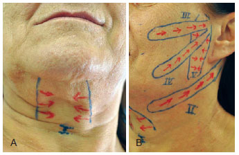
Figure 1 - In A, demarcation of vector I for treatment of the platysma in the midcervical region, by 2 parallel and vertical lines along the bands, even when there is a single visible band. In B, similar demarcation of vector II along the mandible and parallel to it, from the mastoid to the projection of the sublingual salivary gland. The border of vectors III, IV, and V is the same, located in the zygomatic arch and along the helical root, with different orientation, different lengths, and similar widths from 2 to 2.5 cm.
Surgical Procedure
All patients were operated on under sedation in the supine position, with the trunk and head elevated by approximately 30 degrees. Lidocaine (2%) and adrenaline (1:250,000) were infiltrated for local anesthesia. The borders of the skin to be dissected were initially defined by the projection of the helical root with the zygomatic arch, continuing along the malar bone. The demarcation proceeded caudally to the external border of the nasolabial folds, reaching the border of the mandible, and then joining the opposite side. The inferior cervical border was dissected according to a transverse line below the hyoid bone at a distance of approximately 2 to 3 cm. Then, from the earlobe, the demarcation extended along the ear line up to 2 cm from the root of the helix to then bend horizontally 90 degrees in the direction of the scalp, with a variable length of 8 to 10 cm (Figure 2). The skin was then dissected along the demarcated area (Figure 3) and a 4-cm submentonian transverse incision was made along the border of the mandible. The skin dissection was extended caudally to the lower line previously drawn in the cervical region, and a rigorous hemostasis was then achieved. This was followed by a continuous vertical suture of the platysma bands (vector I), "back-and-forth" type in the midline, using 3-0 nonabsorbable nylon suture material, starting 1 cm below the projection of the cricoid cartilage and finishing close to the mandible (Figure 4). Continuous suturing of the platysma was carried out, allowing traction of vector II from the mastoid up to the projection of the parallel sublingual salivary gland approximately 1.5 cm below the jawline (Figure 5). Next, suturing of vectors III and IV, as well as of vector II, was carried out according to their respective directions (Figures 6 and 7). Continuous suture of vector V was then achieved in the molds of vector I, starting from the projection of the angle of the mandible and continuing vertically to the upper implementation of the helix (Figures 8 and 9). Excess skin was then removed by suturing the flaps on both sides to the preauricular and retroauricular areas by using 3-0 absorbable threads. A final periauricular suture of the skin flap was performed with isolated 4-0 and 5-0 nonabsorbable threads. No drains or bandages of any type were used (Figures 10 and 11).
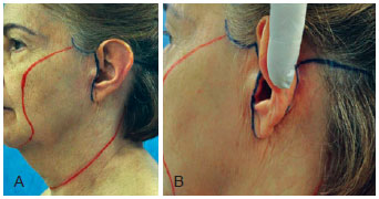
Figure 2 - Lines in red, on the face and neck, mark the borders of a large dissection.
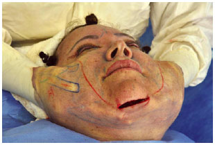
Figure 3 - Both hands below the skin layer illustrate the extent of skin commonly dissected in the face and neck.
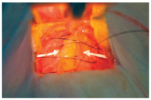
Figure 4 - Vector I for plication of the platysma muscle in the midline, following a common procedure described in the literature for 1 or 2 bands.

Figure 5 - Suturing of vector II from the mastoid to the projection of the sublingual salivary gland, approximately 1.5 to 2 cm below the border of the mandible and parallel to it. Continuous suture with 3-0 nonabsorbable thread, "back-and-forth" type, along the upper line of demarcation and returning to the lower, with a node in the projection of the mastoid.
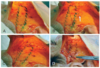
Figure 6 - Suturing of vector III with 3-0 nonabsorbable thread, oriented from the helical root to the midline of the nasolabial folds to the border of the dissected area, along the 2 lines of demarcation in a "back-and-forth" direction, with a node in the region of the helix.
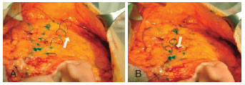
Figure 7 - Continuous suture of vector IV with nonabsorbable thread in a "back-and-forth" direction with a node in the region of the helix, oriented in the midline between the corner of the mouth and the border of the mandible.
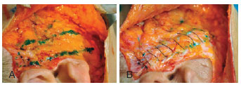
Figure 8 - Continuous suture of vector V in a single direction with 3-0 nonabsorbable nylon thread, starting from the opposite border to the helical root.
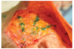
Figure 9 - Orientation of the 3 vectors sutured in the middle segment of the face.
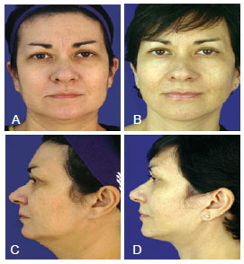
Figure 10 - Female Caucasian patient, 46 years old, submitted to rhytidoplasty and blepharoplasty in the same operation. Maintenance of the mandibular and neck lines with a natural appearance. In A and B, front view, in the preoperative period and 1 year 10 months postoperatively, respectively. In C and D, profile view, in the preoperative period and 1 year 10 months postoperatively, respectively.
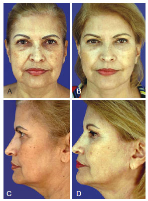
Figure 11 - A 59-year-old patient, submitted to blepharoplasty and rhytidoplasty in the same operation. Natural maintenance of the mandibular line, without recurrence of platysma bands. In A and B, front view, in the preoperative period and 2 years 9 months postoperatively, respectively. In C and D, profile view, in the preoperative period and 2 years 9 months postoperatively, respectively.
Complementary treatment of the eyelids and forehead by using, among other techniques, liposuction, laser, and fat infiltration, although common procedures, are not detailed here, as they are beyond the aim of this article.
RESULTS
All operations were performed using sedation and local anesthesia. Patients were hospitalized in a rehabilitation facility for a period of 1 to 3 days, because of the postoperative care routine.
No drains or bandages of any type were used, because of the sutures in dissected areas, particularly around the auricular region.
Only 1 case of hematoma required subsequent surgery. No nerve lesions or necessity for retouching were observed in the 2 to 3 years following the surgery. This was attributed to the anchoring of the 5 traction and retention vectors.
Figures 12 to 14 illustrate the cases of some patients included in this study.
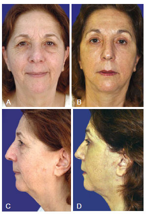
Figure 12 - A 65-year-old patient, submitted to blepharoplasty and rhytidoplasty in the same operation. Natural maintenance of the mandibular line, without recurrence of platysma bands. In A and B, front view, in the preoperative period and 4 years 2 months postoperatively, respectively. In C and D, profile view, in the preoperative period and 4 years 2 months postoperatively, respectively.
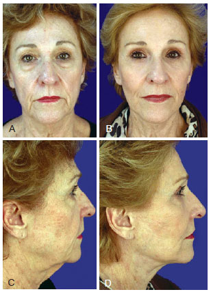
Figure 13 - A 75-year-old patient, submitted to blepharoplasty and rhytidoplasty in the same surgery. Natural maintenance of the mandibular line, without recurrence of platysma bands. In A and B, front view, in the preoperative period and 2 years 1 months postoperatively, respectively. In C and D, profile view, in the preoperative period and 2 years 1 months postoperatively, respectively.

Figure 14 - Patient submitted to blepharoplasty and rhytidoplasty at 79 years old, without treatment of SMAS. In A and B, front view, in the preoperative period and 9 years postoperatively, respectively. In D and E, profile view in the preoperative period and 9 years postoperatively, respectively. In C and F, at 88 years old, new face and neck lift following the procedures described.
DISCUSSION
Suturing SMAS according to 5 vectors with different orientations ensures efficient, reproducible, and satisfactory long-lasting results and motivates us to suggest this procedure for common rhytidoplasties. In the patients who were operated, there was no SMAS dissection or segmental resection with sutures oriented in the cranial or retroauricular areas. Although surgeons acquired thorough knowledge of anatomy, SMAS dissection was often a cause of temporary or permanent facial nerve lesions whose consequences were unpredictable. In cases of temporary lesions, the duration for which the lesions were present raised the concern of the parties involved. The nonmobilization of the SMAS, in addition to reducing surgical trauma, ensures higher safety and long-term efficacy. This should be considered relevant for its indication. Moreover, the procedure using 2 vectors in the cervical region has been demonstrated to be easy and reproducible, delaying the recurrence of platysma bands, commonly observed in a single-maneuver cervical midline suture. No recurrence of the bands was seen in patients followed during the last 5 years.
SMAS plication through vectors III, IV, and V, which are differently oriented, led to a reduction of area, delaying further stretching. Measurements of the dissected skin in the middle segment of the face, according to a transverse and longitudinal orientation, before and after SMAS plication, provided a reduction from 1 to 2 cm of these axes.
The effects of these procedures was better observed in patients older than 60 years, as a result of natural factors that in the elderly might promote a better stretching of the skin and SMAS (Figures 12 to 14). The incidence of edema, ecchymoses, and hematomas was also reduced when compared to surgical procedures using larger dissections. Sutures in the dissected areas limited the hematomas and avoided the use of drains. This favored a rapid recovery, higher efficiency, and better quality of long-lasting results, when compared to traditional procedures described in the literature.
The literature also reports satisfactory results when multiple incisions of platysma bands are performed along the length of the muscle itself11,12. Improvements in surgical procedures leading to technical simplification, quality, and reproducibility of the results is a continuous quest in the history of medicine.
CONCLUSIONS
The replacement of SMAS dissection with suturing according to 5 differently oriented vectors, 2 in the neck and 3 in the face, improved rhytidoplasties. This is because of the quality of the results achieved in patients of different age groups and in secondary surgical procedures. The absence of facial nerve lesions, large reduction in the incidence of hematomas, and short postoperative recovery time motivated the authors to perform and suggest this procedure.
REFERENCES
1. Skoog T. Plastic surgery: new methods and refinements. Philadelphia: WB Saunders; 1974.
2. Mitz V, Payronie M. The superficial musculo-aponeurotic system (SMAS) in the parotid and cheek area. Plast Reconstr Surg. 1976;58(1):80-8.
3. Guerrero-Santos J, Espaillat L, Morales F. Muscular lift in cervical rhytidoplasty. Plast Reconstr Surg. 1974;54(2):127-30.
4. Connell BF. Cervical lifts: the value of platysma muscle flaps. Ann Plast Surg. 1978;1(1):32-43.
5. Stuzin JM, Baker TJ, Gordon HL, Baker TM. Extended SMAS dissection as an approach to midface rejuvenation. Clin Plast Surg. 1995;22(2):295-311.
6. Castro CS. Cirurgia do rejuvenescimento facial. Rio de Janeiro: Medsi; 1998.
7. Aston SJ. Problems and complications in platysma: SMAS cervical rhytidectomy. In: Kaye BL, Gradinger GP, eds. Symposium on problems and complications in aesthetic plastic surgery of the face. St Louis: CV Mosby; 1984. p. 132.
8. Baker DC. Complications of cervicofacial rhytidectomy. Clin Plast Surg. 1983;10(3):543-62.
9. Oswsley JQ. The unfavorable result following face and neck lift. In: Goldwyin RM, ed. Unfavorable result in plastic surgery. Vol II. 2nd ed. Boston: Little, Brown; 1984. p. 59.
10. Pitanguy I, Garcia LC. Filosofia, técnica e complicações das ritidoplastias através da observação e análise de 2.600 casos pessoais consecutivos. Rev Bras Cir. 1972;62:277.
11. Daher JC. Closed platysmoplasty: a new procedure for the treatment of platysma bands without skin dissection. Aesthetic Plast Surg. 2011;35(5):866-77.
12. Gonzalez R. Composite platysmaplasty and closed percutaneous platysma myotomy: a simple way to treat deformities of the neck caused by aging. Aesthetic Surg J. 2009;29(5):344-54.
1. Plastic surgeon at the Hospital Israelita Albert Einstein (Albert Einstein Israelite Hospital) and full member of the Sociedade Brasileira de Cirurgia Plástica (Brazilian Society of Plastic Surgery) - SBCP, Rio Claro, SP, Brazil.
2. Plastic surgeon at the Hospital Israelita Albert Einstein (Albert Einstein Israelite Hospital) and at the Instituto Dante Pazzanese de Cardiologia (Dante Pazzanese Cardiology Institute), full member of SBCP, São Paulo, SP, Brazil.
3. Plastic surgeon, full member of SBCP, São Paulo, SP, Brazil.
Correspondence to:
Nelson Augusto Letizio
Avenida 11, 589 - Centro
Rio Claro, SP, Brazil - CEP 13500-350
E-mail: nletizio@uol.com.br
Article submitted to SGP (Sistema de Gestão de Publicações/ Manager Publications System) of RBCP (Revista Brasileira de Cirurgia Plástica/Brazilian Journal of Plastic Surgery).
Article received: February 6, 2012
Article accepted: May 23, 2012
Study conducted at the Clínica Nelson Augusto Letizio (Nelson Augusto Letizio Clinic), Rio Claro, SP, Brazil.
 Read in Portuguese
 Read in English
 PDF PT
 Print
Print
 Send this article by email
 How to Cite
 Mendeley
 Pocket