
Original Article - Year 2012 - Volume 27 -
Use of the rhomboid flap for the repair of cutaneous defects
Aplicações do retalho romboide em reparações cutâneas
ABSTRACT
BACKGROUND: The plastic surgeon frequently performs reconstructions of diverse types of cutaneous defects; thus, it is essential to be versatile and have knowledge of appropriate techniques for each case. The rhomboid transposition flap, proposed by Alexander Limberg, is an extremely useful flap for a wide range of reconstructive procedures. This study aims to demonstrate the versatility, safety, and applicability of Limberg's flap for reconstruction of cutaneous losses located in a wide variety of body segments.
METHODS: A retrospective analysis of 50 patients with different cutaneous defects that had been reconstructed with the rhomboid flap was performed. A description of the surgical technique and a critical analysis of the results are presented.
RESULTS: The average age of the patients was 59.6 years. Neoplastic lesions accounted for most of the cases (84%). The face was the most frequently affected area, accounting for 36 (72%) cases; it was followed by the lumbosacral region (8%) and by the dorsal and inguinoscrotal regions (6%). Complications were observed in 4 (8%) patients.
CONCLUSIONS: The rhomboid flap provides safe and predictable outcomes, and is the method of choice for most of the defects found.
Keywords: Surgical flaps. Reconstructive surgical procedures/methods. Surgery, plastic/methods.
RESUMO
INTRODUÇÃO: O cirurgião plástico frequentemente defronta-se com a reparação dos mais diversos tipos de defeitos cutâneos; logo, é imprescindível que possua o conhecimento de técnicas versáteis e apropriadas para cada caso. O retalho romboide de transposição, proposto por Alexander Limberg, é um retalho extremamente útil para os mais diversos tipos de reconstrução. O objetivo deste trabalho é demonstrar a versatilidade, a segurança e a aplicabilidade do retalho de Limberg para reconstrução de perdas cutâneas localizadas nos mais diversos segmentos corporais.
MÉTODO: Foi realizada análise retrospectiva de 50 pacientes apresentando defeitos cutâneos, dos mais variados tipos, reconstruídos com o retalho romboide. A descrição da técnica cirúrgica e uma análise crítica dos resultados são apresentadas.
RESULTADOS: A média de idade dos pacientes foi de 59,6 anos. As lesões neoplásicas foram responsáveis pela maioria dos casos (84%). A face foi a área mais envolvida nas reconstruções, totalizando 36 (72%) casos, seguida da região lombossacral (8%), e do dorso e da região inguinoscrotal (6%). Complicações foram observadas em 4 (8%) pacientes.
CONCLUSÕES: O retalho romboide propicia resultados seguros e previsíveis, sendo a alternativa para a maioria dos defeitos encontrados.
Palavras-chave: Retalhos cirúrgicos. Procedimentos cirúrgicos reconstrutivos/métodos. Cirurgia plástica/métodos.
There are a number of studies in the literature on the use of the rhomboid flap in every area of the human body and in various surgical specialties1,2. In spite of several modifications that have already been proposed, it remains an extremely versatile and safe flap3. According to Chasmar4, it can be used in virtually any part of the body, and it is widely used in facial and breast reconstruction, neurosurgery, hand surgery, ophthalmology, and proctology5-8.
This study aims to demonstrate the versatility, safety, and applicability of Limberg's flap for reconstruction of cutaneous losses located at the most varied body segments. The flap fills the defect with tissue of the same thickness and color, has good vascularization, and demonstrates suitable functional and aesthetic outcomes.
METHODS
We performed a retrospective analysis of 50 patients who underwent reconstruction of cutaneous defects with rhomboid flaps; all surgeries were performed at Hospital São Lucas of Pontifícia Universidade Católica of Rio Grande do Sul (Porto Alegre, RS, Brazil) between January 2008 and February 2011. Both the etiology and the location of the defects varied widely.
The patients were assessed by age, etiology, location, and size of the defect (according to measurements obtained during the anatomical-pathological examination, in cases of neoplasia), as well as the presence of postoperative complications. The profiles of the studied patients are described in Table 1.
Surgical technique
The flaps were prepared with consideration of the size and location of the original defect, as well as the force lines and elasticity of the adjacent tissues.
Flap preparation begins with drawing a diamond, with internal angles of 60 and 120 degrees, around the defect resulting from the resection (Figure 1A). This drawing should be prepared, with two equilateral triangles with 60-degree angles lined up from base to base so that all sides of the defect have the same length (which, in practice, is equal to the shortest diagonal). The first side of the flap is an extension outward of the defect, of the shortest diagonal in its own length; the second side of the flap is marked with a line the same length as the first, to the adjacent side of the defect in the diamond, producing an angle of 60 degrees at the flap apex2,9. The final configuration of the flap's scar is foreseeable at all times, as may be seen in Figure 1B.

Figure 1 - Rhomboid flap marking. In A, diamond with internal angles of 60 and 120 degrees and marked flap. In B, final configuration of the scar.
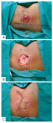
For every defect, four rhomboid flaps can be potentially produced. Then, according to the tension lines and thickness of the skin, and the orientation and location of the defect, the flap that best suits the defect is chosen. As evidenced in Figures 2 to 4, the measurements and production of the flap are performed prior to the resection of the defect's edges, because of the posterior enlargement of the lesions. All edges of the lesion and the flap's edges and base are thus detached, providing suitable approximation of tissues without tension in the closure.

Figure 2 - Patient with lesion secondary to dorsum trauma and difficult scarring of the wound by secondary intention. Resection and cutaneous coverage with Limberg's fasciocutaneous flap. In A, demarcation of the flap prior to resection. In B, resected lesion. In C, at conclusion of operation.

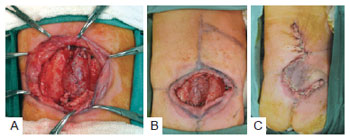
Figure 3 - Patient with wide tumoral lesion in the left temporal-zygomatic region. In A, preoperative appearance. In B, the defect following resection. In C, at conclusion of the operation. In D, appearance at 6 months postoperatively.

Figure 4 - Newborn with meningomyelocele. In A, defect of 8.3 cm × 9.7 cm. In B, marked flap. In C, sutured flap at the site of the defect.
RESULTS
The average age of the patients was 59.6 years and ranged from 2 days to 81 years. Neoplastic lesions accounted for most of the cases (84%). The other cases involved defects deriving from meningomyelocele (8%), Fournier syndrome (4%), necrotizing fasciitis (2%), and trauma (2%). Among the neoplastic lesions, basal cell carcinoma was most frequent (81%), followed by squamous cell carcinoma (14.4%), basal squamous carcinoma (2.4%), and melanocytic nevus (2.4%).
With regard to the affected areas, the face was the most commonly affected, with 36 (72%) cases, followed by the lumbosacral region (8%), and by the dorsal and inguinoscrotal regions (6%). The anterolateral arm, thorax, shoulder, and supraclavicular region each corresponded to 2% of the cases. On the face, the most commonly affected area was the temporal region (25%), followed by the malar region (19.4%). The number of flaps used in facial reconstructions ranged from 1 to 9 (Table 2).

The defects came in different sizes. The largest defect was in the lumbosacral region, due to surgical correction of meningomyelocele, at 8.3 cm × 9.7 cm (Figure 4). In the facial region, the largest defect was 6.2 cm × 5.9 cm and affected the temporal-zygomatic region.
Complications were observed in 4 (8%) patients: epidermolysis in 3 (6%) cases and 1 (2%) case of surgical wound infection. All complications were treated with conservative treatment, and the outcomes were good.
DISCUSSION
This study demonstrates the usefulness of the rhomboid flap, initially proposed by Limberg, for reconstruction of the most varied cutaneous defects, as has been observed by several authors3-5. The flap provides the transference of adjacent tissue to the defect with the same texture and skin color. The position of the scars resulting from the flap transposition is highly foreseeable and can always be taken into account in planning the flap, with the aim of achieving reduced distortion of the underlying tissues and a less apparent scar (Figure 2).
The scar resulting from the flap transposition has acute "broken" angles, making the risk of scar complications, such as trapping and hypertrophy, extremely low. This is, therefore, an attractive option for pediatric patients and/or those with a history of pathological scarring. The flap base, whenever possible, should be inferiorly positioned, in order to facilitate lymphatic drainage of the flap (Figures 2 and 3).
In facial reconstructions, even for small lesions, cutaneous flaps are preferable to primary closure and/or grafting, with the purpose of avoiding distortions of adjacent structures and breaks in scar lines10. The great number of facial reconstructions in the temporal-zygomatic and malar regions (44% of the facial defects) demonstrates the versatility of the rhomboid flap in this area, and that it is the preferable technique in these facial units.
It is possible to argue that resecting extra skin so as to create a parallelogram is not necessary, because it increases the open area. However, considering that most operations are excisions of malignant tumors (84%), such resection provides a greater safety margin (extra tissue can be sent for anatomical-pathological examination), without affecting the final aesthetic-functional outcome11.
The largest defects, in this study, were those deriving from neurosurgical repairs for meningomyelocele correction. Although some authors suggest modifications with the association of two or more rhomboid flaps for large defects on the back3, we believe that the suitable planning of the flap, as well as the maintenance of the paravertebral perforators as proposed by Muneuchi et al.8, enables the closure of large areas with minimal complications. Moreover, the use of a single flap avoids suture lines over the previously repaired dural sac. The transfer of a fasciocutaneous flap of suitable thickness makes the use of muscular flaps unnecessary for closure of selected lesions.
Although it was not part of this case selection, the rhomboid flap has been considered the best option for treating sacrococcygeal pilonidal disease6.
There were few complications in this study, and they did not affect the final result of the surgery; all of them resolved with conservative treatment (dressings and antibiotic therapy), which demonstrates the safety of the rhomboid flap for the most varied reconstructions in the human body.
CONCLUSIONS
Limberg's rhomboid flap provides for closure of small to large defects at a wide range of anatomical sites, with a high level of safety and predictability and a low index of complications. The easy production of the flap design and the resulting strong scar, with no tension in the closure after flap rotation, make it the first option in most reconstructions where the integrity of the skin has been broken.
REFERENCES
1. Tissiani LAL, Alonso N, Carneiro MH, Bazzi K, Rocco M. Versatilidade do retalho bilobado. Rev Bras Cir Plást. 2011;26(3):411-7.
2. Limberg A. Mathematical principles of local plastic procedures on the surface of the human body. Leningrad: Medgis; 1946.
3. Turan T, Kuran I, Ozcan H, Baş L. Geometric limit of multiple local Limberg flaps: a flap design. Plast Reconstr Surg. 1999;104(6):1675-8.
4. Chasmar LR. The versatile rhomboid (Limberg) flap. Can J Plast Surg. 2007;15(2):67-71.
5. Ng SG, Inkster CF, Leatherbarrow B. The rhomboid flap in medial canthal reconstruction. Br J Ophthalmol. 2001;85(5):556-9.
6. Horwood J, Hanratty D, Chandran P, Billings P. Primary closure or rhomboid excision and Limberg flap for the management of primary sacrococcygeal pilonidal disease? A meta-analysis of randomized controlled trials. Colorectal Dis. 2012;14(2):143-51.
7. Silva Neto MP, Adão O, Scandiuzzi D, Chaem LHT. Retalho romboide na reparação mamária imediata pós-quadrantectomia e dissecção axilar. Rev Soc Bras Cir Plást. 2001;16(1):29-34.
8. Muneuchi G, Matsumoto Y, Tamai M, Kogure T, Igawa HH, Nagao S. Rhomboid perforator flap for a large skin defect due to lumbosacral meningocele: a simple and reliable modification. Ann Plast Surg. 2005;54(6):670-2.
9. Limberg AA. Design of local flaps. In: Gibson T, ed. Modern trends in plastic surgery. 2nd ed. London: Butterworth-Heinemann; 1966. p. 38-61.
10. Borges AF. The rhombic flap. Plast Reconstr Surg. 1981;67(4):458-66.
11. Townend J. A template for the planning of rhombic skin flaps. Plast Reconstr Surg. 1993;92(5):968-71.
1. Plastic surgeon, expert member of the Sociedade Brasileira de Cirurgia Plástica (Brazilian Society of Plastic Surgery) - SBCP, Porto Alegre, RS, Brazil.
2. Resident physician in plastic surgery at Hospital São Lucas of Pontifícia Universidade Católica do Rio Grande do Sul (HSL-PUCRS), Porto Alegre, RS, Brazil.
3. Master and full member of the SBCP, preceptor at the Plastic Surgery Department of HSL-PUCRS, Porto Alegre, RS, Brazil.
4. Head of the Plastic Surgery Department of HSL-PUCRS, Porto Alegre, RS, Brazil.
Gustavo Steffen Alvarez
Rua José Francisco Duarte Jr, 52 - ap. 601 - Menino Deus
Porto Alegre, RS, Brazil - CEP 90110-300
E-mail: gusalvarez@terra.com.br
Submitted to SGP (Sistema de Gestão de Publicações/Manager Publications System) of RBCP (Revista Brasileira de Cirurgia Plástica/Brazilian Journal of Plastic Surgery).
Article received: January 18, 2012
Article accepted: February 10, 2012
Study conducted at Hospital São Lucas of Pontifícia Universidade Católica of Rio Grande do Sul (PUCRS), Porto Alegre, RS, Brazil.


 Read in Portuguese
 Read in English
 PDF PT
 Print
Print
 Send this article by email
 How to Cite
 Mendeley
 Pocket
 Twitter