




Case Report - Year 2026 - Volume 41Issue 1
Giant Neurofibroma
Neurofibroma gigante
Vijay Bhatia1, ; Himadri Joshi3; Rajvee Gohil3; Charmi Bodra3; Jay Adeshra3; Harshil Adhiya3; Rakhi Arjariya3
ABSTRACT
Plexiform neurofibromas are benign peripheral nerve sheath tumors considered pathognomonic of neurofibromatosis type 1. They may reach a massive size,making surgical excision technically challenging due to their infiltrative nature and marked vascularity. We herein report the case of a 45-year-old male with a giant congenital plexiform neurofibroma arising from the left gluteal region with extension into the lower limb, presenting with recent rapid enlargement, pain, and skin ulceration. Magnetic resonance imaging (MRI) revealed a large pedunculated lesion involving bilateral S2 to S5 nerve roots, consistent with a plexiform neurofibroma. Surgical excision was successfully performed using temporary vascular control achieved by applying occluding bowel clamps to the large, highly vascular tumor stalk, which ensured controlled division of the pedicle, effective hemostasis, and safe tumor removal. Despite the size and vascularity of the lesion, intraoperative bleeding was manageable, and primary wound closure was achieved. The postoperative course was uneventful, with satisfactory wound healing. This case demonstrates that temporary vascular control of a massive pedunculated stalk using bowel clamps is a simple, effective, and reproducible technique for achieving hemostasis during excision of giant plexiform neurofibromas in anatomically challenging regions.
Keywords: plexiform neurofibroma; neurofibromatosis type 1; peripheral nerve sheath tumor; gluteal region; surgical excision; case report
RESUMO
Os neurofibromas plexiformes são tumores benignos da bainha dos nervos periféricos, considerados patognomônicos da neurofibromatose tipo 1. Podem atingir dimensões gigantescas, tornando a excisão cirúrgica tecnicamente desafiadora devido à sua natureza infiltrativa e à acentuada vascularização. Neste artigo, relatamos o caso de um homem de 45 anos com um neurofibroma plexiforme congênito gigante, originado na região glútea esquerda, com extensão para o membro inferior, que se apresentou com crescimento rápido recente, dor e ulceração cutânea. A ressonância magnética revelou uma grande lesão pediculada envolvendo as raízes nervosas bilaterais de S2 a S5, compatível com neurofibroma plexiforme. A excisão cirúrgica foi realizada com sucesso por meio de controle vascular temporário, mediante a aplicação de pinças intestinais oclusivas no pedículo tumoral volumoso e altamente vascularizado, permitindo a secção controlada do pedículo, hemostasia eficaz e remoção segura do tumor. Apesar do tamanho e da vascularização da lesão, o sangramento intraoperatório foi controlado, e realizou-se o fechamento primário da ferida. Não houve intercorrências no pósoperatório, e a cicatrização foi satisfatória. Este caso demonstra que o controle vascular temporário de um pedículo tumoral maciço, utilizando pinças intestinais, é uma técnica simples, eficaz e reprodutível para alcançar hemostasia durante a excisão de neurofibromas plexiformes gigantes em regiões anatomicamente complexas.
Palavras-chave: neurofibroma plexiforme; neurofibromatose tipo 1; tumor da bainha do nervo periférico; região glútea; excisão cirúrgica; relato de caso
Introduction
Plexiform neurofibromas are benign peripheral nerve sheath tumors and are considered pathognomonic of neurofibromatosis type 1 (NF-1).1, 2 They are usually congenital and slow-growing, and may attain massive proportions over decades. Due to their infiltrative growth pattern, marked vascularity, and proximity to major neurovascular structures, surgical management remains challenging.3, 4 The most common sites are the head and neck, followed by the trunk. Giant plexiform neurofibromas arising from the gluteal region with extension into the lower limb are less common and can significantly impair function and quality of life.5, 6 We report a case of a giant plexiform neurofibroma arising from the left gluteal region in a middle-aged male.
Case Details
A 45-year-old male presented with a long-standing swelling of the left gluteal region. The swelling was congenital in onset and showed slow but progressive enlargement. The patient had not sought medical attention for the swelling, as he had no significant complaints related to it, and he was able to manage his daily activities. His ambulation was preserved with mild difficulty. He had no difficulty passing urine or stool. There was no neurological deficit present. The patient used to wear clothes, such as loose pants or a lungi. Over the past 2-3 months, he noticed a rapid increase in size associated with pain, discomfort, heaviness and a dragging sensation over the left buttock region. An approximately 5 × 4 cm ulcer developed over the swelling within a period of 2 months. Therefore, he came to our hospital for management of left buttock swelling.
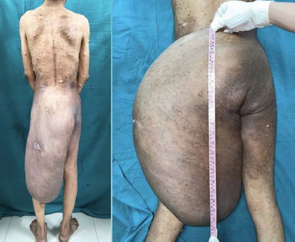
There was a positive family background suggestive of neurofibromatosis, supporting a genetic etiology. On clinical examination, an approximately 57 × 30 cm, diffuse, non-tender soft tissue mass was noted involving the left gluteal region, extending distally into the lower limb (►Fig. 1). The swelling had ill-defined margins, consistent with a plexiform growth pattern. Multiple small, firm, spherical, painless swellings and café-au-lait spots were present throughout the body. The patient weighed 60 kg.
Magnetic resonance imaging revealed a large pedunculated soft-tissue lesion arising from the natal cleft and left gluteal region, extending from the spinal canal and involving the sacral plexus (bilateral S2-S5 nerve roots). The lesion was associated with expansion of the bilateral neural foramina and absence of posterior elements of the sacrum, extending along the limb, with features suggestive of a plexiform neurofibroma. The lesion showed characteristic heterogeneous signal intensity with involvement of multiple tissue planes.
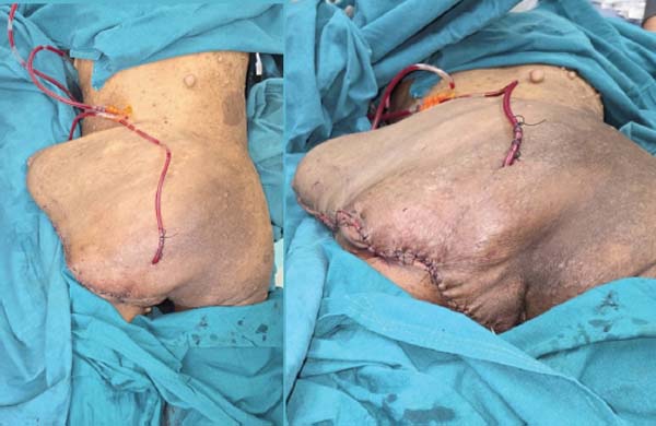
Surgical excision was planned. The neurofibroma was pedunculated with a very large and highly vascular stalk. Due to the excessive thickness and size of the stalk, conventional ligation was not feasible. Therefore, gastric clamps were applied to occlude the stalk. As the stalk was too large to be controlled in a single step, it was divided into two parts, and intestinal occluding clamps were applied on both sides, effectively achieving vascular control and hemostasis. The mass was then excised in a controlled manner. The specimen was sent for histopathological examination. Remaining large skin flaps were closed over two number-14 negative suction drains (►Fig. 2). Patient required 3 units of Packed red blood cells (PRBCs) and 2 units of fresh frozen plasma (FFP) intra-operatively and a total of 8 units of PRBCs during the whole course of treatment. The tumor weighed 11.1 kg. Postoperatively regular dressings were applied to suture line, and wound healed well. The histopathological report of the specimen showed “Massive diffuse soft tissue tumor – neurofibroma.”
Discussion
Plexiform neurofibromas occur in approximately 30–50% of patients with NF-1 and frequently present early in life, though clinical presentation may be delayed due to slow tumor growth and preserved function in early stages.2, 7 The gluteal region is an uncommon site of origin, and tumors arising here may grow to enormous proportions before intervention.6 Ulceration over plexiform neurofibromas is uncommon but may occur due to pressure necrosis and skin compromise in giant lesions.7 MRI is the imaging modality of choice for plexiform neurofibromas, typically showing heterogeneous signal intensity with a characteristic “target sign.”4
Surgical excision remains the mainstay of treatment but is technically demanding due to diffuse infiltration, lack of clear planes, and marked vascularity.5, 8 Massive blood loss is a well-recognized complication in giant plexiform neurofibromas.8
In the present case, temporary vascular control using occluding bowel clamps applied to the large tumor stalk proved to be a simple yet effective method for hemostasis. Dividing the stalk into two segments allowed safe control of the feeding vessels and reduced intraoperative bleeding.
The enormous size of tumor caused progressive stretching of the skin over swelling. This functioned similarly to phenomenon of “delay” and “tissue expansion.” Due to these effects, vascularity of the cutaneous tissue increased and helped in the survival of large skin flaps, which were closed primarily.
Approaches incorporating innovative intraoperative techniques are often required in cases of giant swellings mainly for managing vascular pedicles and minimizing complications.
Conclusion
Giant plexiform neurofibromas originating from the gluteal region are rare and pose significant surgical challenges. A thorough preoperative evaluation and meticulous intra-operative planning, anticipation of blood loss, and innovative intraoperative hemostatic techniques are essential for successful outcomes. Early diagnosis and timely intervention may reduce morbidity and improve quality of life in patients with NF-1.1, 4
References
1. Ferner RE. Neurofibromatosis 1 and neurofibromatosis 2: a twenty first century perspective. Lancet Neurol 2007;6(04):340–351
2. Friedman JM. Neurofibromatosis 1. In: Adam MP, Everman DB, Mirzaa GM, et al., editors. GeneReviews® [Internet]. Seattle (WA): University of Washington; 1998–2023 [updated 2023]
3. Tucker T, Wolkenstein P, Revuz J, Zeller J, Friedman JM. Association between benign and malignant peripheral nerve sheath tumors in NF1. Neurology 2005;65(02):205–211
4. Fisher MJ, Blakeley JO, Weiss BD, et al. Management of neurofibromatosis type 1-associated plexiform neurofibromas. Neuro-oncol 2022;24(11):1827–1844
5. Needle MN, Cnaan A, Dattilo J, et al. Prognostic signs in the surgical management of plexiform neurofibroma: the Children’s Hospital of Philadelphia experience, 1974-1994. J Pediatr 1997; 131(05):678–682
6. Sharma S, Pokhrel B, Khadka N, Rayamajhi S, Man Shrestha J, Lohani I. A 63-kg giant neurofibroma in the right lower extremity and gluteal region of a 22-year-old woman: A case report. Clin Case Rep 2021;9(06):e04152
7. Korf BR. Plexiform neurofibromas. Am J Med Genet 1999;89(01):31–37
8. McCaughan JS Jr, Holloway SM. Surgical management of massive plexiform neurofibromatosis. Ann Plast Surg 1983;10(06):473–480
1. Professor and Chefe, Department of Burns and Plastic Surgery, Smt. NHL Municipal
Medical College, Gujarat University, Ahmedabad, Gujarat, India
2. Consultor, Department of Burns and Plastic Surgery, Sterling Hospital, Gujarat,
India
3. Medico residente, Department of Burns and Plastic Surgery, Smt. NHL Municipal Medical
College, Gujarat University, Ahmedabad, Gujarat, India
Address for correspondence Vijay Bhatia, MBBS, MS, Mch, Department of Burns and Plastic Surgery, Smt. NHL Municipal Medical College, Sterling Hospital, Ahmedabad, Gujarat, 380059, India (e-mail: bhatia101@gmail.com).
Article received: January 06, 2026.
Article accepted: June 01, 2026.
The authors have no conflict of interests to declare.
Editor-in-Chief: Dov Charles Goldenberg.
 Read in Portuguese
 Read in English
 PDF PT
 Print
 Send this article by email
 How to Cite
 Mendeley
 Pocket
 Facebook
 Twitter