


Original Article - Year 2026 - Volume 41Issue 1
Endoscopic Eyebrow Lift with a Modified Double Anchorage
Frontoplastia vídeo-endoscópica com ancoragem dupla modificada
Daniel Nunes1, ; João Ilgenfritz2; Eduardo Zacarias Ramos1; João Pedro Ignácio Santos1; Lucas Vilela1; Pedro Henrique Weber1
ABSTRACT
Introduction Endoscopic forehead lift has become an established technique for rejuvenation of the upper third of the face, allowing elevation of the tail of the eyebrow through a minimally invasive approach. However, the search for an anchoring technique capable of ensuring greater outcome durability has always been a critical concern, particularly given the early recurrence observed in some cases.
Objectives This study aimed to present the outcomes of endoscopic forehead lift using a modified anchoring method.
Methods A retrospective analysis was performed on 44 patients who underwent the procedure, with a minimum follow-up of 12 months. Results: satisfaction rates remained high in both evaluation periods (6 and 12 months), with more than 88% (p<0.001) of the outcomes considered satisfactory or excellent. No relevant complications, such as hematomas, infections, necrosis, or nerve injuries, were observed. The modified double anchoring technique proved to be safe and reproducible, providing long-lasting outcomes and high satisfaction rates.
Conclusion The modified double anchoring technique represents a promising alternative to previously described modalities for fixation of the frontal flap, with the potential to increase the longevity of endoscopic forehead lift outcomes.
Keywords: aesthetics; face; video endoscopy; eyebrow lift; plastic surgery procedures; rejuvenation
RESUMO
Introdução A frontoplastia vídeo-endoscópica consolidou-se como técnica eficaz no rejuvenescimento do terço superior da face, permitindo a elevação da cauda da sobrancelha por meio de uma abordagem minimamente invasiva. Contudo, a busca por uma técnica de ancoragem que garanta maior durabilidade dos resultados sempre foi um ponto crítico, sobretudo diante da recidiva precoce verificada em alguns casos.
Objetivos Apresentar os resultados de frontoplastias vídeo-endoscópicas em que foi empregada uma modificação no método de ancoragem.
Métodos Foram analisados retrospectivamente 44 casos de pacientes submetidas ao procedimento, com seguimento mínimo de 12 meses. Resultados: a taxa de satisfação manteve-se elevada em ambos os momentos de avaliação (6° e 12° meses), com índices superiores a 88% (p < 0,001) de resultados considerados satisfatórios ou excelentes. Não houve intercorrências relevantes como hematomas, infecções, necrose ou lesões nervosas. A ancoragem dupla modificada mostrou-se segura e reprodutível, com resultados duradouros e altos índices de satisfação.
Conclusão A ancoragem dupla modificada configura-se como uma alternativa promissora às modalidades previamente descritas para a fixação do retalho frontal, com potencial de ampliar a longevidade dos resultados das frontoplastias vídeo-endoscópicas.
Palavras-chave: estética; face; videoendoscopia; elevação das sobrancelhas; procedimentos de cirurgia plástica; rejuvenescimento
Introduction
Since the reports by Vasconez et al.1 in 1992, endoscopic forehead lift has become established as an excellent alternative for the treatment of aging signs in the upper third of the face. The treatment of eyebrow ptosis through a minimally invasive approach emerged as a major innovation in the late 2000s and attracted considerable interest among plastic surgeons at that time.2
However, like any new procedure, despite its initial success, the technique eventually faced some resistance, especially because it required specialized training, specific equipment, and longer operative times, ultimately resulting in higher procedural costs.3 Nevertheless, concerns regarding outcome durability discouraged many interested surgeons.4
In a significant proportion of cases, eyebrow ptosis recurred in the medium term, particularly in more complex cases managed by less experienced surgeons.4 Experts attributed this recurrence to inadequate forehead dissection or improper fixation of the advanced tissues during surgery.5
In fact, in the initial approaches, dissection was limited and fixation was either not performed or consisted only of cutaneous fixation, most commonly using sutures.6 In an attempt to achieve better outcomes, Ramirez et al.7 advocated more extensive dissections extending beyond the lateral periorbital region. In contrast, authors such as Romo et al.,8 Gladstone et al.,9 and Graça Neto et al.,10 advocated more durable fixation methods, using, for example, screws, Endotine®, and fixation of the flap to the deep temporal fascia with nonabsorbable sutures, respectively.
In 1996, McKinney et al.11 proposed fixation of the frontal flap using sutures passed through bony tunnels. The authors advocated placement of a single 3-0 polydioxanone suture through each bony tunnel, fixed with a Reverdin needle at the lateral aspect of the eyebrow.11 Despite improved outcomes, recurrence rates in more complex cases involving heavier flaps and lower eyebrows remained concerning.4
Since then, several fixation techniques have been described for endoscopic forehead lift procedures, and the current understanding is that a true “gold standard” method has not yet been established, with the search for the ideal alternative still ongoing.12 Thus, the present study was designed to present the double anchoring method used by the authors and to evaluate patient satisfaction rates.
Methods
The medical records of all female patients who underwent endoscopic forehead lift for primary elevation of the eyebrow tail between January 2018 and February 2024 were retro-spectively reviewed. The study was duly approved by the Research Ethics Committee at the private clinic of one of the study authors.
Data regarding each patient’s assessment of the surgical outcome at the 6- and 12-month postoperative follow-up visits were collected. Following a modification of the method described by Salles et al.,13 the nursing staff at the authors’ clinic asked patients to classify the surgical outcome as unsatisfactory, satisfactory, or excellent.
The evaluation of patient satisfaction levels regarding endoscopic forehead lift with modified double anchoring was performed using the McNemar test. Comparison between the evaluation periods regarding the median satisfaction score (0 = unsatisfactory outcome, 1 = satisfactory outcome, and 2 = excellent outcome) was performed using the nonparametric Wilcoxon test, since the data did not pass the Shapiro-Wilk normality test. Comparison between the percentage of women whose outcomes were considered unsatisfactory and the percentage whose outcomes were considered satisfactory or excellent was performed using the binomial test. The remaining results of this study are presented as descriptive statistics or in the form of tables and graphs. Statistical analysis was performed using SPSS software, version 23.0, adopting a significance level of 5%.14
The endoscopic forehead lift was based on the classic technique described by Isse,15 with the following modifications to the fixation technique.
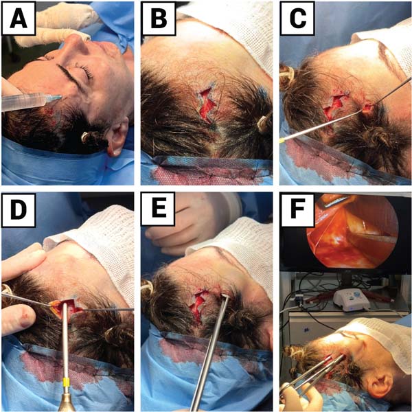
The procedure was performed under general anesthesia, with the patient in the supine position and slight flexion of the head. Antisepsis was performed using a 2% chlorhexidine degerming solution, followed by placement of sterile drapes. The surgeon stood behind the patient and infiltrated the lateral two-thirds of the frontal region and the temporoparietal region with approximately 350 mL of a solution containing 2% lidocaine with epinephrine 1:200,000. Four incisions were then made: two paramedian and two in the temporal region. After release of the subperiosteal and interfascial planes with endoscopic assistance, the temporal crest and the periorbital periosteum were released from the 12 o’clock to the 3 o’clock position (►Fig. 1).
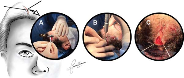
After dissection of the desired area, bony tunneling was performed through the paramedian incisions for cranial fixation of the dissected temporofrontal flap. Using the Beltec LB100® drill and a 3-mm spherical burr with an umbrella drill guard protector, bony tunnels (one in each paramedian opening) were created very close to the distal end of the incision while the scalp was pulled caudally (►Fig. 2).

The double anchoring technique described here was based on a modification of the classic technique described by McKinney et al.11 A braided 2-0 mononylon suture (Biosut®, TC-40, 3/8 curvature) was passed through the cortical bony tunnel, maintaining the needle end in the occipital region. Using a Casagrande needle from Richter®, the non-needle end of the suture was passed subperiosteally for approximately 2 cm towards the medial region of the eyebrow. At this point, the needle perforated the skin, and the suture was exteriorized (►Fig. 3A–C).
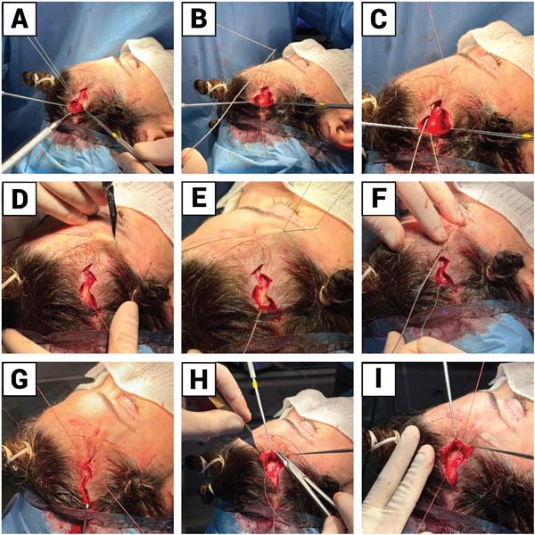
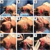
The suture was removed from the needle, and the needle was withdrawn through the paramedian incision. The end of the suture, now exiting through the frontal skin, was then passed again through the needle, which re-entered through the same orifice where the suture exited and, through the subcutaneous plane in a parallel trajectory of approximately 1 cm, carried the suture to the more lateral region of the forehead (close to the hair-bearing area), where it was exteriorized again (►Fig. 3D–E).
The suture was removed from the needle, and the needle exited through the entry orifice in the frontal region. Once again, the suture, now exiting near the scalp, was threaded through the Casagrande needle, which was then passed through the subcutaneous plane until it exited through the paramedian incision, bringing the suture with it. In this way, a mixed loop – partially subperiosteal and partially subcutaneous – was created and subsequently secured to the bony tunnel (►Fig. 3F).
While the assistant posteriorly retracted the dissected temporal flap using a Gilles hook, the braided 2-0 mononylon suture was secured to the bony tunnel with a surgical knot, advancing the frontal flap posteriorly. The vector of elevation was more medial than in the classic technique, thereby promoting greater elevation of the temporal region, enhancing elevation of the eyebrow tail, and minimizing excess skin in the area (►Fig. 3G).
After this first suture and before cutting the thread, a second anchoring was performed. The needle was passed through the frontal flap at the most anterior point possible of the paramedian incision, incorporating the reticular dermis, subcutaneous tissue, and periosteum. This suture was then secured with a surgical knot to the distal stump of the suture that had already been fixed to the bony tunnel during the first anchoring. This maneuver created an additional fixation point, helping maintain the frontal flap in its new position and minimizing recurrence of tissue descent (►Fig. 3H–I). The skin was then sutured with simple 3-0 mononylon sutures.
In more complex cases, in which the expected recurrence rate was higher, an additional suture could be passed through the bony tunnel to create a third anchoring point.
Results
During the study period, 54 patients underwent the described procedure. Of these, eight were excluded for not attending all postoperative follow-up visits or for not responding to the assessment regarding outcome quality, while two were excluded because they underwent early reoperation (one presented with significant eyebrow asymmetry, and the other was dissatisfied with the limited eyebrow elevation).
Thus, 44 women who underwent endoscopic forehead lift surgery were evaluated. The age of the patients included in this study ranged from 37 to 75 years, with a mean age of 53.82 ± 9.52 years. Postoperative recovery occurred without complications, and no patient developed hematoma, infection, superficial necrosis, cerebrospinal fluid fistula, or nerve injuries.
Patient satisfaction with the forehead lift is shown in ►Table 1 and ►Figure 4.
| Satisfaction level | Time after surgery | p value | |
|---|---|---|---|
| 6 months (n = 44) | 12 months (n = 44) | ||
| Unsatisfactory outcome | 6.8% (n = 3) | 11.4% (n = 5) | 0.675 |
| Satisfactory outcome | 40.9% (n = 18) | 43.2% (n = 19) | |
| Excellent outcome | 52.3% (n = 23) | 45.5% (n = 20) | |
| Median score | 2 (0 to 2) | 1 (0 to 2) | 0.344 |
Regarding the total score: 0 = unsatisfactory outcome; 1 = satisfactory outcome; 2 = excellent outcome. Results are presented as relative frequency (absolute frequency) or median (minimum–maximum). P-values refer to the McNemar test (satisfaction level) or the Wilcoxon test (median score).

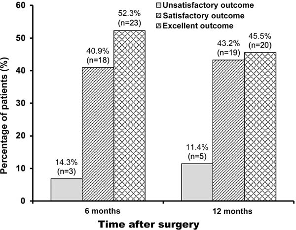
No significant change in patient satisfaction levels was observed between the 6- and 12-month postoperative evaluations (McNemar test, p = 0.675; Wilcoxon test, p = 0.344).
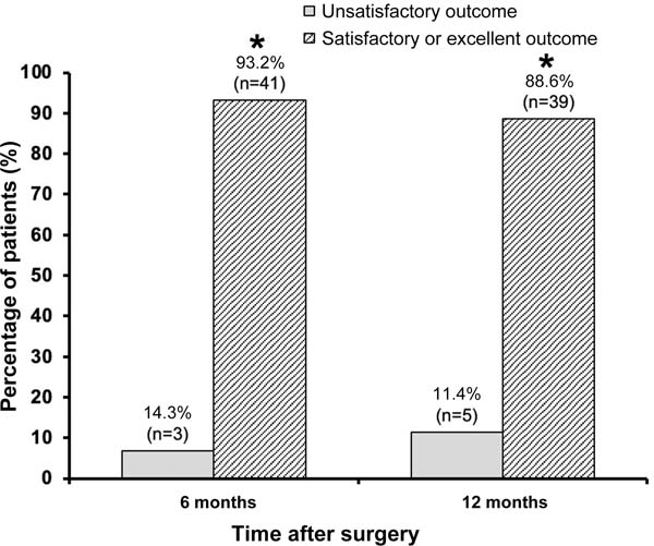
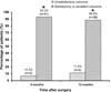
At both 6 and 12 months after surgery, the percentage of women who considered the outcome satisfactory or excellent (6 months: 93.2%, n = 41; 12 months: 88.6%, n = 39) was significantly higher than the percentage of those who considered the outcome unsatisfactory (6 months: 6.8%, n = 3; 12 months: 11.4%, n = 5) (binomial test, p < 0.001 for both time points), as demonstrated in ►Table 2 and ►Figure 5.
| Satisfaction level | Time after surgery | |
|---|---|---|
| 6 months (n = 44) | 12 months (n = 44) | |
| Unsatisfactory outcome | 6.8% (n = 3) | 11.4% (n = 5) |
| Satisfactory or excellent outcome | 93.2% (n = 41) | 88.6% (n = 39) |
| P-value | <0.001 | <0.001 |
Results are presented as relative frequency (absolute frequency). P-values refer to the binomial test.

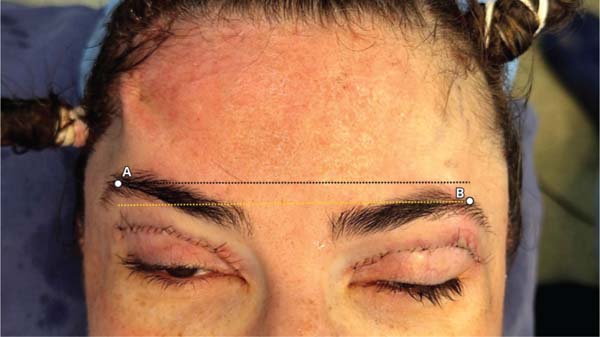
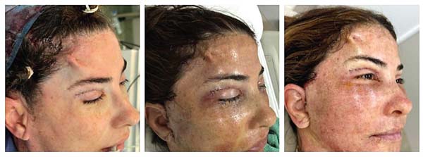
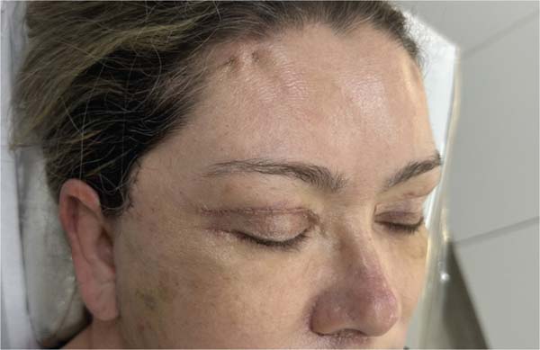
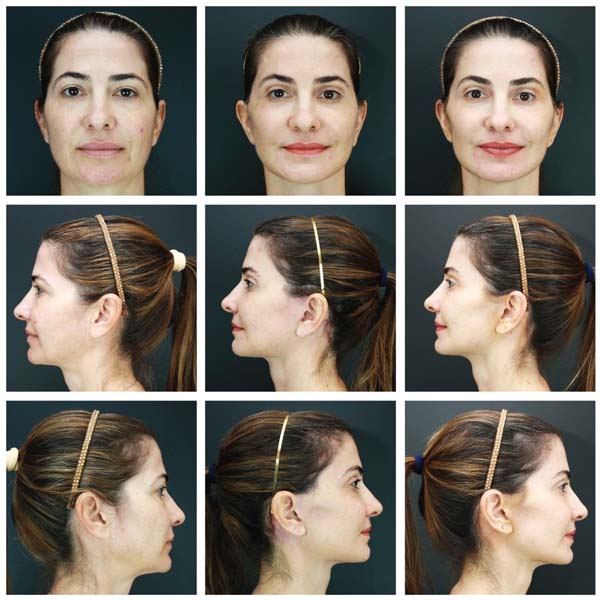
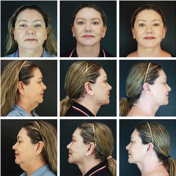
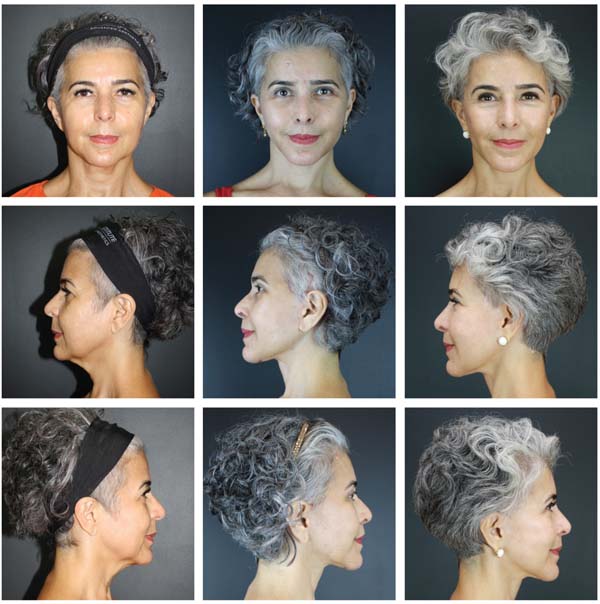
►Figure 6 presents the intraoperative view of a patient undergoing elevation of the right eyebrow compared with the untreated left side. ►Figure 7 depicts images obtained at the end of surgery and at 24 and 72 hours after the procedure. ►Figure 8 shows the 12-day postoperative outcome of a patient who underwent the technique. ►Figures 9 and 10 illustrate the preoperative findings as well as the 6- and 12-month postoperative outcomes.


Discussion
The literature describes numerous technical alternatives for fixation of the advanced frontal flap in endoscopic forehead lift procedures, and the absence of consensus regarding the ideal approach reflects both the complexity of the topic and the distance still to be traveled before establishing a definitive standard.16–18
In recent years, as long-term maintenance of outcomes became the primary objective, a shift in preference regarding fixation methods has been observed.17,19,20 The need for more effective anchoring has been increasingly emphasized.17–22 Authors such as Guyuron23 and Ramirez24 reported improved outcomes and lower recurrence rates after adopting more effective fixation techniques. Others, like Rohrich et al.,5 Romo et al.,8 and Kennedy et al.,25 also emphasized the importance of more durable fixation methods in modern endoscopic forehead lift procedures.
In this context, the literature describes options that are not widely used in Brazil, such as fixation with screws (absorbable or nonabsorbable) and the Endotine® device,26 which present limitations related to cost, technical complexity, possible device palpability, and reports of foreign body reactions.3,27 In addition, recurrence of eyebrow tail ptosis has also been reported with the use of these methods.27
A simpler yet more robust anchoring alternative is fixation of the frontal flap with sutures anchored in bony tunnels.28 This option, first described by McKinney et al.11 in 1996, has gained increasing acceptance in recent years.18,21 Using surgical drills and, ideally, protective devices such as the “umbrella drill guard,” a cortical bony tunnel is created, through which a suture of varying material and caliber is passed, consistent with the reports of McKinney et al.,11 Malata et al.,12 Graf et al.,16 Mowlavi et al.,18 and Kim et al.21
Different fixation methods using cortical tunnels have been described, the most common being direct single fixation of the frontal flap using a suture passed through the bony tunnel. McKinney et al.11 advocated fixation using sutures passed through the periosteal edge and connected to the previously created bony tunnel, allowing appropriate eyebrow tension and elevation while providing firm fixation with an adequate vector force. Although this fixation method is widely used and considered reliable, it has also been criticized because of eyebrow descent, the need for specific training, and the requirement for specialized surgical instruments and equipment.4,15,22
To improve the outcomes of bony fixation, some authors recommended the creation of two bony tunnels in each paramedian incision in order to increase the fixation points and distribute the pressure exerted during the forehead lift.2,21,29 Despite the initial success reported by the authors, no significant difference in maintenance of the outcomes was observed with the creation of double tunnels.21 Increased operative time, the need for larger paramedian incisions, and greater bone trauma are important considerations that discouraged adoption of this method by most specialists.
Using suture fixation without bony tunnels, Borille et al.2 observed a decrease in eyebrow elevation height during the first 6 months after surgery, with a tendency toward stabilization after 12 to 24 months. The authors associated this occurrence with loss of tensile strength of the mononylon used for fixation.
In a recently published meta-analysis, Caceres et al.28 demonstrated that forehead lift with bony tunnel fixation presented the lowest complication profile, high satisfaction rates, and greater outcome durability when compared with the four most common fixation categories (Endotine®, sutures, screw fixation, and bony tunnel fixation). In the proposed algorithm, bony tunnel fixation was indicated as the gold standard both for patients with thin or fragile skin and for patients older than 50 years, provided that the surgeon has the requisite experience and familiarity with the technique.
In our series, we concur with the trend proposed in the most recent literature and advocate the creation of only one bony tunnel in each hemisphere, combined with a modification of the fixation technique – the modified double anchoring technique. With high satisfaction rates even after 12 months of postoperative follow-up, the modified double anchoring technique presented here emerges as a promising alternative, offering durability, safety, and high satisfaction rates.
Conclusion
Endoscopic forehead lift with modified double anchoring provided satisfactory and long-lasting outcomes using a simple and accessible fixation method. The high level of patient satisfaction achieved with this technique makes it a promising alternative for frontal flap fixation in endoscopic forehead lift procedures.
REFERENCES
1. Core GB, Vasconez LO, Ashen C, Yamamoto Y, Gamboa M. Coronal face-lift with endoscopic techniques. Plast Surg Forum 1992; 15:227-8.
2. Borille G, Santos PV Junior, Rocha GSP, et al. Prognostico da videocirurgia da região frontal com técnica modificada de fixação. Rev Bras Cir Plást 2023;29:447-450
3. Patrocínio LG, Reinhart RJ, PatrocínioTG, Patrocínio JA, Guimarães TR, Patrocínio TG. Endoscopic frontoplasty: 3-year experience. Braz J Otorhinolaryngol 2006;72(05):624-630
4. Chiu ES, Baker DC. Endoscopic brow lift: a retrospective review of 628 consecutive cases over 5 years. Plast Reconstr Surg 2003;112 (02):628-633, discussion 634-635
5. Rohrich RJ, Beran SJ. Evolving fixation methods in endoscopically assisted forehead rejuvenation: controversies and rationale. Plast Reconstr Surg 1997;100(06):1575-1582, discussion 1583-1584
6. Keller GS, Mashkevich G. Endoscopic forehead and brow lift. Facial Plast Surg 2009;25(04):222-233
7. Ramirez OM. Endoscopic techniques in facial rejuvenation: an overview. Part I. Aesthetic Plast Surg 1994;18(02):141-147
8. Romo T III, Zoumalan RA, Rafii BY. Current concepts in the management of the aging forehead in facial plastic surgery. Curr Opin Otolaryngol Head Neck Surg 2010;18(04):272-277
9. Gladstone GJ, Nesi FA, Black EH, Eds. Oculoplastic surgery atlas: cosmetic facial surgery. New York: Springer-Verlag; 2005
10. Graça Neto L, Araújo LRR, Graça ACM. Subperiosteal endoscopic frontoplasty with temporal fixation by sutures. Rev Bras Cir Plást 2022;37(01 ):3-8
11. McKinney P, Celetti S, Sweis I. An accurate technique for fixation in endoscopic brow lift. Plast Reconstr Surg 1996;97(04): 824-827
12. Malata CM, Abood A. Experience with cortical tunnel fixation in endoscopic brow lift: the “bevel and slide” modification. Int J Surg 2009;7(06):510-515
13. Salles AG, Ferreira MC, Remigio AFN, Gemperli R. Escala para avaliação de resultados de cirurgia estética do abdome. Rev Bras Cir Plást 2011;26(01):147-150
14. Rowe P. Essential statistics for the pharmaceutical sciences. Chichester: John Wiley & Sons; 2007
15. Isse NG. Endoscopic facial rejuvenation: endoforehead, the functional lift. Case reports. Aesthetic Plast Surg 1994;18(01):21-29
16. Graf R, Pace D, Araújo LR. Cirurgia endoscópica da fronte e terço médio da face: oito anos de experiência. Rev Bras Cir Plást 2005; 20(03):197-203
17. Newman JP, LaFerriere KA, Koch RJ, et al. Fixação de sutura transcalvária para elevações endoscópicas de sobrancelhas e testa. Arch Otolaryngol Head Neck Surg 1997;123(03):313-317
18. Mowlavi A, Pham S, Lee R, Huynh P, Wilhelmi B. Cortical thickness parameters for endoscopic browlift fixation. Aesthet Surg J 2012; 32(05):547-551
19. Vasconez LO, Core GB, Gamboa-Bobadilla M, Guzman G, Askren C, Yamamoto Y. Endoscopic techniques in coronal brow lifting. Plast Reconstr Surg 1994;94(06):788-793
20. Matarasso A, Matarasso SL. Endoscopic surgical correction of glabellar creases. Dermatol Surg 1995;21(08):695-700
21. Kim M, Lee N, Tark W, Lee W, Roh T, Baek W. Dual cortical tunneling method for endoscopic forehead lift. Arch Aesthetic Plast Surg 2022;28(03):85-89
22. Karimi N, Kashkouli MB, Sianati H, Khademi B. Techniques of eyebrow lifting: a narrative review. J Ophthalmic Vis Res 2020;15 (02):218-235
23. Guyuron B, Michelow BJ. Refinements in endoscopic forehead rejuvenation. Plast Reconstr Surg 1997;100(01):154-160
24. Ramirez OM. Endoscopic forehead and face-lift: Step by step. Oper Tech Plast Reconstr Surg. 1995;2(02):129-136
25. Kennedy BD, Pogue MD. Fixation techniques for endoscopic browlift. J Oral Maxillofac Surg 1999;57(05):588-594
26. Honig JF, Frank MH, Knutti D, et al. Video-endoscopic-assisted brow lift: comparison of the eyebrow position after fixation with Endotine tissue vs suture fixation. J Craniofac Surg 2008;19(04): 1140-1147
27. Heffelfinger RN, Blackwell KE, Rawnsley J, Keller GS. A simplified approach to midface aging. Arch Facial Plast Surg 2007;9(01):48-55
28. Caceres H, Raza SN, Katta T, et al. Fixation techniques in endoscopic brow lifts: a comprehensive review. Aesthetic Plast Surg 2025. Forthcoming PubMed
29. Badin AZ, Bittencourt LM, Balderrama CR. Fixação lateral da sobrancelha no lifting endoscópico da testa: resultados a longo prazo com suturas percutâneas de nylon trançado. Aesthetic Plast Surg 2010;34(01):78-87
1. School of Medicine, Fundação Universidade Federal de Mato Grosso do Sul, Campo
Grande, Mato Grosso do Sul, Brazil
2. Clínica João Ilgenfritz de Cirurgia Plástica, Campo Grande, Mato Grosso do Sul,
Brazil
Data Availability Data will be available upon request to the corresponding author.
Address for correspondence Daniel Nunes, Rua Marco Túlio Murando Garcia 51, Chácara Cachoeira, Campo Grande – MS – ZIP Code: 79040-010 (e-mail: dermatoeplastica@gmail.com).
Article received: January 19, 2026.
Article accepted: April 10, 2026.
Conflict of Interest: The authors declare no conflicts of interest.
Editor-in-chief: Dov Charles Goldenberg
 Read in Portuguese
 Read in English
 PDF PT
 Print
Print
 Send this article by email
 How to Cite
 Mendeley
 Pocket
 Facebook
 Twitter