


Original Article - Year 2022 - Volume 37 -
Low-level laser therapy in the management of skin wound healing
Terapia a laser de baixa potência no manejo da cicatrização de feridas cutâneas
Ana Carolina Vasconcellos Guedes Otsuka1* ; Clarissa Lima Vilela Moreira1; Eduardo Willian Pasquarelli1; Karin Cristina Periz Pavani1; Patricia Pereira dos Anjos1; Soraya Yumi Hashimoto1; Marcon Censoni de Avila e Lima1; João Pedreira Duprat Neto2
ABSTRACT
Introduction: Tissue repair and skin wound healing are complex processes that involve a series of dynamic events, including coagulation, inflammation, granulation tissue formation, wound contraction and tissue remodeling. The need to care for patients with difficulties in wound healing is a growing challenge and requires innovative strategies. One approach that stands out in the treatment of these injuries is low-power laser therapy. This study aims to demonstrate the applicability and results of a low-power laser therapy protocol in the management of skin wound healing.
Methods: This is a retrospective case review study reporting the use of laser in the treatment of skin wounds in a single center.
Results: We evaluated the results of the implementation of this low-power laser therapy protocol and reported five cases of patients with skin wounds that did not improve with conventional treatment. The lesions were located in different places, with lesions on the trunk, upper limb and lower limb. The five cases showed a good response to laser therapy, evolving with wound healing in a short period of time, with an average healing time of 48 days.
Conclusion: It is concluded that low power laser therapy when applied to skin wounds suggests a beneficial, promising action and has the potential to increase the therapeutic options available to the surgeon, however as we report a number of five cases, further studies are needed for check laser efficiency on wounds.
Keywords: Low-level light therapy; Wound healing; Cell proliferation; Laser therapy; Skin Diseases.
RESUMO
Introdução: O reparo tecidual e a cicatrização de feridas cutâneas são processos complexos que envolvem uma série de eventos dinâmicos, incluindo coagulação, inflamação, formação de tecido de granulação, contração da ferida e remodelação tecidual. A necessidade de cuidar de pacientes com dificuldades na cicatrização de feridas é um desafio crescente e requer estratégias inovadoras. Uma abordagem que se destaca no tratamento dessas lesões é a laserterapia de baixa potência. Este estudo tem como objetivo demonstrar a aplicabilidade e os resultados de um protocolo de laserterapia de baixa potência no manejo da cicatrização de feridas cutâneas.
Métodos: Trata-se de estudo retrospectivo de revisão de casos relatando o uso de laser no tratamento de feridas cutâneas em um centro único.
Resultados: Avaliamos os resultados do uso de laserterapia de baixa potência e relatamos cinco casos de pacientes com feridas cutânea que não apresentaram melhora com o tratamento convencional. As lesões eram localizadas em sítios diversos, com lesão no tronco, membro superior e membro inferior. Os cinco casos apresentaram uma boa resposta com a terapia a laser, evoluindo com a cicatrização das feridas em um curto período de tempo, com período médio de cicatrização de 48 dias.
Conclusão: Conclui-se que a laserterapia de baixa potência quando aplicada sobre feridas cutâneas sugere uma ação benéfica, promissora e tem um potencial para aumentar as opções terapêuticas disponíveis para o cirurgião, porém, como relatamos um número de cinco casos, há necessidade de mais estudos para verificar a eficiência do laser em feridas.
Palavras-chave: Terapia com luz de baixa intensidade; Cicatrização; Proliferação de células; Terapia a laser; Pele.
INTRODUCTION
Tissue repair and wound healing are complex processes involving dynamic events, including coagulation, inflammation, granulation tissue formation, wound contraction and tissue remodeling1,2. The need to care for patients with wound healing difficulties is a growing challenge and requires innovative strategies. One approach that stands out in treating these lesions is low-level laser therapy3. Thus, in recent decades, numerous studies and research have been conducted to investigate the use of laser therapy in favor of tissue repair1,2.
Low-Level Laser Therapy (LLLT) is conducted at low irradiation intensities, and its biological effects are secondary to the direct effects of photonic radiation without thermal reactions. LLLT was first introduced by the work of Mester et al.4 and is known to deliver direct biostimulating light energy to the body’s cells, thereby enhancing normal cell function and tissue repair4,5.
Therapeutic or low-power lasers are used to accelerate tissue repair processes due to their biomodulatory effects on cells. They activate or inhibit physiological, biochemical and metabolic processes through photophysical or photochemical effects. These biomodulatory phenomena promote the therapeutic effects of morphodifferentiation and cell proliferation, tissue neoformation, revascularization, reduced edema, greater cell regeneration, and increased local microcirculation and vascular permeability6.
Many benefits are associated with biomodulation with LLLT. According to Belkin & Schwartz7, the effects require a threshold of exposure to irradiation; depending on the desired effect, the required fluency, or energy dose, is calculated. The threshold is specific, and each tissue responds differently to a given wavelength (l).
The methodology is simple, low cost and can be integrated as an adjunct to therapy for conventional treatments or used alone as an alternative method in some conditions. The therapeutic effects are anti-inflammatory, analgesic and tissue repair inducer. The substantial increase in interest in LLLT has been noted due to the significant number of satisfactory results6.
Several parameters must be adjusted to apply LLLT, such as wavelength, light power, light type, energy density, power, and total time. The effectiveness of treatment also depends on many factors, such as the location and nature of the wound and the patient’s physiological state8. Due to these diverse variables, using a protocol to guide LLLT is very important in clinical practice.
This work aims to demonstrate the applicability of a low-level laser therapy protocol in managing cutaneous wound healing. Report the protocol previously created in this institution by nurses who are members of the skin cancer group (GOPE) and demonstrate the results obtained in practice.
OBJECTIVE
This study aims to demonstrate the applicability and results of a low-level laser therapy protocol in managing cutaneous wound healing.
METHODS
This is a retrospective case review study reporting the use of the low-level laser therapy protocol in managing cutaneous wound healing in the ambulatory dressing service of the A.C.Camargo Cancer Center. It covers patients who had a skin wound that did not resolve with conventional treatment, the origin of the wounds being of various causes, from skin necrosis due to extravasation of intravenous medication to dehiscence of the surgical wound after tumor exeresis.
All reported cases are of skin wounds and occurred in the years 2016, 2017, 2018 and 2019. The protocol was previously created by nurses who are members of this institution’s skin cancer group (GOPE), following safety standards for the patient, family and multi-professional team. There was approval from the local Research Ethics Committee for this study, number RC 100/21.
Inclusion criteria for using the laser therapy protocol were: patients with wounds with bone/tendon exposure, necrosis, and chronic or complex wounds that did not respond to conventional treatment. Exclusion criteria were: malignant neoplastic wound, pregnancy, patient in serious health condition, metallic prosthesis at the wound site, black skin, tattoo at the time of chemotherapy infusion, and patient using a cardiac pacemaker.
The characteristics and variables of the low-level laser are applied according to the particularities of each lesion. In lesions with cutaneous involvement with partial tissue loss, the variables were: type of light: red, energy density (DE): 80J/cm2, energy (E): 2.1J, power (P): 100mw, time per point: 22 seconds, technique: point, mode: continuous. In lesions with necrotic tissue (coagulation/liquefaction) or contaminated wound: type of light: red, DE: 220J/cm2, E: 6.1J, P: 100mw, application time per point: 2 minutes per point, technique: punctual, mode: pulsed (100pps). In painful lesions: type of light: infrared/red, DE: 80J/cm2, E: 2.1J, P: 100mw, application time per point: 22 seconds, technique: punctual, mode: continuous. The application of the laser is performed using the punctual technique, observing a distance of 2 cm per point along the entire length of the wound.
Low-level laser application routine
Materials needed are a procedure mask, goggles, non-sterile procedure gloves, tip shield, clear film, and polyhexamethylene biguanide (PHMB) solution for surface cleaning. The outpatient nurse performs the application of the laser after evaluation, and a conventional dressing is performed after the laser.
The procedure begins with the orientation of the patient and family about the procedure and its purpose; positioning the patient comfortably according to the location of the wound; removing the previous dressing, cleaning the wound efficiently, removing any and all products existing in the lesion and peripheral area (ointments, secondary dressings of any kind); offer protective eyewear to the patient, family member and any other individual who remains in the room during laser application; protect the laser tip with suitable plastic; protect the wound with a transparent film; proceed with the application of the laser in the wound according to the type of lesion; punctual application, with a distance of 2 cm from each application.
The application of low-level red or infrared laser does not cause discomfort to the patient. Any pain complaint should be investigated before proceeding with the treatment. The minimum interval time between applications of 48 hours must be respected. The laser in black skin has an increased response; therefore, irradiate with lower doses.
RESULTS
We evaluated the results of implementing this low-level laser therapy protocol and reported five cases of patients with skin wounds that did not improve with conventional treatment. The lesions were located in different sites, with lesions on the trunk, upper limbs and lower limbs. The lesions had an average healing time of 48 days.
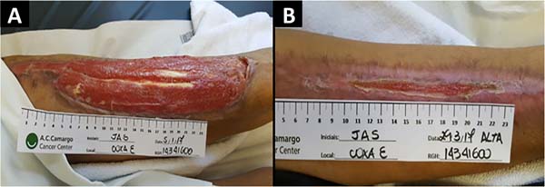
Case 1: female patient, 64 years old, smoker. She presented a dehiscence of the surgical wound in the left thigh region. Thirteen sessions of low-level laser therapy were performed; the initial lesion measuring 31x5.5cm in diameter evolved with 98% of the wound’s healing, uniform appearance, and high-quality tensile strength of the scar. Closure of the lesion at 83 days (Figure 1).

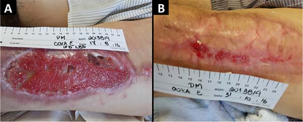
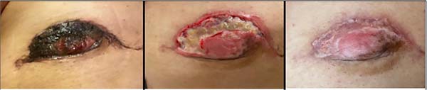
Case 2: male patient, 81 years old, with multiple skin carcinomas. He evolved with total loss of the partial skin graft in the left thigh. Low-level laser therapy was started once a week. Significant retraction of the lesion margins of up to 2 cm was observed on one of the lateral edges of the wound. There was a progressive retraction of the wound margins and complete healing in 60 days, completing 11 sessions of laser applications (Figure 2).
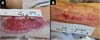
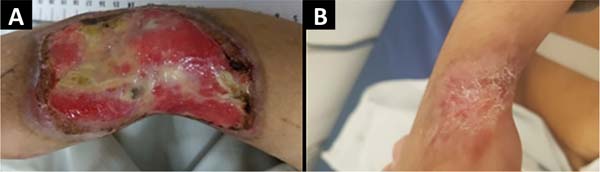
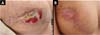
Case 3: female patient, 35 years old, presented skin necrosis after extravasation of intravenous medication in the dorsum region of the right hand with tendon exposure. Patient submitted to surgical debridement and vacuum dressing; after 1 month of negative pressure therapy, the lesion presented 80% of granulation tissue. Currently, 12 laser therapy sessions are performed twice weekly, maintaining outpatient follow-up, with complete skin wound healing in 30 days (Figure 3).

Case 4: The female patient, 34 years old, has surgical wound dehiscence in the right breast. Eighteen sessions of laser therapy were performed twice a week, initial lesion measuring 4.5x2.5cm in diameter - resolution of the skin wound in 45 days (Figure 4).
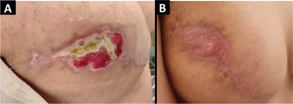
Case 5: female patient, 39 years old, evolved in the postoperative period with necrosis in the region of the nipple-areolar complex (NAC) of approximately 5 cm. After debridement of skin necrosis at the dressing outpatient clinic, she underwent seven sessions of low-level laser therapy, with wound healing in approximately 25 days of evolution (Figure 5).
DISCUSSION
The pathophysiology of laser therapy on healing is a process not fully elucidated so far. There are theories such as the absorption of light by specific proteins (porphyrins and flavoproteins) in the respiratory chain, thus increasing the concentration of intracellular oxygen and stimulating the synthesis of RNA (ribonucleic acid) and DNA (deoxyribonucleic acid)9. Another possibility would be the photoexcitation of chromophores in the cytochrome C oxidase molecule, leading to increased cellular metabolism and greater production of factors related to healing. Despite the incomplete understanding of how the laser affects the healing process, its modulating and pro-scarring effect is notably widespread and reported in the scientific literature10.
Black skin is a contraindication to the use of laser therapy in the protocol mentioned in this study. There is a dearth and limitation of information and research in the literature on lasers on darker ethnic skin types. The reason for this is the increased risk of transient and permanent side effects (e.g., blisters, depigmentation and scarring)11.
Through the institutional protocol created by nurses from the Skin Oncology Group (GOPE) at Hospital AC Camargo, we discussed the results of five cases based on laser therapy as the main form of treatment for cutaneous wounds, wounds that did not respond well to treatments with conventional dressings.
The first and second cases are about dehiscence of a donor area in the lateral face of the left lower limb, one being of a patient already with probable microcirculation alteration due to smoking, and the second of an elderly patient with comorbidities and associated atheromatous alteration in the risk factors. We noticed an important contraction of the wound, associated with a process of delicate cell proliferation, with a totally satisfactory result considering the extension and complexity of the wound. The modulatory property of laser therapy is clearly present here, given that extensive wounds, when not treated with tissue replacement such as grafts, tend to end up with gross and limiting retractions, very commonly seen in large burns.
When the evaluation focuses on treating complex lesions in institutions with little economic power, that is, the reality of the vast majority of Brazilian services, implementing a device with low operational cost can work as an adjuvant to cases that would previously only be solved with expensive maintenance dressings (such as the negative pressure dressing) and a greater number of visits to the service, making laser therapy an option to be observed with special appreciation by health managers12.
In the third case reported in the study, we found a lesion on the dorsal aspect of the wrist and right hand. A lesion that, in addition to being extensive, presents greater technical complexity of coverage with grafts due to the difficulty of keeping the region immobile for adhesion and prolonged immobilization of the hand can lead to joint impairment due to inactivity. Another point that stands out in case 3 is the short time interval until the complete resolution of the wound, as well as the wound’s quality not limiting the limb’s range of motion. The favorable characteristics related to the patient’s pathological history probably potentiated the effectiveness of the activation of the healing cascade by the laser and more sessions with shorter intervals between them.
In the fourth and fifth cases reported, healing of the patient’s breasts occurs after dehiscence and necrosis, respectively, with an abundance of fibrin, with the possibility of reconstruction loss due to prosthesis exposure. Lesions with dehiscence and necrosis are critical points in treating breast cancer, as they can delay the chain of adjuvant procedures such as radiotherapy. In this case, laser therapy demonstrated a very satisfactory healing potential, leading to the complete healing of an extensive and deep area relatively quickly and simply.
An important point to be discussed is not performing laser therapy in potentially malignant lesions. The laser seems to favor a greater substrate of genomically altered cells, indirectly accelerating the gain of additional mutations in the natural process of carcinogenesis13,14.
According to the study by Avci et al.15, low-level laser therapy is a rapidly growing technology used to treat a wide variety of conditions requiring healing stimulation, relief of pain and inflammation, and restoration of function.
Although the skin is the organ that is naturally exposed to light more than any other organ, it still responds well to red and near-infrared wavelengths. Photons are absorbed by mitochondrial chromophores in skin cells, with consequent transport of electrons, adenosine triphosphate (ATP) release of nitric oxide, increase in blood flow. Stem cells can be activated, allowing for increased tissue repair and healing15.
Pinto et al.16 conducted a study on a patient undergoing myocardial revascularization who developed a dehiscence of the surgical wound in the lower limb on the 15th postoperative day (PO). Conventional treatment was initially performed in the outpatient clinic without clinical improvement. On the 30th PO day, only laser therapy was applied around the wound edge punctually. The lesion responded with granulation tissue, decreased inflammatory process and analgesia from the first application. In this study, laser therapy was shown to play an important role as a healing facilitator through a non-invasive, effective and safe therapy16.
CONCLUSION
It is concluded that low-level laser therapy, when applied to skin wounds, suggests a beneficial, promising action and can potentially increase the therapeutic options available to the surgeon; however, as we reported five cases, further studies are needed to verify the efficiency of laser in wounds. The presence of a nursing staff trained in low-level laser therapy is a cornerstone of the entire treatment.
REFERENCES
1. Gutknecht N, Eduardo CP. A odontologia e o laser: atuação do laser na especialidade odontológica. São Paulo: Quintessence; 2004. p. 25-43.
2. Rodrigo SM, Cunha A, Pozza DH, Blaya DS, Moraes JF, Weber JB, et al. Analysis of the systemic effect of red and infrared laser therapy on wound repair. Photomed Laser Surg. 2009;27(6):929-35.
3. Chaves ME, Araújo AR, Piancastelli AC, Pinotti M. Effects of low-power light therapy on wound healing: LASER x LED. An Bras Dermatol. 2014;89(4):616-23.
4. Mester E, Korényi-Both A, Spiry T, Tisza S. The effect of laser irradiation on the regeneration of muscle fibers (preliminary report). Z Exp Chir. 1975;8(4):258-62.
5. Li S, Wang C, Wang B, Liu L, Tang L, Liu D, et al. Efficacy of low-level light therapy for treatment of diabetic foot ulcer: A systematic review and meta-analysis of randomized controlled trials. Diabetes Res Clin Pract. 2018;143:215-24.
6. Henriques ACG, Cazal C, Castro JFL. Ação da laserterapia no processo de proliferação e diferenciação celular: revisão da literatura. Rev Col Bras Cir. 2010;37(4):295-302.
7. Belkin M, Schwartz M. New biological phenomena associated with laser radiation. Health Phys. 1989;56(5):687-90.
8. Chung H, Dai T, Sharma SK, Huang YY, Carroll JD, Hamblin MR, et al. The nuts and bolts of low-level laser (light) therapy. Ann Biomed Eng. 2012;40(2):516-33. DOI: 10.1007/s10439-011-0454-7
9. Kreisler M, Christoffers AB, Willershausen B, d’Hoedt B. Low-level 809 nm GaAlAs laser irradiation increases the proliferation rate of human laryngeal carcinoma cells in vitro. Lasers Med Sci. 2003;18(2):100-3.
10. Karu TI, Pyatibrat LV, Kalendo GS. Photobiological modulation of cell attachment via cytochrome c oxidase. Photochem Photobiol Sci. 2004;3(2):211-6.
11. Battle EF Jr, Hobbs LM. Laser therapy on darker ethnic skin. Dermatol Clin. 2003;21(4):713-23.
12. Follador W, Secoli SR. A farmacoeconomia na visão dos profissionais da saúde. In: Nita ME, Campino ACC, Secoli SR, Sarti FM, Nobre M, Costa AM, et al., eds. Avaliação de tecnologias em saúde. Porto Alegre: Artmed; 2010. p. 248-68.
13. Pinheiro AL, Carneiro NS, Vieira AL, Brugnera A Jr, Zanin FA, Barros RA, et al. Effects of low-level laser therapy on malignant cells: in vitro study. J Clin Laser Med Surg. 2002;20(1):23-6.
14. Mognato M, Squizzato F, Facchin F, Zaghetto L, Corti L. Cell growth modulation of human cells irradiated in vitro with low-level laser therapy. Photomed Laser Surg. 2004;22(6):523-6.
15. Avci P, Gupta A, Sadasivam M, Vecchio D, Pam Z, Pam N, et al. Low-level laser (light) therapy (LLLT) in skin: stimulating, healing, restoring. Semin Cutan Med Surg. 2013;32(1):41-52.
16. Pinto NC, Pereira MHC, Stolf NAG, Chavantes MC. Laser de baixa intensidade em deiscência aguda de safenectomia: proposta terapêutica. Rev Bras Cir Cardiovasc. 2009;24(1):88-91.
1. A.C.Camargo Cancer Center, São Paulo, SP, Brasil
2. A.C.Camargo Cancer Center, Departamento Oncologia Cutânea, São Paulo, SP, Brasil
ACVGO Analysis and/or data interpretation, Conception and design study, Conceptualization, Final manuscript approval, Writing - Original Draft Preparation.
CLVM Contribution: Final manuscript approval, Writing - Review & Editing.
EWP Final manuscript approval, Formal Analysis, Writing - Original Draft Preparation.
KCPP Conception and design study, Final manuscript approval, Writing - Review & Editing.
PPA Conception and design study, Final manuscript approval, Writing - Review & Editing.
SYH Conception and design study, Final manuscript approval, Writing - Review & Editing.
MCAL Final manuscript approval, Project Administration, Supervision.
JPDN Final manuscript approval, Project Administration, Supervision.
Corresponding author: Ana Carolina Vasconcellos Guedes Otsuka Av. da Aclimação, 314, São Paulo, SP, Brazil. Zip code: 01531-000, E-mail: ac.otsuka@gmail.com
Article received: 2021/10/6.
Article accepted: 2022/4/7.
Conflicts of interest: none.
 Read in Portuguese
 Read in English
 PDF PT
 Print
Print
 Send this article by email
 How to Cite
 Mendeley
 Pocket