


Original Article - Year 2022 - Volume 37 -
Vascular clamp of mononylon thread in experimental microsurgery
Clamp vascular de fio de mononylon em microcirurgia experimental
Balduino Ferreira de Menezes1,* ; Murilo Sgarbi Secanho1; Matheus Scuracchio Fernandes1; Laisa Brandão Carvalho1; Fausto Viterbo1
ABSTRACT
Introduction: Reconstructive microsurgery is now an inseparable and essential branch of plastic surgery. The training is long, has a relatively high financial cost and requires a lot of the proponents. To improve this equation in favor of the formation of new microsurgeons in Brazil, it is essential to facilitate access to experimental training, using simple materials. Huaraca described a technique using a simple 5-0 mononylon thread to replace the vascular clamp, which is an indispensable instrument for microsurgical anastomosis and is generally expensive. The objective is to compare the Huaraca technique with mononylon thread and the traditional metal clamp during vascular microsurgical anastomosis.
Methods: Six Wistar rats whose both femoral arteries were randomly selected for end-to-end suture after complete section, with one side performed with usual vascular clamp and the contralateral with Huaraca technique, at the same surgical time and by the same surgeon.
Results: In both situations, the patency rate was 67% after 72 hours, with an average time of 26 minutes with the Huaraca technique and 18 minutes with the traditional clamp (p=0.001).
Conclusion: Despite the longer execution time, the Huaraca technique is a simple and low-cost measure that can replace the traditional vascular clamp.
Keywords: Reconstructive surgical procedures; Microsurgery; Anastomosis, surgical; Nylons; Learning curve; Models, animal.
RESUMO
Introdução: A microcirurgia reparadora é ramo hoje indissociável e imprescindível na cirurgia plástica. O treinamento é longo, custo financeiro relativamente alto e exige muito dos proponentes. Para melhorar essa equação a favor da formação de novos microcirurgiões no Brasil, é fundamental facilitar o acesso ao treinamento experimental, utilizando materiais simples. Huaraca descreveu uma técnica utilizando um simples fio mononylon 5-0 para substituir o clamp vascular, que é instrumento indispensável da anastomose microcirúrgica e geralmente de alto custo. O objetivo é comparar a técnica de Huaraca com fio de mononylon e o clamp metálico tradicional durante anastomose microcirúrgica vascular.
Métodos: Seis ratos da raça Wistar cujas duas artérias femorais foram aleatoriamente selecionadas para sutura término-terminal após secção completa, sendo um dos lados realizado com clamp vascular habitual e o contralateral com técnica de Huaraca, no mesmo tempo cirúrgico e pelo mesmo cirurgião.
Resultados: Em ambas as situações, a taxa de patência foi de 67% após 72 horas, sendo que o tempo médio foi de 26 minutos com a técnica de Huaraca e de 18 minutos com o clamp tradicional (p=0,001).
Conclusão: Apesar do tempo de execução mais longo, a técnica de Huaraca é medida simples e de baixo custo que pode substituir o clamp vascular tradicional.
Palavras-chave: Procedimentos cirúrgicos reconstrutivos; Microcirurgia; Anastomose cirúrgica; Nylons; Curva de aprendizado; Modelos animais
INTRODUCTION
Plastic surgery can be considered general surgery in its maximum technical refinement. The constant search for evolution and ways to make what previously did not seem possible into something feasible is one of its guidelines. Thus, microsurgery appears in this specialty, which, using magnification optics, can transfer tissues and correct defects that seem irreversible1.
In the latter case, the technical requirement is high, and the training is long. Especially in developing countries, the issue of time versus cost becomes a major obstacle2.
The complexity of these procedures is high and requires great practical refinement, with maximum precision so that errors and catastrophic losses for the patient do not occur3.
Understanding and executing each reconstruction possibility is a fundamental skill for the plastic surgeon, especially the repairer4.
Clinical training, although essential, when it can be done initially in an experimental way, is much safer and involves less risk to patients5-7.
The metallic vascular clamp is considered one of the limiting factors to the simultaneous training of more than one resident. Delicate material, normally expensive for training laboratories, easily loses its precision over time, mainly due to manipulation of students at the beginning of their training - the most common in this type of activity.
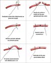
As a way of solving this type of obstacle, in 2009, at the same institution, Walter Huaraca, under the guidance of Professor Fausto Viterbo, proposed a simple and easy-to-perform technique: a single 5.0 mononylon thread to create two false knots in the proximal and distal of the area that will be sectioned for the training of end-to-end vascular suture, with traction capacity for approximation of the edges of the vessels, managing to replace the metallic vascular clamp8.
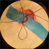
The technical sequence begins with a false knot distal to the femoral artery, with minimal local pressure, and the same thread is used for another false knot, now proximal, approximately 1 cm from the previous one. The artery is sectioned at the central point, maintaining thread tension to facilitate microsurgical anastomosis. After this, the wire clamp is removed, pulling it through the distal end. The appearance after mounting the wire clamp and before sectioning is shown in Figure 1.

OBJECTIVE
The main objective is to evaluate the replicability of the technique proposed by Huaraca in 2009, comparing the clamp made with mononylon wire with the traditional metallic clamp, demonstrating a way to reduce the cost of training without harming the evolution of the treatment.
METHODS
Six male Wistar rats available in the experimental microsurgery laboratory of the Hospital das Clínicas, Faculdade de Medicina de Botucatu were selected for end-to-end suturing of the femoral artery itself, one side being performed with the usual metallic vascular clamp and the contralateral with the technique of Huaraca, in the same surgical time and by the same surgeon. The suture sides were chosen at random. The surgeries were performed in December 2020.
Schedule
The animals’ weight, vessel diameters, number of stitches, and surgical time were measured in the experiment, and the final patency was evaluated after 30 minutes by an independent researcher.
After 72 hours of the initial procedure, another researcher reassessed the rats for weight checking and final arterial patency.
Patency assessment
The patency was considered patent when there was a sign of flow distal to the anastomosis, considering the vessel color, pulsation and according to Acland’s maneuver - performed with two clamps to assess vascular refilling in a proximal to distal direction.
Surgical technique
The standard anesthesia procedure in both groups was ketamine 80 mg/kg and xylazine 10 mg/kg intraperitoneally, followed by local anesthesia with 7 mg/kg lidocaine in the inguinal regions. This was done after weighing and shaving the inguinal region of the animals.
Intraoperatively, tramadol 5 mg/kg intramuscularly, enrofloxacin (10 mg/kg) and ketoprofen (5 mg/kg) subcutaneously were used. A dose of 100 mg/kg of sodium dipyrone subcutaneously was applied daily in the days following the initial procedure for pain control.
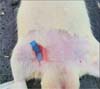
The standard incision was performed obliquely in the inguinal region of the rat, and the femoral artery was dissected as proximally as possible, where its diameter was measured at the estimated section site (Figure 2).

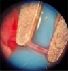
After arterial dissection, it was clamped with a wire (Huaraca technique) or a metallic clamp (Figure 3), chosen at random. Then, the artery was sectioned with microsurgical scissors, its lumen was washed with saline and had its adventitial layer was also removed with microsurgical scissors. Then, the anastomosis was performed with a 10-0 mononylon with a cylindrical needle.

The muscle and skin were closed in planes on all sides with separate stitches of 5.0 mononylon thread - the same used in the Huaraca technique.
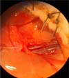
After 72 hours, the rats were weighed again, and the skin and muscle sutures were reopened to reassess vascular patency also by direct observation of the flow (Figure 4), and the skin and muscle were resutured with black 5.0 mononylon stitches.

The technique for making the wire clamp proposed by Huaraca is outlined in Figure 5.

RESULTS
The six rats selected in the study were between 60 and 90 days old, with a weight variation between 242 and 307 g, with an average of 270.8 g. After 72 hours, they had an average weight gain of 3 g.
The vessels had a diameter of 0.9 to 1 mm, with a mean of 0.95 mm on the right and 0.93 mm on the left.
The number of sutures per anastomosis ranged from 5 to 6 on both sides, with a mean of 5.6 on the right and 5.5 on the left.
The time to perform the anastomosis included developing the Huaraca technique, both for the passage of the stitches and creation of the wire clamp and the final removal to check for patency and bleeding. This execution time added 3 to 5 minutes to the final procedure, with an average of 4.16 minutes.
The mean anastomosis time with the Huaraca technique was 26.8 minutes versus 18.83 minutes using the conventional metallic clamp (p=0.001). The number of points per minute was 0.26 with the former and 0.25 with the latter (p=0.85).
The failure rate (thrombosis) - absence of final patency after 72 hours - was 33% in both groups (p=1.00).
Some of these data are summarized in Tables 1 and 2 below:
| PATENCY | ||||
|---|---|---|---|---|
| Huaraca | Metal | p | ||
| DIAMETER | AVERAGE | 0.95 | 0.93 | 0.60 |
| SD | 0.05 | 0.05 | ||
| TIME | AVERAGE | 26.83 | 18.83 | 0.001 |
| SD | 3.66 | 2.40 | ||
| POINT/MIN | AVERAGE | 0.26 | 0.25 | 0.85 |
| SD | 0.08 | 0.04 | ||
SD: Standard Deviation.

| HUARACA | Metal | p |
|---|---|---|
| 0.33 | 0.33 | 1.00 |

DISCUSSION
Microsurgery has multiple clinical applications: useful in the treatment of severe trauma in which the simplest reconstructive stair and elevator methods are not sufficient, such as local grafts and flaps, especially when there are large fractures and exposure of noble structures; essential in limb reimplantation, especially fingers; fundamental tool in large head and neck tumors, in which it can enable the patient to carry out their usual activities.
Training is essential, and the procedure’s success will hardly be achieved with simple trial and error directly in the clinic since lost flaps cause great harm to the patient, especially to the donor area, which cannot be reversed in some cases. Therefore, each free flap must be meticulously performed, with close to 100% average success rates.
The technique proposed by Huaraca is very useful when it is observed that the thread used is the same that will be needed for the final suture of the rat, as well as it is low cost, easily found, and with a small learning curve for its execution. This method is especially important when the number of surgeons undergoing simultaneous training requires multiple metal clamps.
Live animals are considered the gold standard in this type of training since it creates a simulation very close to humans’ practice. However, the ethical problems involved demand replacement, whenever possible, with other materials, such as synthetic or cadaveric ones9. Our choice was based on the availability of these animals in our institution and the more realistic nature of the procedure.
Several studies have shown that the ideal is to reach patencies around 95% on an experimental basis before starting the clinical treatment of patients. We consider our patency rate satisfactory since the average diameter of the vessels covered in our study is also lower in percentage than many experiments with rates close to 100%10.
The average time to perform the anastomosis varies in the literature. The average anastomosis time in our study was considered satisfactory and shorter than many other previously published studies, but with fewer stitches per anastomosis11.
The patencies reached were slightly lower than the rates found in the literature for this type of end-to-end arterial anastomosis12-15.
CONCLUSION
As proposed by Huaraca, the wire clamp proves to be a safe alternative to the traditional metallic clamp, being a useful tool in the daily routine of the experimental laboratory in microsurgery, expanding the range of possibilities for surgeons in the learning phase.
Despite increasing the surgical time, it has good applicability concerning the gold standard, which is more expensive and can facilitate training in various medical residency services in Brazil, encouraging continued and more comprehensive education in this noble area of Medicine.
REFERENCES
1. http://www.scielo.br/scielo.php?script=sci_arttext&pid=S1983-51752012000100002&lng=en&nrm=iso https://doi.org/10.1590/S1983-51752012000100002
2. http://www.scielo.br/scielo.php?script=sci_arttext&pid=S0102-86502002000300008&lng=en&nrm=iso http://dx.doi.org/10.1590/S0102-86502002000300008
3. Acland RD. Manual Prático de Microcirurgia. São Paulo: Rocca; 1981.
4. Lascar I, Totir D, Cinca A, Cortan S, Stefanescu A, Bratianu R, et al. Training program and learning curve in experimental microsurgery during the residency in plastic surgery. Microsurgery. 2007;27(4):263-7. DOI: 10.1002/micr.20352
5. Daniel RK, Entin MA. Practical microsurgery. Clin Plast Surg. 1976;3(1):39-47.
6. Lima DA, Galvão MSL, Cardoso MM, Leal PRA. Rotina de treinamento laboratorial em microcirurgia do Instituto Nacional do Câncer. Rev Bras Cir Plást. 2012;27(1):141-9.
7. Karl P, Tilgner-Peter A. Mikrogefäss- und Nervenchirurgie im Tierexperiment und ihre Bedeutung für die plastische Chirurgie [Experimental microsurgery of blood vessels and nerves in laboratory animals and their importance for plastic surgery]. Z Exp Chir. 1979;12(6):368-78. German.
8. Huaraca W. Nueva técnica de clampeamiento de arterias con mononylon 5.0. en microcirurgía. Estudio experimental em ratones. Rev Univ Guayaquil. 2010;108(3):27-30.
9. Javid P, Aydın A, Mohanna PN, Dasgupta P, Ahmed K. Current status of simulation and training models in microsurgery: A systematic review. Microsurgery. 2019;39(7):655-68. DOI: 10.1002/micr.30513 Epub 2019 Sep 12 PMID: 31513303
10. http://www.scielo.br/scielo.php?script=sci_arttext&pid=S0102-86502014001400002&lng=en https://doi.org/10.1590/S0102-86502014001400001
11. Cigna E, Curinga G, Bistoni G, Spalvieri C, Tortorelli G, Scuderi N. Microsurgical anastomosis with the ‘PCA’ technique. J Plast Reconstr Aesthet Surg. 2008;61(7):762-6. DOI: 10.1016/j.bjps.2008.04.003 Epub 2008 May 12 PMID: 18468969
12. Naides A, Noland R, Lu JG, Akelina Y, Marboe C, Strauch RJ. Histological Changes in the Rat Femoral Artery Following the Use of the Empty-and-Refill Test. J Reconstr Microsurg. 2018;34(4):270-6. DOI: 10.1055/s-0037-1621727
13. Adams WP Jr, Ansari MS, Hay MT, Tan J, Robinson JB Jr, Friedman RM, et al. Patency of different arterial and venous end-to-side microanastomosis techniques in a rat model. Plast Reconstr Surg. 2000;105(1):156-61. DOI: 10.1097/00006534-200001000-00026
14. Barrera-Ochoa S, Gallardo-Calero I, López-Fernández A, Romagosa C, Vergés R, Aguirre-Canyadell M, et al. Effect of Previous Irradiation on Vascular Thrombosis of Microsurgical Anastomosis: A Preclinical Study in Rats. Plast Reconstr Surg Glob Open. 2016;4(11):e1073. DOI: 10.1097/GOX.0000000000001073 PMID: 27975009 PMCID: PMC5142475
15. Salgarello M, Lahoud P, Selvaggi G, Gentileschi S, Sturla M, Farallo E. The effect of twisting on microanastomotic patency of arteries and veins in a rat model. Ann Plast Surg. 2001;47(6):643-6. DOI: 10.1097/00000637-200112000-00011 PMID: 11756835
1. Hospital das Clínicas, Faculty of Medicine of Botucatu, Department of Plastic Surgery,
Botucatu, SP, Brazil.
| COLLABORATIONS | |
|---|---|
| BFMN | Analysis and/or data interpretation, Conception and design study, Data Curation, Final manuscript approval, Formal Analysis, Methodology, Project Administration, Realization of operations and/or trials, Writing - Original Draft Preparation, Writing - Review & Editing |
| MSS | Analysis and/or data interpretation |
| MSF | Realization of operations and/or trials |
| LBC | Data Curation |
| FV | Supervision |

Corresponding author: Balduino Ferreira de Menezes Neto, Avenida Professor Mário Rubens Guimarães Montenegro, S/N - Jardim São Jose, Botucatu - SP, Brazil, Zip Code 18618-970, E-mail: balduinofmneto@gmail.com
Article received: January 27, 2021.
Article accepted: April 23, 2021.
Conflicts of interest: none.
 Read in Portuguese
 Read in English
 PDF PT
 Print
Print
 Send this article by email
 How to Cite
 Mendeley
 Pocket