


Case Report - Year 2019 - Volume 34 -
Use of the "spaghetti" technique for surgical treatment of lentigo maligna
Uso da técnica em "spaghetti" para tratamento cirúrgico do lentigo maligno
ABSTRACT
Lentigo maligna (LM) is a melanoma in situ that commonly presents as a macula with progressive and irregularly pigmented growth, especially in the face of elderly people with sun-damaged skin. This melanoma in situ has a risk (30-50%) of progression to lentigo maligna melanoma. Complete surgical excision of the lesion requires margins of at least 10 mm, even for lesions in situ. However, when the growth of LM occurs in areas of aesthetic or functional implications (face, neck, and soles), the excision is often reduced to preserve important anatomic structures and for cosmetic purposes. Moreover, the peripheral margins may be clinically ill-defined and not always pigmented, and thus, such cases are associated with underestimated extension and risk of insufficient resection. The "spaghetti" technique, described by Gaudy Marqueste, is a strategic surgical approach based on sampling of a range of "spaghetti-like" strips to determine the margins of the lesion prior to removal of the tumor. After the pathological confirmation of neoplasia-free margins, the main central lesion is resected, allowing reconstruction of the defect in the same procedure, as an alternative to Mohs micrographic surgery.
Keywords: Lentigo; Melanoma; Excision Margins; Reconstructive surgical procedures; Nose
RESUMO
O lentigo maligno (LM) é uma forma de melanoma in situ que mais comumente se apresenta como uma mácula de crescimento lentamente progressivo, pigmentada, na face de idosos com pele danificada pelo sol. Esse melanoma in situ tem um risco (30% a 50%) de progressão para lentigo maligno melanoma. A excisão cirúrgica completa da lesão requer margens de pelo menos 10mm, mesmo para lesões in situ. Porém, quando o crescimento de LM ocorre em áreas de implicações estéticas ou funcionais (face, pescoço, solas), a excisão é frequentemente reduzida para preservar estruturas anatômicas importantes e por razões cosméticas. Além disso, as margens periféricas podem ser clinicamente mal definidas e nem sempre pigmentadas, com extensão subestimada e risco de ressecção insuficiente. A "técnica de espaguete", descrita por Gaudy Marqueste, é uma cirurgia estratégica baseada na amostragem de uma faixa de tecido "spaghetti-like" para determinar as margens da lesão antes da remoção do tumor. Após a confirmação anatomopatológica de margens livres de neoplasia, a lesão principal central é ressecada, permitindo a reconstrução do defeito no mesmo procedimento, sendo uma alternativa à cirurgia micrográfica de Mohs.
Palavras-chave: Lentigo; Melanoma; Margens de excisão; Procedimentos cirúrgicos reconstrutivos; Nariz
INTRODUCTION
Lentigo maligna (LM) is a melanoma in situ that commonly presents as a macula with progressive and irregularly pigmented growth, especially in the face of elderly people with sun-damaged skin1. The risk of progression to lentigo maligna melanoma varies from 30-50% in this type of lesion2.
Treatment options for LM include surgical excision with oncologic margins or non-surgical treatments such as cryotherapy, curettage, electrocautery, laser, radiotherapy, fluorouracil, and imiquimod. However, these non-surgical methods have high recurrence rates (20-100%)3.
Complete surgical excision of the melanoma in situ requires circumferential margins of at least 5 mm4. In cases of LM in areas with substantial aesthetic or functional implications (face, neck, and soles of the feet), surgical margins are usually reduced to preserve important anatomic structures. In addition, clinical margins of the LM may be poorly defined and not always pigmented, and thus, such cases are associated with underestimated extension, which increases the risk of inadequate resection,5 and the appropriate treatment is challenging.
The “spaghetti technique”, described by Gaudy Marqueste, is a surgical technique based on the sampling of a narrow band of tissue just beyond the lesion to determine the surgical margins before resection of the tumor lesion. This tissue band is referred for conventional histopathological examination and, if the margins are free of tumor tissue, the central lesion is then resected, minimizing tissue loss; thus, being an alternative to Mohs micrographic surgery5.
CASE REPORT
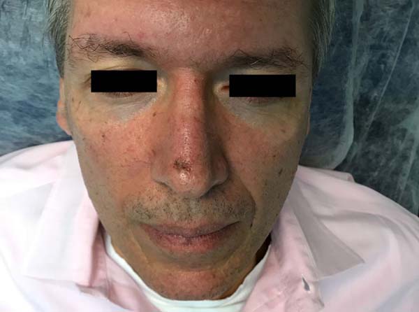
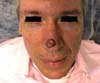
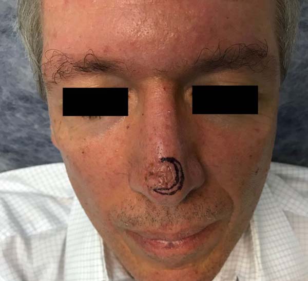
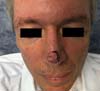
The medical records of patient M.B.M.C., a 54-year-old man with skin phototype III on the Fitzpatrick scale, who presented with a pigmented lesion with irregular edges and coloration and progressive growth located on the nasal tip of approximately 1.5 cm in diameter, were reviewed (Figure 1).

The patient underwent incisional biopsy of the lesion in April 2018 with a diagnosis of melanoma in situ type lentigo maligna.
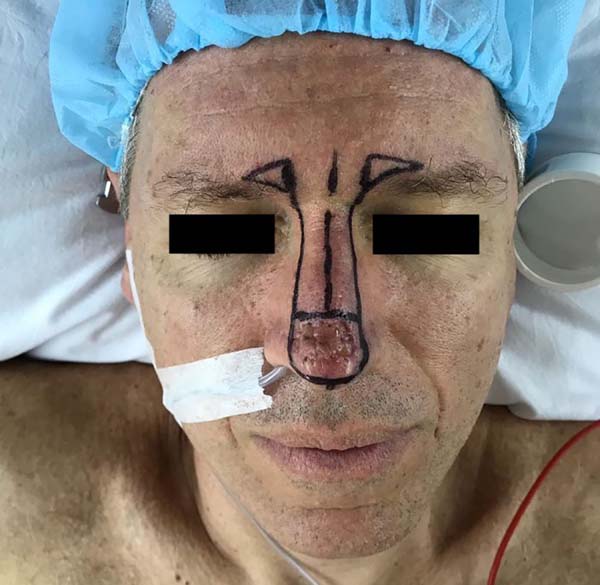
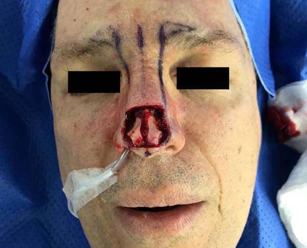
The “spaghetti technique” was then used to determine the surgical margins before complete resection of the lesion (Figure 2). Tumor limits were defined with the aid of confocal dermoscopy and subsequently a band of skin beyond the tumor with a margin of 1 mm was resected (Figures 3 and 4).


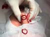

The surgical piece was marked with a nylon 5.0 suture at the 12 o’clock position to guide the histopathological examination, which subdivided it into 4 quadrants (12 - 3 hours, 3 - 6 hours, 6 - 9 hours, and 9 - 12 hours).
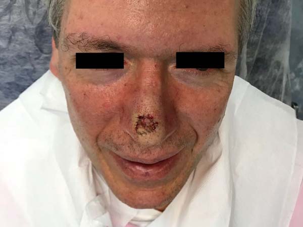
The result of the examination revealed neoplastic involvement in the margins of the 3 to 6 o’clock quadrant. The margins were enlarged, and the material was forwarded again for analysis (Figure 5).
The resection of the central lesion was performed after 15 days of expansion of margins that proved to be free of neoplastic involvement by histopathological examination. During the resection, the scar tissue peripheral to the central lesion was encompassed and local reconstruction was performed with a Rintala flap during the same surgery (Figures 6 to 8).


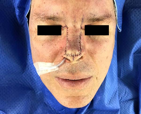

The patient recovered well, without any signs of recurrence during a six-month follow-up period.
DISCUSSION
The “spaghetti technique” is an easy and safe method to control surgical margins in the case of lentigo maligna, especially when the lesion is in an area where there is a greater risk of impairment of vital structures.
One of the precautions that must be taken during the resection of the tumor lesion is englobing the scar area from previous surgeries to prevent local recurrence.
The skin suturing after resection of the circumferential margin in the «spaghetti» technique should be done in a careful manner with minimal passage of the needle to the healthy tissue.
Other techniques, such as the “square” technique and the “perimeter” technique, have used the concept of pathological control of margins before total resection of LM. In these techniques, a geometric resection shape (square, triangle, or pentagon) is determined to facilitate the analysis of margins while maintaining the central lesion intact. The objective is to verify the periphery of this geometric figure before resection6,7.
The advantage of the «spaghetti» technique is the higher preservation of adjacent tissues when compared to that with the other techniques mentioned.
Mohs micrographic surgery is also an option in these cases, but a trained team must perform the operation. In addition, by using paraffin blocks, the “spaghetti” technique is more reliable than those techniques using freezing5.
The disadvantages of the technique consist of the requirement of at least two surgical operations, which may increase the risk of complications of the surgical wound. Conversely, it may be more comfortable for the patient, since it prevents the permanence of open wounds and allows the removal of the tumor with immediate reconstruction. Confocal dermoscopy is a tool that can be used to assist the delimitation of the tumor margins, but may not be accessible.
In this case, the “spaghetti” technique proved to be a good option in the treatment of the LM as a simple and reproducible method, ensuring the control of margins and lower morbidity for the patient.
COLLABORATIONS
|
GSS |
Analysis and/or data interpretation, conception and design study, data curation, writing - original draft preparation, writing - review & editing. |
|
FBH |
Writing - review & editing. |
|
CHSTS |
Writing - review & editing. |
|
LADS |
Writing - review & editing. |
|
FHSP |
Supervision, writing - review & editing. |
|
IDAOSF |
Supervision, writing - review & editing. |
|
CSS |
Supervision, writing - review & editing. |
|
ERB |
Conception and design study, final manuscript approval, supervision, writing - review & editing. |
REFERENCES
1. Huilgol SC, Selva D, Chen C, Hill DC, James CL, Gramp A, et al. Surgical margins for lentigo maligna and lentigo maligna melanoma: the technique of mapped serial excision. Arch Dermatol. 2004;140(9):1087-92. PMID: 15381549 DOI: https://doi.org/10.1001/archderm.140.9.1087
2. Weinstock MA, Sober AJ. The risk of progression of lentigo maligna to lentigo maligna melanoma. Br J Dermatol. 1987;116(3):303-10. PMID: 3567069 DOI: https://doi.org/10.1111/j.1365-2133.1987.tb05843.x
3. Cohen LM. Lentigo maligna and lentigo maligna melanoma. J Am Acad Dermatol. 1997;36(6 Pt 1):913.
4. Zitelli JA, Brown CD, Hanusa BH. Surgical margins for excision of primary cutaneous melanoma. J Am Acad Dermatol. 1997;37(3 Pt 1):422-9. PMID: 9308558
5. Gaudy-Marqueste C, Perchenet AS, Taséi AM, Madjlessi N, Magalon G, Richard MA, et al. The "spaghetti technique": an alternative to Mohs surgery or staged surgery for problematic lentiginous melanoma (lentigo maligna and acral lentiginous melanoma). J Am Acad Dermatol. 2011;64(1):113-8. PMID: 21167406 DOI: https://doi.org/10.1016/j.jaad.2010.03.014
6. Johnson TM, Headington JT, Baker SR, Lowe L. Usefulness of the staged excision for lentigo maligna and lentigo maligna melanoma: the "square" procedure. J Am Acad Dermatol. 1997;37(5 Pt 1):758-64.
7. Mahoney MH, Joseph M, Temple CL. The perimeter technique for lentigo maligna: an alternative to Mohs micrographic surgery. J Surg Oncol. 2005;91(2):120-5. PMID: 16028282 DOI: https://doi.org/10.1002/jso.20284
1. Hospital dos Defeitos da Face, Cruz Vermelha de
São Paulo, São Paulo, SP, Brazil
2. A.C. Camargo - Cancer Center, São Paulo, SP,
Brazil.
Corresponding author: Gabriela Suemi Shimizu Rua Sabará, nº 210 - Higienópolis - São Paulo, SP, Brazil Zip Code 01239-010 E-mail: gabrielashimizu@gmail.com
Article received: August 6, 2018.
Article accepted: November 11, 2018.
Conflicts of interest: none.
 Read in Portuguese
 Read in English
 PDF PT
 Print
Print
 Send this article by email
 How to Cite
 Mendeley
 Pocket