


Case Report - Year 2015 - Volume 30 -
Use of a carbon dioxide laser in the treatment of delayed polymethylmetacrylate reaction in the face: a case report
Uso do laser de CO2 no tratamento da reação tardia de polimetilmetacrilato na face: relato de caso
ABSTRACT
The use of synthetic implants as dermal facial fillers may cause adverse reactions, which may occur owing to inappropriate techniques or the intrinsic nature of the product. Polymethylmetacrylate (PMMA) microspheres is one of the materials used. This is a report of a case of nodular reaction in the face of a female patient, after 15 years of Artecoll® implant use, a product composed of microspheres of bovine collagen and PMMA. She was treated with a carbon dioxide laser. The delayed effect caused by the application of synthetic implants in the face, such as Artecoll®; the limiting factors of conventional resection with a scalpel and scissors; and the option for the use of carbon dioxide laser are outlined. Highlighted are the ablative and precise functions in the removal of compromised tissue, the preservation of healthy and noble tissues, the high hemostatic potential, and low morbidity, with smaller scars, edema, ecchymosis, and rapid postoperative recovery.
Keywords: Laser; Adverse reaction; Polymethylmetacrylate.
RESUMO
O uso de implantes sintéticos para o preenchimento na face pode acarretar reações adversas. Estas podem ocorrer da má técnica ou decorrentes do produto. Entre os materiais utilizados, está o polimetilmetacrilato (PMMA) na forma de microesferas. Trata-se do relato de um caso de reação nodular na face de paciente do sexo feminino, após 15 anos do uso de implante de Artecoll®, produto composto por microesferas de colágeno bovino e PMMA. Foi tratada com laser de gás carbônico. Evidenciou-se o efeito tardio provocado da aplicação de implantes sintéticos na face como o Artecoll®, os fatores limitantes para ressecção convencional com bisturi e tesoura, e a opção da utilização do laser de CO2. Destacando-se a função ablativa e precisa na remoção do tecido comprometido, a preservação dos tecidos sadios e nobres, o grande potencial hemostático e a baixa morbidade, com menor cicatriz, edema, equimose e a rápida recuperação pós-operatória.
Palavras-chave: Laser; Reação adversa; Polimetilmetacrilato.
Several synthetic products have been used for facial filling, in conjunction with rhytidoplasties, for atrophy, scars, and facial irregularities, with the objective of restoring lost volume. Usually, the filler products are injectable or introduced surgically. Depending on the characteristics of the product, including chemical composition and degradation, the materials can have permanent effects or induce side effects such as migration, formation of granulomas, and allergic reactions, both immediate and in the long term1-3
The most commonly used synthetic products are dimethylsiloxane, polyacrylamide, and polymethylmetacrylate (PMMA). Polymethylmetacrylate (PMMA), commercially known as Artecoll®, which is used in this case, was developed in Germany in 1994 and consists of 20% homogeneous polymethylmetacrylate microspheres in a solution with 3.5% partially denatured bovine collagen and 0.3% lidocaine. Its microspheres measure from 30 to 40 microns in size. It is used as an injectable microimplant for subdermal filling for facial wrinkles and connective tissue defects2. According to Vargas et al.1, in a study conducted on adverse reactions to filler materials in 31 patients, the so-called permanent fillers may cause complications and deformities of varying degrees, and thus should be used cautiously. The follow-up for delayed complications is long and difficult, both for the patient and the plastic surgeon. This author described a conservative treatment method as the first choice, reserving the surgical indications for cases with anatomic deformities, tissue necrosis, visible nodules, and subcutaneous granulomas.
Christensen et al.4 published a study on adverse reactions to filler products, describing reactions to PMMA after 6 years and to silicone gel after 28 years of application. The removal of foreign material in the face presents a risk of lesions of noble structures, such as the terminal branches of the facial nerve. In addition, the removal of lesions with imprecise limits with a scalpel or scissors, or the complete resection of the nodule or granuloma can be risky or impossible and even worsen the irregularities in the atrophic face. The use of laser in the ablative mode is a common practice in plastic surgery procedures and in other fields such as ophthalmology. It offers, among other advantages, accurate incisions associated with better hemostasis, the absence of bleeding in the surgical edges, smaller and eutrophic scars, and attenuation of edema and ecchymosis. It also provides a better approach to naturally difficult areas when treated with conventional dissection methods, which is the intended goal in facial procedures5-7.
The purpose of this report is to describe a case of the use of a carbon dioxide laser in the treatment of delayed foreign-body reaction caused by Artecoll®, polymethylmetacrylate (PMMA), and bovine collagen, which were used as facial fillers in a patient with idiopathic facial atrophy.
CASE REPORT
A 57-year-old female patient with idiopathic facial atrophy was admitted to our hospital. She reported receiving facial filling with Artecoll® 15 years ago, with good immediate progression. Five years prior to her visit, a relapse was noticed, with clear facial atrophy and six nodulations on the face located in the nasogenian region, four on the right and two on the left. The nodules were, on average, 1.2 cm in diameter and were palpable in the aforementioned anatomical regions but without signs of infection or inflammation (Figure 1).

Figure 1. Above, appearance 15 years ago, before the implantation of Artecoll®. Below, in the immediate post-implantation stage.
Surgical Technique
Antisepsis was performed under local alcoholic chlorhexidine anesthesia with 2% lidocaine associated with adrenaline 1:200,000 IU. Incision was performed by using a No. 15 scalpel blade, addressing only the dermis in the affected regions. Dissection was performed under direct visualization with a carbon dioxide Ultrapulse 5000 laser (Coherent Medical Group, Palo Alto, California, USA), using a 0.2-mm focal point and piece for dissection and resection of two tumor growths located in the deep dermis, measuring 1.2 cm in diameter on average (Figures 2 and 3). The use of the carbon dioxide ultrapulse laser at 250 MJ and 20 watts in ultrapulse mode, with a pulse duration of 300 ms and interval of 900 ms, prevented the thermal effect on the neighboring tissues. The evaluation of the withdrawal of the material was by direct visualization because Artecoll® is pearl white and therefore totally distinct from other tissues of the face by direct visualization.
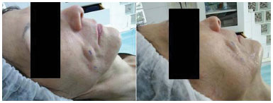
Figure 2. Preoperative aspect of the patient with two nodules in each nasogenal sulcus, marked in blue for incision of the skin, before anesthetic infiltration.

Figure 3. LASER application technique showing the end piece and the light incidence; the bidigital maneuver for projection of the implanted material; implanted material being externalized in the face; comparative example measuring 1.2 cm.
These lesions were totally removed and presented a pseudocapsule and filling with whitish material composed of polymethylmetacrylate. The two other lesions with imprecise dissection limits located in the deep dermis were vaporized with a collimated 3-mm diameter end piece for 2 minutes. The skin was sutured with nylon 6-0 in simple separated sutures, which were removed on the seventh postoperative day. A satisfactory evolution was observed 2 weeks after the surgical procedure and at 6 months of follow-up (Figure 4).

Figure 4. Front view and profile of the immediate postoperative period and after 1 week.
DISCUSSION
Implantable synthetic materials are used in the deep dermis for facial filling and may present adverse effects related to the form of application or the intrinsic characteristics of the product1-4,8. Cohen and Holmes9 found that among 251 subjects who received application of Artecoll® during 5 years, 2.2% had complications. Since its introduction on the market in 1994, a 0.01% poor progression rate has been reported. The most common complications described were sclerosing granuloma located mainly in the nasogenian ridges. More recently, formation of biofilm and proliferation of microorganisms such as Candida albicans when used in the form of bone cement10 have also been discussed. On histological examination, granulomas are differentiated from nodules and reveal an infiltrate of macrophages, giant cells, fibroblasts, and collagen fibers, but with few inflammatory cells. In this case report, we identified solid lesions with a nodular aspect.
According to the authors, these products are composed of non-biodegradable microspheres that are polymerized and encapsulated to prevent the displacement of the material. Normally, they do not cause intense foreign-body reactions; however, their clinical appearance and permanence can be lengthy or definitive. The time between the injection and the first foreign-body reaction is, in general, between 6 and 24 months, but some reports indicate that onset occurs 10 years after its application, as was described in the present case.
The pathogenesis of these reactions is still unknown8. The carbon dioxide ultrapulse laser at 250 MJ and 20 watts, with a pulse duration of 300 ms and interval of 900 ms, as used, prevented the thermal effect in neighboring tissues. With these parameters, the laser led to the vaporization of tissue and by direct visualization, for Artecoll®, which presents PMMA microspheres (20%/ volume), 30-50 micron diameter, suspended in 3.5% of bovine collagen solution and water (80%/volume) in its formulation, which was therefore possible to be vaporized by the laser. The fact that it is absorbed mainly by water does not mean that it has no action in other areas, which is also useful for vaporizing benign and/or malignant tumors11, cartilage, bones, and other tissues, by direct action. Moreover, in a study on thermal damage caused in fresh cadavers using the conventional electric scalpel and carbon dioxide laser, it was observed that the laser causes less deep thermal damage12.
Laser is an acronym that stands for light amplification by stimulated emission of radiation. The laser radiation uses the wavelengths of the invisible and visible electromagnetic spectrum13 for outpatient surgical or medical treatments. The physical properties of the lasers are reflection, ability of the laser beam to focus on the tissue and reflect the boundary layer that depends on the optical property, and vascularization of the tissue; absorption, defined as the ability of radiation to be converted into heat, causing a local increase in temperature and, depending on the heat produced, the tissue will coagulate or vaporize. The absorption capacity is closely linked to the presence of chromophores in tissues, among these hemoglobin and water contained in the tissue that absorbs 90% of the light from the carbon dioxide laser. The density of energy absorbed determines the direct action of the laser on the tissue photovaporization or carbonization.
The ablative and vaporization functions of lasers are applied in several medical fields, including blepharoplasty in plastic surgery5-7. In the present case, the authors demonstrated the application of this technique to the resection of the nodules and for the vaporization of lesions where resection was not possible. Since 2006, the 120-watt laser with a collimated beam has been available. The application of a carbon dioxide laser has become the treatment of choice for small oral tumors and oropharyngeal carcinomas14. Animal models and the demonstration of practical proof have been useful in the evaluation of the characteristics of the laser, including ablation rates, efficacy of ablation with power adjustment, hemostatic properties, extension in the tissue, and their practical application in urologic, ophthalmic, plastic, and other surgeries13. The laser was operated at a wavelength that transforms solid lesions into vapor. The technique used in this case was ablative for removal of the delimited lesions and photo-selective vaporization on lesions with imprecise limits.
Given the aforementioned findings, we call attention to the delayed effect caused by the application of synthetic implants in the face, the limiting factors for conventional resection with scalpel and scissors, and the option of using the carbon dioxide laser. We highlight the ablative and precise function in removal of compromised tissue, the preservation of healthy and noble tissues, the high hemostatic potential, and low morbidity, with smaller scars, edema, ecchymosis, and rapid postoperative recovery.
REFERENCES
1. Vargas AF, Amorim NG, Pitanguy I. Complicações tardias dos preenchimentos permanentes. Rev Bras Cir Plást. 2009;24(1):71-81.
2. Franco T, Passy S, Correa WEM. Preenchimentos. Rev Soc Bras Cir Plást. 2005;20(3):190-3.
3. Rosa SC, Macedo JLS. Reações adversas a substâncias de preenchimento subcutâneo. Rev Soc Bras Cir. Plást. 2005;20(4):248-52.
4. Christensen L, Breiting V, Janssen M, Vuust J, Hogdall E. Adverse reactions to injectable soft tissue permanent fillers. Aesthetic Plast Surg. 2005;29(1):34-48. DOI: http://dx.doi.org/10.1007/s00266-004-0113-6
5. Seckel BR, Cetrulo CL Jr, Wang KK, Hagan RR. Cutaneous Healing of CO2 Laser Incisions. Presented at the Annual Meeting of the American Society for Laser Medicine and Surgery, San Diego, USA; 1998.
6. Seckel BR, Kovanda CJ, Cetrulo CL Jr, Passmore AK, Meneses PG, White T. Laser blepharoplasty with transconjunctival orbicularis muscle/septum tightening and periocular skin resurfacing: a safe and advantageous technique. Plast Reconstr Surg. 2000;106(5):1127-41. DOI: http://dx.doi.org/10.1097/00006534-200010000-00024
7. Kang DH, Koo SH, Choi JH, Park SH. Laser blepharoplasty for making double eyelids in Asians. Plast Reconstr Surg. 2001;107(7):1884-9. DOI: http://dx.doi.org/10.1097/00006534-200106000-00040
8. Lemperle G, Romano JJ, Busso M. Soft tissue augmentation with artecoll: 10-year history, indications, techniques, and complications. Dermatol Surg. 2003;29(6):573-87. DOI: http://dx.doi.org/10.1097/00042728-200306000-00004
9. Cohen SR, Holmes RE. Artecoll: a long-lasting injectable wrinkle filler material: Report of a controlled, randomized, multicenter clinical trial of 251 subjects. Plast Reconstr Surg. 2004;114(4):964-76. DOI: http://dx.doi.org/10.1097/01.PRS.0000133169.16467.5F
10. Queiroz JR, Fissmer SF, Koga-Ito CY, Salvia AC, Massi M, Sobrinho AS, et al. Effect of diamond-like carbon thin film coated acrylic resin on candida albicans biofilm formation. J Prosthodont. 2013;22(6):451-5. DOI: http://dx.doi.org/10.1111/jopr.12029
11. Teixeira V, Reis JP, Tellechea Ó, Vieira R, Figueiredo A. Verruciform xanthoma: report of two cases. Dermatol Online J. 2012;18(5):10.
12. Hanby DF, Gremillion G, Zieske AW, Loehn B, Whitworth R, Wolf T, et al. Harmonic scalpel versus flexible CO2 laser for tongue resection: a histopathological analysis of thermal damage in human cadavers. World J Surg Oncol. 2011;9:83. DOI: http://dx.doi.org/10.1186/1477-7819-9-83
13. Teichmann HO, Herrmann TR, Bach T. Technical aspects of lasers in urology. World J Urol. 2007;25(3):221-5. DOI: http://dx.doi.org/10.1007/s00345-007-0184-5
14. Jerjes W, Hamdoon Z, Hopper C. CO2 lasers in the management of potentially malignant and malignant oral disorders. Head Neck Oncol. 2012;4:17. DOI: http://dx.doi.org/10.1186/1758-3284-4-17
1. Sociedade Brasileira de Cirurgia Plástica, São Paulo, SP, Brazil
2. Sociedade Brasileira de Laser em Medicina e Cirurgia, São Paulo, SP, Brazil
3. Universidade do Vale do Paraíba, São José dos Campos, SP, Brazil
4. Clínica Lucano de Cirurgia Plástica, São Paulo, SP, Brazil
5. Santa Casa de Misericórdia de São Paulo, São Paulo, SP, Brazil
6. Hospital Sarah Brasília, Brasília, DF, Brazil
7. Universidade Santa Cecília, Santos, SP, Brazil
Institution: Clínica Lucano de Cirurgia Plástica, São Paulo, SP, Brazil.
Corresponding author:
Claudio Roncatti
Rua Alameda dos Guaramomis, 460, Moema
São Paulo, SP, Brazil Zip Code 04076-010
E-mail: cursos@clinica-lucano.com.br
Article received: June 5, 2012.
Article accepted: May 19, 2013.
 Read in Portuguese
 Read in English
 PDF PT
 Print
Print
 Send this article by email
 How to Cite
 Mendeley
 Pocket